Abstract
Abstract 4108
In Philadelphia-negative chronic myeloproliferative neoplasms (MPN) increased microvascular density, bizarre vessel architecture and increased number of pericytes are distinct histopathological features; apart from the characteristic proliferation of myeloid cell lines and the progressive accumulation of connective tissue in the bone marrow. Pericytes express several markers such as CD146, CD271, Smooth Muscle Actin (SMA), Desmin, Platelet-derived growth factor receptor beta and Neuron-glial 2. However, these markers are also expressed in other cell types of which some are related to vascular structures. Immunofluorescence labelling is the golden standard for detection of co-expressed cellular antigens, but due to the crowded cellular environment in bone marrow and excessive autofluorescence, identification of cell types by light microscopy is preferred.
This is a methodological study aiming to identify pericyte marker profiles by light microscopy in bone marrow biopsies, contributing to our understanding of the pathogenesis of MPN.
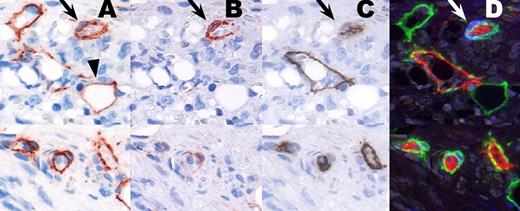
Formalin fixed, decalcified and paraffin-embedded blocks of bone marrow trephine specimens from normal donor (n=1) and patients with primary myelofibrosis (PMF) (n=3) were included. Specimens were subjected to an immunohistochemical sequential multi-labelling and erasing technique (SE-technique), inspired by the work of Glass et al. 2009 (J Histochem Cytochem). Briefly, antigens of interest in the first and/or second layer were detected with an immunoperoxidase system and visualised with Amino-Ethyl-Carbazole (AEC). After imaging, erasing of AEC with 96% alcohol and blocking of immunoreagents, the slides were stained with a traditional double immuno-labelling procedure. We successfully applied up to four layers of antibodies using CD146, SMA, CD34, CD271, and Ki67 in different combinations; either displayed as single or single followed by traditional double sequential staining runs (figure 1). In addition to the conventional light microscopy analysis we applied a Photoshop color palette, where the different immunohistochemical reactions in the staining sequence were assigned to the different color channels creating a single composite image. The SE-technique significantly improves morphological studies especially in bone marrow trephines with the cells of interest intermingled with other cells. Additionally, the SE-technique makes it possible to detect more than two antigens regardless of immunoglobulin type or animal host.
To our knowledge, the SE-technique described in this study, is the first to multi-label antigens, identifying vessel and pericyte architecture in bone marrow trephines at light microscopic level. This technique may unravel novel aspects of the composition of the microvessel structures in patients with PMF and related neoplasms.
The SE-technique displayed as single staining images and Photoshop color palette combined image. Panel A-D shows an identical area in the different steps of the method. A) CD146. Positive pericytes (arrow). B) SMA. Positive pericytes (arrow). C) CD34. Negative pericytes (arrow). D) Combined image with CD146 (green channel), CD34 (red channel), and SMA (blue channel). Coexpression of CD146/CD34 is seen as yellow reaction deposit, and coexpression of CD146/SMA as cyan reaction deposit (arrow). Note, as a negative control, the CD146 positive fat cell in A (arrowhead) - negative in B and C, and SMA positive pericyte in B (arrow) – negative in C.
No relevant conflicts of interest to declare.
Author notes
Asterisk with author names denotes non-ASH members.


This feature is available to Subscribers Only
Sign In or Create an Account Close Modal