Abstract
Abstract 4214
Deep venous thrombosis is an important cause of morbidity and mortality in clinical medicine. There has been extensive research dedicated to the clinical aspects of venous thrombosis, especially with regards to its diagnosis and treatment. However, animal models studying this phenomenon are scarce and, in most cases, very crude, relying on sacrificing animals to excise the formed thrombi. Developing an in vivo murine model of venous thrombosis, detecting and monitoring thrombi non-invasively as is done in humans can be a powerful tool given our ability to genetically modify the murine genome. Therefore, we developed such a murine model using the Vevo770®, a microimaging ultrasound system previously developed to study the arterial circulation of mice. Two different thrombosis models were employed to generate clots in the inferior vena cava (IVC) of wild type C57Bl6 mice: 1) ligation of the IVC to generate venous stasis and 2) application of Ferric Chloride (FeCl3) to the outer layer of the IVC to injure the endothelium. Using both of these techniques, adequate thromboses were generated in the IVCs of mice as determined pathologically. Other mice were allowed to recover after surgery, and the development of venous thrombosis was assessed by ultrasonography using the Vevo 770®. In order to assess the precision of clot measurements using this novel technique, we then sacrificed the mice and excised the clots. In both models, the measurement of the clot pathologically correlates favorably (R2= 0, 9116 for the ligation model, and R2 = 0,905 for the FeCl3 model) with measurements done by ultrasonography (n=20 for the ligation model, and n=5 for the FeCl3 injury model). In the ligation model, a thrombus develops less than an hour after ligation of the IVC, and the size of the clot increases over time. For example, five hours after the ligation of the IVC, a clot develops and has a cross sectional area of 4,5 mm2. The clot size increases significantly (p=0.001) over time to 6.2 mm2 at 24 hours post ligation (n=20). Treatment of these mice with an anticoagulant (dalteparin at a dose of 200 u/kg) prior to the procedure prevented the development of IVC thrombosis as determined by ultrasonagraphy. These data suggest that the Vevo770® can be used as a reliable technique for the non-invasive assessment of venous thrombosis in mice. Developing a murine model for thrombosis using more accurate, and clinically more relevant techniques such as ultrasonography, is a step towards better understanding the pathophysiology of venous thromboembolism.
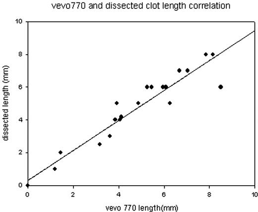
Clot length correlation using histology and ultrasonography, 24 hrs post ligation of the IVC in 20 mice. R2= 0,9116.
Clot length correlation using histology and ultrasonography, 24 hrs post ligation of the IVC in 20 mice. R2= 0,9116.
No relevant conflicts of interest to declare.
Author notes
Asterisk with author names denotes non-ASH members.


This feature is available to Subscribers Only
Sign In or Create an Account Close Modal