Recently, we reported a novel system whereby human pericytes are recruited to endothelial cell (EC)–lined tubes in 3-dimensional (3D) extracellular matrices to stimulate vascular maturation including basement membrane matrix assembly. Through the use of this serum-free, defined system, we demonstrate that pericyte motility within 3D collagen matrices is dependent on the copresence of ECs. Using either soluble receptor traps consisting of the extracellular ligand-binding domains of platelet-derived growth factor receptor β, epidermal growth factor receptor (EGFR), and ErbB4 receptors or blocking antibodies directed to platelet-derived growth factor (PDGF)–BB, or heparin-binding EGF-like growth factor (HB-EGF), we show that both of these EC-derived ligands are required to control pericyte motility, proliferation, and recruitment along the EC tube ablumenal surface. Blockade of pericyte recruitment causes a lack of basement membrane matrix deposition and, concomitantly, increased vessel widths. Combined inhibition of PDGF-BB and HB-EGF–induced signaling in quail embryos leads to reduced pericyte recruitment to EC tubes, decreased basement membrane matrix deposition, increased vessel widths, and vascular hemorrhage phenotypes in vivo, in support of our findings in vitro. In conclusion, we report a dual role for EC-derived PDGF-BB and HB-EGF in controlling pericyte recruitment to EC-lined tubes during developmental vascularization events.
Introduction
Recent work from our laboratory as well as others has demonstrated a critical role for pericytes in supporting endothelial cell (EC) tube formation and stabilization over time in 3-dimensional (3D) matrix environments.1,,,,,,,,,–11 This pericyte-regulated influence is mediated through increased expression of molecules such as tissue inhibitor of metalloproteinase (TIMP)-3 and basement membrane proteins, including laminin isoforms, collagen type IV, nidogens, perlecan, and fibronectin, derived from both pericytes and ECs.8,12,,–15 We have recently shown that as a consequence of vascular guidance tunnel formation (ie, matrix conduits proteolytically generated in conjunction with EC lumen and tube formation), pericytes recruit to the EC tubes within these tunnel spaces and induce basement membrane formation along the EC ablumenal surface.8,16,–18 In addition, the appearance of the basement membrane during in vivo vascular development was shown to correlate with the arrival of pericytes to blood vessels in the quail chorioallantoic membrane (CAM).8 Over time, formation of the vascular basement membrane allows for increased tube stabilization and has been shown to restrict EC vessel diameter.8,14,15
Although significant interest has been paid to understanding pericyte accumulation along EC tubes, many questions remain in this area.5,7,12,19,,,–23 It has been shown that platelet-derived growth factor (PDGF)-BB, a growth factor produced by the endothelium, is a mural cell mitogen and chemoattractant.22,24,–26 EC-specific PDGF-BB knockout mice show a significant loss in the number of pericytes supporting microvascular beds, leading to associated vascular defects.4,6,21,22,25,27,28 However, there is still a proportion of pericytes associated with the vessels (∼ 50% less than control).5,6,22,25,28 Recently, epidermal growth factor (EGF) family members have been implicated during vessel development, particularly in the context of vascular smooth muscle coverage of vessels suggesting a possible role in pericyte recruitment to vessels as well.7,19,20,29
Here, using a novel model of EC-pericyte tube coassembly in 3D collagen matrices, we address the role of specific growth factors during the pericyte recruitment process in this defined model (only 2 cell types present and under serum-free conditions).4,8,30 We hypothesized that it is the combinatorial effects of EC-derived PDGF-BB with an abundant EC-derived EGF family member, such as HB-EGF, that regulates pericyte recruitment to EC tubes during development and postnatal vascularization events. We present the novel finding that pericyte motility in 3D collagen matrices depends on the copresence of ECs, in that they are unable to initiate sustained directional motility without ECs. We show that this pericyte response is due to EC production of PDGF-BB and HB-EGF, as blockade of both factors results in marked decreases in pericyte motility as well as inhibition of pericyte recruitment to EC-lined tubes. Furthermore, there is a marked decrease in vascular basement membrane matrix deposition, as well as an increase in EC vessel width, as a consequence of inhibited EC-pericyte associations. Blockade of both PDGF-BB– and HB-EGF–induced signaling in vivo during quail vascular development leads to reduced pericyte recruitment to EC tubes, decreased vascular basement membrane assembly, and increased vessel width, with concomitant vessel disruption that is apparent due to the appearance of vascular hemorrhage, supporting the phenotypes observed in vitro. Overall, this work shows that the EC-derived factors, PDGF-BB and HB-EGF, together control pericyte recruitment to newly formed EC-lined tubes during vascular development.
Methods
Reagents
Recombinant human stromal cell–derived factor 1 (SDF-1)α, [chemokine (C-X-C motif) ligand 12 (CXCL12)], stem cell factor (SCF) (Kit ligand), interleukin 3 (IL-3), basic fibroblast growth factor (bFGF), α-IL-6, α-PDGF-BB, α-HB-EGF, epidermal growth factor receptor (EGFR)-Fc, ErbB4-Fc, platelet-derived growth factor receptor (PDGFR)β-Fc, and neurotrophic tyrosine kinase receptor, type 2 (TrkB)–Fc were purchased from R&D Systems. CD31 antibody was purchased from Dako. Antibodies to collagen IV (Millipore), fibronectin (Sigma-Aldrich), and laminin (Sigma-Aldrich) were purchased. In vivo staining antibodies were purchased from Developmental Studies Hybridoma Bank (under the auspices of the National Institute of Child Health and Human Development (NICHD) and maintained by The University of Iowa, Department of Biology): Endothelial Cell Surface (QH1, F. Dieterlen) and Fibronectin (B3/D6, D.M. Fambrough). Imatinib was purchased from Cayman Chemical, and gefitinib (Iressa) was purchased from Sigma-Aldrich. Hoechst 33342 dye was purchased from Invitrogen. siRNAs to the following sequences were purchased from Invitrogen and resuspended following the manufacturer's directions at a concentration of 20μM: PDGFRβ: #1: CAAAGATGTAG-AGCCGTTTCCGCTC; GAGCGGAAACGGCTCTACATCTTTG; #2: GAGGTGGAT-TCTGATGCCTACTATG; CATAGTAGGCATCAGAATCCACCTC, EGFR: #1: CGGGCTGCCTCGGGATTATTTAGAT; ATCTAAATAATCCCGAGGCAGCCCG; #2: CATTCGGAGAGGACGGTCACGTTTA; TAAACGTGACCGTCCTCTCCGAA-TG; ErbB4: #1: CAGGCTACGTGTTAGTGGCTCTTAA; TTAAGAGCCACTAAC-ACGTAGCCTG; #2: ACAGACTGCTTTGCCTGCATGAATT; AATTCATGCAGGC-AAAGCAGTCTGT. Luciferase-GL2 duplex was purchased from Dharmacon to use as a control and resuspended in 1× siRNA buffer following manufacturer's instructions to a concentration of 20μM.
Cell culture
Human umbilical vein ECs (HUVECs) were purchased from Lonza and used from passages 3-6 cultured in Supermedia (base M199, GIBCO), bovine hypothalamus extract, and 20% fetal bovine serum. Human brain vascular pericytes (HBVPs) were purchased from ScienCell and cultured in Dulbecco modified Eagle medium (DMEM)/10% fetal bovine serum and used from passages 2-12. Both cell types were cultured on gelatin-coated flasks. Green fluorescent protein (GFP)-HBVP lines were prepared as follows: Enhanced GFP was amplified from the pEGFP-N2 vector (CLONTECH) using upstream primer (UP): (5′-CACCATGGTGAGCAAGGGCGAGG-3′) and the downstream primer (DN): (5′-AGTCTAGATTACTTGTACAGCTCGTCCATGC-3′). The insert was cloned into the pLenti6/V5 TOPO vector (Invitrogen). Individual clones were verified via polymerase chain reaction (PCR) and sequence analysis. GFP lentiviruses were produced according to the manufacturer's protocol, and GFP-pericytes were selected using Blasticidin (Invitrogen) at a final concentration of 15 μg/mL.
Vasculogenic tube assembly assays
ECs (at 2 × 10⁶ cells/mL) were suspended with pericytes (0.4 × 10⁶ cells/mL) in 2.5 mg/mL collagen type I matrices and incubated at 37°C in culture media containing reduced serum supplement and fibroblast growth factors (FGF)-2 at 40 ng/mL. Stem cell factor, stromal cell–derived factor 1α, and IL-3 were added at 200 ng/mL in the collagen type I matrix. Cultures were allowed to assemble over time (72-120 hours) and fixed to be processed for further analysis. Three percent glutaraldehyde or 2% paraformaldehyde in phosphate-buffered saline (PBS), maintained at pH 7.5 for at least 1 hour, were used as general fixatives before either staining with 0.1% toluidine blue in 30% methanol for nonfluorescence visualization and photography or being immunostained for fluorescence visualization, respectively.
Immunostaining of cultures
Cultures were fixed in 2% paraformaldehyde for at least 1 hour before the addition of blocking solution: PBS containing 1% bovine serum albumin (BSA) and serum to block the secondary; no detergents were used in staining protocols to detect basement membrane proteins. Cultures were incubated with primary antibody overnight at 4°C and then were washed in PBS and incubated for 2 hours with secondary antibody. Final washes were performed over several hours, and the cultures were examined by immunofluorescence microscopy.
Injection of quail embryos
Fertilized quail eggs were purchased from Ozark Egg Company and set at 37°C to initiate development. Embryos and CAM tissue were collected at predetermined time points for immunostaining analysis. Injection of drugs or antibodies was done at 72 hours of development through a small hole created at the apex of the egg. The eggs were reincubated for another 72 hours at 37°C, and the tissues isolated. Embryos and CAM tissue were fixed in 2% paraformaldehyde for further analysis. Whole-mount embryo images were obtained using a Leica MZ95 dissecting microscope with a Plan 1.0× lens.
Microscopy, imaging, and data analysis
Time-lapse videomicroscopy and fluorescence imaging were performed using a fluorescence inverted microscope (Eclipse TE2000-E; Nikon) and the analysis software MetaMorph 7.0 (Molecular Devices). Data analysis was done by tracing EC-tube area or vessel width in individual high-powered fields or measuring immunostaining intensity of entire high-powered fields using MetaMorph software. To assess the number of pericytes associated with tubes, bright field images of EC vessels were overlaid with the corresponding GFP field of pericytes. The number of GFP-pericytes associated or not with EC tubes was then counted and averages per high-power field assessed.
Lenses used for imaging and time lapse included a CFI plan-Fluor 10× with a numerical aperture of 0.30 or 40× with an numerical aperture of 0.60. Time-lapse imaging of living cells was performed with a temperature-controlled chamber (Solent Scientific) set to 37°C with a continuous flow of 5% CO2. Images were captured every 10 minutes in single z-planes with a monochromatic camera (CoolSNAP HQ; Photometrics) and a 6.45 × 6.45-μm pixel pitch (Photometrics) using MetaMorph software (Molecular Devices). After image acquisition, stacks of each stage position were assembled using MetaMorph software.
For quantification of EC branch points and EC vessel width in vivo, randomly selected sections of CAM were isolated from treated versus control embryos. Sections were then immunolabeled using the EC-specific marker, QH1. Random, representative images were obtained and width measurements made across individual vessel diameters. Measurements made from multiple vessels and regions were then averaged for the final data presented. EC branch points were quantified, using the same images obtained for width measurements, by counting the number of intersecting vessel points. Data from multiple images and regions were then averaged for the final data presented.
Statistical analysis
Statistical analysis of selected data were performed using Microsoft Excel Office 2003 (Microsoft). Analysis of variance was used to compare means of 2 or more groups. Statistical significance was set at a minimum with P < .05. Student t tests were used when analyzing 2 groups within a single experiment. Randomly selected sections were used for all analyses with a minimum n ≥ 5 per single experiment, and ≥ 3 corroborating experimental replicates in total.
Results
ECs are necessary to induce pericyte motility in 3D collagen matrices
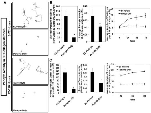
Models of EC-pericyte tube coassembly in 3D collagen matrices have been developed by our laboratory as a means to study the interactions between the 2 cell types (supplemental Figure 1A, available on the Blood Web site; see the Supplemental Materials link at the top of the online article) during vasculogenic tube assembly events.8 These models allow specific questions to be addressed regarding the behavior of both ECs and pericytes, and here, we specifically assess pericyte motility in 3D collagen matrices in response to EC-derived cues (Figure 1). We show that ECs are required for pericytes to migrate and invade in 3D collagen matrices in a sustained, directional manner (Figure 1). To assess pericyte motility in 3D collagen matrices, human brain microvascular pericytes were nuclear GFP–labeled to allow tracking of their movement using real-time video analysis (supplemental Videos 1-2). The labeled pericytes were either cocultured with ECs or cultured alone in 3D over a period of 0-72 hours (Figure 1A-B; supplemental Videos 1-2) and from 72-120 hours (Figure 1A,C). Over these time frames, average pericyte migratory velocities were calculated, as well as measurements made of the average distance from the origin that the pericytes moved to assess directional motility responses. In the absence of ECs, pericytes were unable to move in a directed manner (ie, there was no chemotactic and/or chemokinetic cue provided to induce pericyte motility away from the cells' point of origin; Figure 1). In contrast, when ECs were present with the pericytes in 3D, pericyte motility was markedly induced and there was evidence for directional motility toward the endothelium (Figure 1). Without ECs, pericytes displayed significantly reduced velocity, and the cells tended to move in a circular, nondirectional fashion. This latter phenotype was observed from either 0-72 or 72-120 hours of culture (Figure 1). Furthermore, in the absence of ECs, pericyte proliferative ability was reduced during both the 0-72-hour and the 72-120-hour time frames (Figure 1B-C). The presence of ECs stimulated pericyte proliferation by 3-fold, which occurred predominantly in the first 72 hours (Figure 1B-C). These analyses demonstrate a critical requirement for EC-derived factors in controlling both pericyte motility and proliferation in 3D collagen matrices during EC-pericyte tube coassembly events.
ECs are required to induce pericyte motility and proliferation in 3D collagen matrices during EC-pericyte tube coassembly. Nuclear GFP–labeled pericytes (nuc-pericytes) were incorporated into 3D collagen matrices in the presence or absence of ECs and allowed to undergo morphogenesis over a period of 120 hours. Real-time video analysis was performed to assess pericyte motility and proliferative rates over 0-72 hours or 72-120 hours. (A) Representative images of tracking analysis that were done with nuc-pericytes over 0-72 or 72-120 hours demonstrates the requirement of ECs to induce pericyte motility in 3D matrices. (B-C) From the tracking data generated in A, the average distance pericytes moved from their point of origin and the average pericyte velocity was measured for both 0-72 hours (B) or 72-120 hours (C). Both of these measures display impaired pericyte motility and decreased pericyte velocity over time. Furthermore, in the absence of ECs, the pericyte proliferative rate is reduced over both time periods, as quantified by the total number of pericytes per high-powered field over time (B-C). n ≥ 5; P ≤ .01.
ECs are required to induce pericyte motility and proliferation in 3D collagen matrices during EC-pericyte tube coassembly. Nuclear GFP–labeled pericytes (nuc-pericytes) were incorporated into 3D collagen matrices in the presence or absence of ECs and allowed to undergo morphogenesis over a period of 120 hours. Real-time video analysis was performed to assess pericyte motility and proliferative rates over 0-72 hours or 72-120 hours. (A) Representative images of tracking analysis that were done with nuc-pericytes over 0-72 or 72-120 hours demonstrates the requirement of ECs to induce pericyte motility in 3D matrices. (B-C) From the tracking data generated in A, the average distance pericytes moved from their point of origin and the average pericyte velocity was measured for both 0-72 hours (B) or 72-120 hours (C). Both of these measures display impaired pericyte motility and decreased pericyte velocity over time. Furthermore, in the absence of ECs, the pericyte proliferative rate is reduced over both time periods, as quantified by the total number of pericytes per high-powered field over time (B-C). n ≥ 5; P ≤ .01.
To identify possible EC-derived factors that might control this pericyte motility response, Western blot analysis was performed using EC-only cultures to determine the production of cytokines over time (eg, PDGF-BB and EGF family members; supplemental Figure 1B). Previous work has demonstrated that human umbilical vein ECs predominantly make HB-EGF and neuregulin-1 in the EGF family of proteins,31 and here, we demonstrate that HB-EGF, along with PDGF-BB, are made in abundant amounts by ECs undergoing morphogenesis (supplemental Figure 1B). To address the degree of PDGFRβ, EGFR, or ErbB4 phosphorylation events, we performed EC-only, pericyte-only, and EC-pericyte cocultures and collected cell lysates at varying time points (supplemental Figure 1C). Strong PDGFRβ phosphorylation was observed in the EC-pericyte cocultures but not in EC-only or pericyte-only cultures (supplemental Figure 1C). An increase in EGFR (ErbB1) and ErbB4 phosphorylation was seen in the EC-pericyte cocultures over EC-only or pericyte-only cultures (supplemental Figure 1C). Thus, it appears that pericytes are responsive to the EC-derived cytokines, PDGF-BB and HB-EGF, during the tube coassembly process.
Soluble PDGFRβ and HB-EGF receptor traps lead to decreased recruitment of pericytes to EC tubes and reduced pericyte numbers in EC-pericyte cocultures
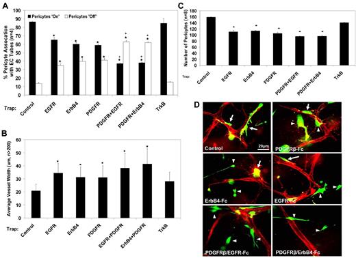
To determine the potential role of PDGF-BB and EGF family members during tube coassembly in 3D collagen matrices, the soluble receptor traps, PDGFRβ-Fc, EGFR-Fc, ErbB4-Fc, and TrkB-Fc (as a control), were added to block the function of PDGF-BB, HB-EGF, and other factors such as brain-derived neurotrophic factor (BDNF), which are endogenously produced from either human ECs and/or pericytes. Human brain microvascular pericytes stably expressing GFP were allowed to coassemble with ECs over a period of 72 hours under these varying soluble receptor trap conditions, and the cultures were fixed and processed for further analysis (supplemental Figure 1A). The influence of the soluble receptor traps on pericyte recruitment was analyzed by quantitating the number of pericytes associated with the vessels (ie, Pericytes “On”) versus the number of nonassociated pericytes (ie, Pericytes “Off”) (Figure 2A-D). EC tube width was also measured as an indicator of proper EC-pericyte tube coassembly. This was done in accordance with our previous work demonstrating that EC tubes have greater widths in the absence of pericytes and fail to deposit basement membrane matrix (supplemental Figure 2A).8 In addition, total pericyte numbers were quantified at this 72-hour time point.
Addition of soluble PDGFRβ, EGFR, and ErbB4 receptor traps leads to decreased pericyte recruitment and proliferation. GFP-pericytes were allowed to coassemble with ECs for a period of 72 hours, at which time cultures were fixed for further analysis. (A) Addition of soluble PDGFRβ, EGFR, and ErbB4 receptor traps (50 μg/mL) as individual molecules leads to approximately 60% of pericytes being associated with EC tubes and 40% being nonassociated. However, under conditions in which these soluble proteins are added in combination, only 30%-40% of pericytes are associated with EC tubes, and 60%-70% are nonassociated. Data are reported as percent pericyte association. (B) After treatment with soluble protein inhibitors, there is a dramatic increase in EC tube width in all conditions with marked disruption of pericyte recruitment. (C) Addition of these soluble protein inhibitors also leads to decreased pericyte proliferation, as quantified by assessing the total number of GFP-pericytes per high-powered field. (D) Representative images of EC-pericyte coassembly are shown in which pericytes are GFP-labeled and ECs are CD31-immunostained red. Associated pericytes are denoted by arrows; nonassociated pericytes are denoted by arrowheads. Bar equals 20 μm. n ≥ 5; P ≤ .01. *Significance from control conditions. +Significance from individual factor addition.
Addition of soluble PDGFRβ, EGFR, and ErbB4 receptor traps leads to decreased pericyte recruitment and proliferation. GFP-pericytes were allowed to coassemble with ECs for a period of 72 hours, at which time cultures were fixed for further analysis. (A) Addition of soluble PDGFRβ, EGFR, and ErbB4 receptor traps (50 μg/mL) as individual molecules leads to approximately 60% of pericytes being associated with EC tubes and 40% being nonassociated. However, under conditions in which these soluble proteins are added in combination, only 30%-40% of pericytes are associated with EC tubes, and 60%-70% are nonassociated. Data are reported as percent pericyte association. (B) After treatment with soluble protein inhibitors, there is a dramatic increase in EC tube width in all conditions with marked disruption of pericyte recruitment. (C) Addition of these soluble protein inhibitors also leads to decreased pericyte proliferation, as quantified by assessing the total number of GFP-pericytes per high-powered field. (D) Representative images of EC-pericyte coassembly are shown in which pericytes are GFP-labeled and ECs are CD31-immunostained red. Associated pericytes are denoted by arrows; nonassociated pericytes are denoted by arrowheads. Bar equals 20 μm. n ≥ 5; P ≤ .01. *Significance from control conditions. +Significance from individual factor addition.
Quantification of the number of pericytes associated with EC tubes under control conditions reveals that at 72 hours of assembly, approximately 85% of pericytes are tube associated. Under conditions of PDGFRβ-Fc, EGFR-Fc, or ErbB4-Fc addition, only approximately 60% of pericytes are associated with EC tubes. In contrast, when combinations of PDGFRβ-Fc and EGFR-Fc or PDGFRβ-Fc and ErbB4-Fc proteins are incorporated into the EC-pericyte coculture, only 35%-40% of pericytes are associated with EC tubes (Figure 2A). Furthermore, under conditions where pericyte recruitment is inhibited, there is a significant increase in EC tube width (Figure 2B) and a decrease in the proliferative rate of the pericytes, suggesting a mitogenic role for PDGF-BB and EGF family members on pericytes during this tube coassembly process (Figure 2C). Representative images of EC (red) pericyte (green) overlays corresponding to each of these conditions are shown. These images demonstrate associated pericytes (denoted by arrows) versus nonassociated pericytes (denoted by arrowheads) in these 3D culture systems (Figure 2D). These same soluble receptor traps were added to a second model of EC-pericyte tube coassembly using bovine pericytes, demonstrating that both PDGF and EGF family members are involved in bovine pericyte recruitment to EC tubes, just like with human pericytes (supplemental Figure 2B).
To confirm the data obtained using the receptor traps, pericyte-specific siRNA suppression protocols were used. Two single siRNA sequences were generated to each of the following receptors: PDGFRβ, EGFR, and ErbB4. Human pericytes were then treated with the siRNAs to suppress gene expression of these proteins and then used in the EC-pericyte coculture 3D assay system. After 72 hours of EC-pericyte tube assembly, the number of pericytes associated with EC tubes and EC tube widths were assessed. Suppression of each of the receptors in pericytes leads to a significant reduction in pericyte association with EC tubes and a significant increase in EC tube width within these cultures (supplemental Figure 3A,C), supporting the data obtained using soluble protein receptor fragments. Importantly, suppression of these same genes in ECs instead of pericytes (using the same siRNAs) does not lead to this result, confirming that their function during this tube coassembly process occurs selectively with pericytes (supplemental Figure 3B-C).
HB-EGF– and PDGF-BB–specific blocking antibodies markedly decrease pericyte recruitment to EC tubes, as well as EC-regulated pericyte motility responses
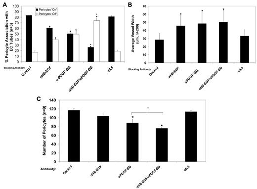
Because both EGFR-Fc and ErbB4-Fc addition lead to a blockade of pericyte recruitment (Figure 2), HB-EGF represents the likely target of these traps since it is known to interact with both receptors,32 whereas EC-derived neuregulin-1 would be selectively targeted by the ErbB4-Fc trap and not the EGFR-Fc trap. To further address this question, blocking antibodies directed to HB-EGF, a member of the EGF family of growth factors produced in large quantity by ECs (supplemental Figure 1B), and PDGF-BB were used alone or in combination to assess their influence on pericyte recruitment to tubes and pericyte motility responses in 3D collagen matrices. A blocking antibody directed to IL-6 was used as a control. Under control and α-IL-6 conditions, approximately 85% of pericytes are associated with EC tubes, and the remaining 15% of pericytes are nonassociated (Figure 3A). When blocking antibodies to HB-EGF or PDGF-BB were added to the cultures individually, there was a significant decrease (20%-30%) in the number of pericytes associated with EC tubes (Figure 3A). However, when the 2 antibodies were combined, nearly 80% of pericytes were left nonassociated with EC tubes, an almost complete reversal of the control condition (Figure 3A). In all cases there was an associated decrease in pericyte proliferation (Figure 3C) and an increase in EC tube width (Figure 3B) that was secondary to vessel remodeling events without evidence for increased EC proliferation (supplemental Figure 4A-D).
PDGF-BB– and HB-EGF–specific neutralizing antibodies lead to decreased pericyte recruitment to EC tubes and decreased pericyte proliferation. ECs and GFP-pericytes were allowed to coassemble for a period of 72 hours in the presence or absence of neutralizing antibodies to PDGF-BB and HB-EGF. A control neutralizing antibody directed to IL-6 was also used. Each of the antibodies was added at 50 μg/mL. After 72 hours, cultures were fixed for analysis of pericyte recruitment, EC tube width, and pericyte proliferation. (A) Under conditions of either HB-EGF or PDGF-BB neutralization individually, there is a 20%-30% decrease in the number of pericytes associated with EC tubes. When neutralizing antibodies are added to HB-EGF and PDGF-BB in combination, nearly 80% of pericytes are nonassociated with EC tubes. Data are reported as percent pericyte association. (B) EC tube width was measured in conjunction with pericyte association, demonstrating a dramatic width increase of EC tubes in conditions of disrupted pericyte recruitment. (C) Blockade of PDGF-BB and HB-EGF leads to decreased pericyte proliferation, as measured by assessing the total number of GFP-pericytes per high-powered field. n ≥ 5; P ≤ .01. *Significance from control conditions. +Significant from individual factor addition.
PDGF-BB– and HB-EGF–specific neutralizing antibodies lead to decreased pericyte recruitment to EC tubes and decreased pericyte proliferation. ECs and GFP-pericytes were allowed to coassemble for a period of 72 hours in the presence or absence of neutralizing antibodies to PDGF-BB and HB-EGF. A control neutralizing antibody directed to IL-6 was also used. Each of the antibodies was added at 50 μg/mL. After 72 hours, cultures were fixed for analysis of pericyte recruitment, EC tube width, and pericyte proliferation. (A) Under conditions of either HB-EGF or PDGF-BB neutralization individually, there is a 20%-30% decrease in the number of pericytes associated with EC tubes. When neutralizing antibodies are added to HB-EGF and PDGF-BB in combination, nearly 80% of pericytes are nonassociated with EC tubes. Data are reported as percent pericyte association. (B) EC tube width was measured in conjunction with pericyte association, demonstrating a dramatic width increase of EC tubes in conditions of disrupted pericyte recruitment. (C) Blockade of PDGF-BB and HB-EGF leads to decreased pericyte proliferation, as measured by assessing the total number of GFP-pericytes per high-powered field. n ≥ 5; P ≤ .01. *Significance from control conditions. +Significant from individual factor addition.
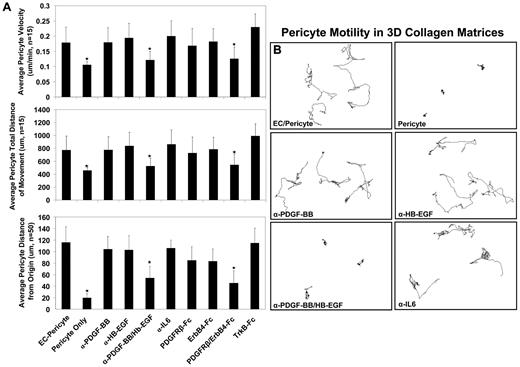
Cell tracking analysis of pericyte motility was performed in the presence of each of the blocking antibodies as well as soluble protein receptor traps. The average pericyte velocity, average pericyte total distance of movement, and average pericyte distance from the origin were measured to further assess the roles of PDGF-BB and HB-EGF individually or in combination. When both PDGF-BB and HB-EGF were inhibited, either through the use of blocking antibodies or soluble receptor protein fragments, all measures of pericyte motility were strongly inhibited over 0-72 hours of culture (Figure 4A). Representative images are shown, revealing the migration patterns of pericytes under these different conditions (Figure 4B).
Blockade of PDGF-BB and HB-EGF, using neutralizing antibodies or soluble protein fragments, leads to abrogated EC-induced pericyte motility responses in 3D collagen matrices. Nuc-pericytes were allowed to coassemble with ECs either in the presence or absence of neutralizing antibodies to PDGF-BB and HB-EGF or soluble protein fragments to PDGFRβ and the HB-EGF receptor, ErbB4. Real-time video analysis was carried out to determine the effect of these molecules on pericyte motility. (A) The movement of nuc-pericytes was tracked, using MetaMorph software, with measures of pericyte average velocity, average total distance of movement, and average distance from the origin shown. As demonstrated, disruption of PDGF-BB and HB-EGF signaling in combination leads to blockade of each of the measurements made as markers of pericyte motility in the presence of ECs. (B) Representative images of the nuc-pericyte tracking analysis are shown to demonstrate the inability of pericytes to migrate under conditions of combined PDGF-BB and HB-EGF inhibition. n ≥ 5; P ≤ .01.
Blockade of PDGF-BB and HB-EGF, using neutralizing antibodies or soluble protein fragments, leads to abrogated EC-induced pericyte motility responses in 3D collagen matrices. Nuc-pericytes were allowed to coassemble with ECs either in the presence or absence of neutralizing antibodies to PDGF-BB and HB-EGF or soluble protein fragments to PDGFRβ and the HB-EGF receptor, ErbB4. Real-time video analysis was carried out to determine the effect of these molecules on pericyte motility. (A) The movement of nuc-pericytes was tracked, using MetaMorph software, with measures of pericyte average velocity, average total distance of movement, and average distance from the origin shown. As demonstrated, disruption of PDGF-BB and HB-EGF signaling in combination leads to blockade of each of the measurements made as markers of pericyte motility in the presence of ECs. (B) Representative images of the nuc-pericyte tracking analysis are shown to demonstrate the inability of pericytes to migrate under conditions of combined PDGF-BB and HB-EGF inhibition. n ≥ 5; P ≤ .01.
These same phenotypes were observed during analysis carried out during a 72-120-hour time frame as well (supplemental Figure 5). Since pericytes have reached the EC tubes by 72 hours (supplemental Figure 1A; Figures 2-3), this latter result suggests that pericyte motility along the EC ablumenal tube surface (a process that we have previously reported through the use of real-time video analysis from 72-120 hours of culture8 ) also depends on both PDGF-BB and HB-EGF. The blocking effect appears less prominent, suggesting the possibility that additional factors might affect pericyte motility at this later stage of tube maturation. Overall, these data strongly support the concept that pericyte motility in 3D collagen matrices requires ECs—with the EC-induced response mediated through the production of PDGF-BB and HB-EGF—to activate pericyte PDGFRβ and EGFRs and control the directed motility and recruitment of pericytes to EC-lined tubes, as well as to activate motility along them (Figures 1,Figure 2,Figure 3–4; supplemental Figures 1-5).
Blockade of pericyte recruitment to developing EC tubes leads to decreased basement membrane matrix deposition in vitro and in vivo
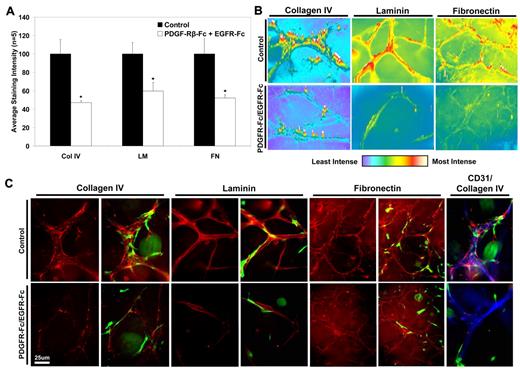
As we have previously reported, pericyte association with endothelial tubes leads to vascular basement membrane matrix deposition, a key step in vascular maturation and stabilization (supplemental Figure 2A).8 To determine whether a blockade of pericyte recruitment to EC tubes alters basement membrane deposition, EC-pericyte cocultures were allowed to assemble over 72 hours either under control conditions or in the presence of combined PDGFRβ and EGFR soluble receptor trap treatment (Figure 5A). Cultures were then fixed and prepared for immunostaining analysis to assess the extracellular deposition of the basement membrane proteins collagen IV, fibronectin, and laminin. Cultures were immunostained in the absence of detergent to ensure staining of only extracellular proteins.8 Under conditions where pericyte recruitment is inhibited (ie, PDGFRβ-Fc/EGFR-Fc soluble receptor traps), there is a significant decrease in the staining intensities for collagen IV, laminin, and fibronectin (Figure 5A-C). Representative images of basement membrane protein deposition are shown for further comparison with the indicated basement membrane components in red and the corresponding GFP field showing the relationship of the pericytes; the final panels show triple overlays of the EC marker, CD31 (blue), collagen IV (red), and pericytes (green) to display the relationship between all 3 components (Figure 5C). Intensity mapping of this immunostaining was performed to illustrate the differences in basement membrane matrix deposition in the control versus treatment conditions (Figure 5B). The loss of basement membrane matrix deposition surrounding EC tubes directly correlates with the increased EC tube widths measured under the conditions of decreased pericyte recruitment shown above (Figures 2–3; supplemental Figure 4). These data demonstrate the requirement of physical contact between ECs and pericytes through the recruitment process in order to induce vascular basement membrane assembly.
Blockade of pericyte recruitment to EC tubes leads to decreased basement membrane matrix deposition. EC/GFP-pericyte cocultures were established for 3 days and fixed for immunostaining analysis. Detergent-free immunostaining methods were used to assess the extracellular deposition of the key basement membrane components: collagen IV (Col IV), laminin (LM), and fibronectin (FN). (A) Quantification of immunostaining intensity is shown, demonstrating the decrease in basement membrane protein deposition under conditions of abrogated pericyte recruitment. (B) Intensity mapping of the representative images from panel C is shown to demonstrate the sites of highest basement membrane protein deposition, based on immunostaining patterns. (C) Representative images of the immunostained EC-pericyte cocultures are shown, with each of the basement membrane proteins in red and GFP-pericytes in green, under control versus PDGFRβ-Fc/EGFR-Fc–treated cultures. As shown by these images, there is decreased basement membrane protein deposition in cultures that have inhibited pericyte recruitment. The final column (CD31/Collagen IV) displays representative overlays of collagen IV (red), with the EC marker, CD31 (blue), and the corresponding pericytes (green) to show the relationship between the 3 structures. Bar equals 20 μm; n ≥ 5; P ≤ .01.
Blockade of pericyte recruitment to EC tubes leads to decreased basement membrane matrix deposition. EC/GFP-pericyte cocultures were established for 3 days and fixed for immunostaining analysis. Detergent-free immunostaining methods were used to assess the extracellular deposition of the key basement membrane components: collagen IV (Col IV), laminin (LM), and fibronectin (FN). (A) Quantification of immunostaining intensity is shown, demonstrating the decrease in basement membrane protein deposition under conditions of abrogated pericyte recruitment. (B) Intensity mapping of the representative images from panel C is shown to demonstrate the sites of highest basement membrane protein deposition, based on immunostaining patterns. (C) Representative images of the immunostained EC-pericyte cocultures are shown, with each of the basement membrane proteins in red and GFP-pericytes in green, under control versus PDGFRβ-Fc/EGFR-Fc–treated cultures. As shown by these images, there is decreased basement membrane protein deposition in cultures that have inhibited pericyte recruitment. The final column (CD31/Collagen IV) displays representative overlays of collagen IV (red), with the EC marker, CD31 (blue), and the corresponding pericytes (green) to show the relationship between the 3 structures. Bar equals 20 μm; n ≥ 5; P ≤ .01.
To determine the possible dual role of PDGF-BB and HB-EGF in regulating pericyte recruitment and pericyte-induced tube maturation events in vivo, the use of chemical inhibitors and blocking antibodies was used. Imatinib, or Gleevec, was used for its ability to inhibit PDGFRβ,33,34 while gefitinib, or Iressa, was used to inhibit EGFR20 ; additionally, the blocking antibodies to PDGF-BB and HB-EGF used for the in vitro studies (Figure 3) were separately administered (Figures 6–7). These reagents were all delivered individually or in combination to quail embryos at 72 hours of development versus vehicle controls. Embryos were then incubated with the reagents until 144 hours of development when phenotypes were assessed. Under control conditions at this time, we observed evidence for pericyte association with EC-lined tubes (Figure 6) and extracellular deposition of fibronectin as an indicator of basement membrane matrix assembly (Figure 7). Embryos treated with the combination of imatinib/gefitinib or the combination of PDGF-BB/HB-EGF–blocking antibodies showed a reproducible cranial and abdominal hemorrhage phenotype that was not observed in control embryos (Figure 6A-B), a phenotype commonly associated with disrupted vascular beds during development. To assess pericyte association with developing quail EC tubes under these varying conditions, quail CAMs from control versus individual (imatinib, gefitinib, anti-PDGF-BB, and anti-HB-EGF) and double treatment (imatinib/gefitinib and anti-PDGF-BB/HB-EGF) embryos were double-stained with anti-QH-1 antibodies (to visualize ECs, green) and anti-PDGFRβ antibodies (to visualize pericytes, red). As shown in Figure 6C-D, there is a significant increase in the number of pericytes that are not associated with EC tubes (ie, “nonassociated” pericytes) in the gefitinib/imatinib and anti-PDGF-BB/HB-EGF treatment groups compared with the control and the individual reagent conditions.
PDGFRβ and EGFR inhibition through the use of chemical inhibitors or neutralizing antibodies in vivo leads to a blockade of pericyte recruitment to EC tubes and concomitant cranial and abdominal hemorrhage phenotypes in developing quail embryos. Two chemical inhibitors and 2 neutralizing antibodies were identified based on their ability to interfere with PDGFR signaling (imatinib and α-PDGF-BB) and EGFR signaling (gefitinib and α-HB-EGF) and administered individually or in combination to quail at 72 hours of embryonic development at a doses of 100nM for the chemical inhibitors and 20 μg/mL for the neutralizing antibodies. The quail were then allowed to develop for 144 hours, at which time the eggs were cracked and the embryos assessed for vascular phenotypes. (A-B) Embryos treated with individual reagents developed mild cranial hemorrhages, while those embryos treated with both gefitinib/imatinib or α-PDGF-BB/HB-EGF, to block PDGFR and EGFR signaling simultaneously, led to more severe hemorrhage phenotypes (Table A). (C-D) CAM tissue from control, gefitinib/imatinib double treatment, and α-PDGF-BB/HB-EGF double treatment embryos was isolated and double stained for the quail EC-specific marker QH1 (green) and PDGFRβ (red). (C) Representative images are shown demonstrating pericyte association with microvascular beds. Arrows denote representative nonassociated pericytes. (D) The number of nonassociated pericytes per high-powered field was quantified, showing an increase in the number of nonassociated pericytes with blood vessels in vivo after treatments to inhibit PDGFR and EGFR signaling. n ≥ 5; P ≤ .01.
PDGFRβ and EGFR inhibition through the use of chemical inhibitors or neutralizing antibodies in vivo leads to a blockade of pericyte recruitment to EC tubes and concomitant cranial and abdominal hemorrhage phenotypes in developing quail embryos. Two chemical inhibitors and 2 neutralizing antibodies were identified based on their ability to interfere with PDGFR signaling (imatinib and α-PDGF-BB) and EGFR signaling (gefitinib and α-HB-EGF) and administered individually or in combination to quail at 72 hours of embryonic development at a doses of 100nM for the chemical inhibitors and 20 μg/mL for the neutralizing antibodies. The quail were then allowed to develop for 144 hours, at which time the eggs were cracked and the embryos assessed for vascular phenotypes. (A-B) Embryos treated with individual reagents developed mild cranial hemorrhages, while those embryos treated with both gefitinib/imatinib or α-PDGF-BB/HB-EGF, to block PDGFR and EGFR signaling simultaneously, led to more severe hemorrhage phenotypes (Table A). (C-D) CAM tissue from control, gefitinib/imatinib double treatment, and α-PDGF-BB/HB-EGF double treatment embryos was isolated and double stained for the quail EC-specific marker QH1 (green) and PDGFRβ (red). (C) Representative images are shown demonstrating pericyte association with microvascular beds. Arrows denote representative nonassociated pericytes. (D) The number of nonassociated pericytes per high-powered field was quantified, showing an increase in the number of nonassociated pericytes with blood vessels in vivo after treatments to inhibit PDGFR and EGFR signaling. n ≥ 5; P ≤ .01.
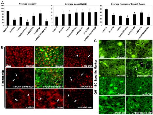
Blockade of EC-pericyte interactions in vivo leads to decreased basement membrane deposition and increased EC vessel width. (A) Quail CAM tissue from the controls, gefitinib/imatinib-treated quail embryos and α-PDGF-BB/HB-EGF–treated embryos, was isolated and immunostained for the basement membrane component fibronectin. Quantification of immunostaining intensity of extracellular basement membrane protein deposition displays a decrease in deposition under conditions of inhibited pericyte recruitment, most severely in conditions of combined PDGFR and EGFR inhibition. (B) Representative images of the fibronectin stains are shown, with arrows highlighting areas of decreased levels of extracellular basement membrane protein deposition. Overlays of QH1 staining (EC marker, green) versus fibronectin (red) are included for control versus α-PDGF-BB/HB-EGF treatments. (C) Measurements of EC tube width were done (from QH1 stains of EC tubes), demonstrating increased EC vessel width under conditions of inhibited pericyte recruitment to EC tubes. Furthermore, there was a decrease in the number of EC branch points in CAMs treated with these chemical inhibitors or blocking antibodies, further implicating direct vascular phenotypes. (D) Representative images of CAM tissue stained with the quail EC specific marker, QH1, are shown demonstrating the increased vessel width and decreased branch point phenotypes. Arrowheads indicate the “membrane-ruffled appearance” that was particularly observed in the gefitinib/imatinib condition and correlated with strongly reduced fibronectin deposition and increased vessel widths. n ≥ 5; P ≤ .01. *Significance from control conditions. +Significance from individual factor addition.
Blockade of EC-pericyte interactions in vivo leads to decreased basement membrane deposition and increased EC vessel width. (A) Quail CAM tissue from the controls, gefitinib/imatinib-treated quail embryos and α-PDGF-BB/HB-EGF–treated embryos, was isolated and immunostained for the basement membrane component fibronectin. Quantification of immunostaining intensity of extracellular basement membrane protein deposition displays a decrease in deposition under conditions of inhibited pericyte recruitment, most severely in conditions of combined PDGFR and EGFR inhibition. (B) Representative images of the fibronectin stains are shown, with arrows highlighting areas of decreased levels of extracellular basement membrane protein deposition. Overlays of QH1 staining (EC marker, green) versus fibronectin (red) are included for control versus α-PDGF-BB/HB-EGF treatments. (C) Measurements of EC tube width were done (from QH1 stains of EC tubes), demonstrating increased EC vessel width under conditions of inhibited pericyte recruitment to EC tubes. Furthermore, there was a decrease in the number of EC branch points in CAMs treated with these chemical inhibitors or blocking antibodies, further implicating direct vascular phenotypes. (D) Representative images of CAM tissue stained with the quail EC specific marker, QH1, are shown demonstrating the increased vessel width and decreased branch point phenotypes. Arrowheads indicate the “membrane-ruffled appearance” that was particularly observed in the gefitinib/imatinib condition and correlated with strongly reduced fibronectin deposition and increased vessel widths. n ≥ 5; P ≤ .01. *Significance from control conditions. +Significance from individual factor addition.
Furthermore, quail CAM tissues from treated versus control embryos were immunostained in the absence of detergent, to assess the extracellular deposition of basement membrane matrix proteins as an indicator of vascular maturation events.8 As shown in Figure 7, quantification of staining intensity for each of the treatment conditions (gefitinib, imatinib, and gefitinib/imatinib, as well as anti-PDGF-BB, anti-HB-EGF, and anti-PDGF-BB/HB-EGF) reveals decreased extracellular protein deposition of fibronectin surrounding these vessels in vivo compared with the control (Figure 7A-B), corroborating the results observed in vitro (Figure 5). Furthermore, there is a decrease in the number of EC branch points in the double treatment conditions (suggesting vascular remodeling defects) and an increase in the average EC vessel width measured that corresponds to the loss of basement membrane deposition (Figure 7C-D). We also observe that the vessels in the gefitinib/imatinib group, in particular, show a “membrane-ruffled” appearance (Figure 7D arrowheads), which may represent additional evidence for reduced tube stabilization (eg, continued EC activation) when pericyte recruitment and consequent basement membrane matrix assembly is blocked. Overall, our results in vivo support the concept that dual PDGFR and EGFR signaling events play a fundamental role in pericyte-induced vessel maturation, including basement membrane matrix deposition and accompanying restrictions in vessel diameter.
Discussion
EC-derived PDGF-BB and HB-EGF control pericyte recruitment to EC-lined tubes in 3D collagen matrices
In this work, we demonstrate a requirement for the EC-derived factors, PDGF-BB and HB-EGF, in mediating pericyte recruitment to developing EC-lined tubes (supplemental Figure 1; Figure 2). ECs were shown to be required to induce pericyte motility and invasive functions in 3D collagen matrices, as pericytes seeded as single cells in the absence of ECs failed to move in a directional manner from their point of origin (Figure 1A-C). Initially, PDGFRβ-Fc-, EGFR-Fc-, and ErbB4-Fc-soluble extracellular domains were added as growth factor traps to assess the broad potential role of PDGF and EGF family members in directing pericyte motility and recruitment. As individual inhibitors, a blockade of each receptor suppressed pericyte recruitment by approximately 30%. However, when administered in combination, the number of pericytes associated with EC tubes was nearly completely reversed from control conditions (Figure 2). These treatments also led to decreased pericyte proliferation during these events and increased EC vessel width (Figure 2). The specific EC-derived ligands activating these receptors were determined through the use of blocking antibodies (Figure 3). PDGF-BB– and HB-EGF–blocking antibodies phenocopied the results obtained through the use of soluble receptor traps, leading to nearly complete blockade of pericyte recruitment to EC tubes and increases in EC tube width (Figure 3). Furthermore, the addition of these blocking antibodies or soluble protein inhibitors in combination lead to inhibited pericyte motility, shown by decreases in average pericyte velocity, decreases in total distance moved, and decreases in the average distance from the origin (Figure 4). Similar results were obtained whether the analysis was performed from 0-72 or 72-120 hours of culture (Figure 4; supplemental Figure 5). Thus, both of these factors appear to be major regulators of pericyte motility at early and later times of pericyte interactions with developing and maturing EC-lined tubes.
Pericytes were determined to be the primary target of these inhibitors through the use of siRNA suppression technologies in ECs versus pericytes. EC suppression of PDGFRβ, EGFR, and ErbB4 did not affect average EC vessel area, vessel width, or the number of pericytes associated with EC tubes (supplemental Figure 3B). In contrast, the same siRNAs delivered to pericytes led to a marked decrease in the number of pericytes associated with EC tubes, as well as increases in EC tube width as a consequence of this failed recruitment (supplemental Figure 3A). In addition, the addition of blocking antibodies to HB-EGF did not affect EC tube formation but did effect pericyte recruitment to these tubes. Since ECs do express EGFRs, including ErbB1, ErbB2, and ErbB4, it is possible that HB-EGF does affect EC behavior in some specific manner during these events, as we do show that ErbB1 phosphorylation is detectable in EC-only cultures (supplemental Figure 1). Interestingly, HB-EGF is shed from the EC surface in an ADAM17-dependent manner and this has been shown to influence EC motility on 2-dimensional surfaces.7,29,35 EC-specific but not pericyte-specific knockout of ADAM-17 has recently been reported to modulate pathologic angiogenic responses,7 a process known to be affected by EC-pericyte interactions.
EC-derived PDGF-BB and HB-EGF control pericyte recruitment to EC tubes, leading to basement membrane matrix assembly and restriction of EC vessel diameter
As a consequence of decreased pericyte recruitment to EC tubes, there is a concomitant decrease in the extracellular deposition of basement membrane proteins both in vitro and in vivo (Figures 5 and 7). Thus, this work strongly confirms our previous conclusions that pericyte recruitment to EC-lined tubes leads to vascular basement membrane matrix assembly.8 Through the use of the chemical inhibitors gefitinib and imatinib to block EGFR and PDGFRβ, respectively, and by blocking antibodies to PDGF-BB and HB-EGF, we observed inhibition of pericyte-induced EC tube maturation events in vivo within the developing quail vasculature (Figure 7). Similar to our findings in vitro (Figure 5), blockade of EGFR and PDGFRβ in developing embryos leads to decreased pericyte recruitment (Figure 6), decreased vascular basement membrane assembly, as well as an associated increase in EC tube width (Figure 7). Further evidence for these conclusions was the high frequency of vascular hemorrhage events, a phenotype consistent with abnormalities in vessel stabilization, in the embryos treated with both chemical inhibitors or both blocking antibodies (Figure 6A-B).
Mechanisms of pericyte recruitment to microvascular tubes and its potential relevance in human disease states
Interestingly, in diseases associated with microvascular dysfunction, such as diabetes or oxygen-induced retinopathies, there tends to be a simultaneous decrease in pericyte coverage with the progression of the disease state.7,36,37 This may be caused by decreased recruitment, decreased pericyte proliferation, decreased pericyte survival, or abnormalities in maintaining pericyte association with microvessels after they are recruited. These abnormal EC-pericyte interactions may result in incomplete or immature vascular basement membranes, which may underlie the signaling abnormalities within the microvascular wall in these disease states. Our findings here suggest that 2 EC-derived growth factors, PDGF-BB and HB-EGF, play a critical role in the pericyte recruitment process that leads to vessel maturation and stabilization. The presence of ECs was necessary to induce pericyte motility and proliferation events, and these events are required to promote appropriate pericyte coverage of vessels and continuous basement membrane assembly along the EC ablumenal surface, a fundamental step in vascular stabilization.8 The experimental approach that we have presented here will be extremely useful in identifying and elucidating the functional role of additional regulators of these events. Furthermore, the model system allows characterization of signaling pathways that are necessary for ECs and pericytes to coassemble into tube networks and control vascular maturation events. It will be important in future work to assess how EC-derived PDGF-BB and HB-EGF together affect microvascular dysfunction in the context of diseases such as diabetes and cancer, where dysfunctional EC-pericyte interactions play an important pathogenetic role.
The online version of this article contains a data supplement.
The publication costs of this article were defrayed in part by page charge payment. Therefore, and solely to indicate this fact, this article is hereby marked “advertisement” in accordance with 18 USC section 1734.
Acknowledgments
This work was supported by National Institutes of Health grant HL87308 to G.E.D. and AHA grant 09PRE2140028 to A.N.S.
National Institutes of Health
Authorship
Contribution: A.N.S. designed and performed research, analyzed data, and wrote the paper; A.E.S. performed research; K.M.M. performed research; and G.E.D. designed research, analyzed data, and wrote the paper.
Conflict-of-interest disclosure: The authors declare no competing financial interests.
Correspondence: George E. Davis, Mulligan Professor of Medical Research, Department of Medical Pharmacology and Physiology, School of Medicine, MA415 Medical Sciences Bldg, University of Missouri at Columbia, Columbia, MO 65212; e-mail: davisgeo@health.missouri.edu.


This feature is available to Subscribers Only
Sign In or Create an Account Close Modal