Abstract
Sox6 belongs to the Sry (sex-determining region Y)–related high-mobility-group–box family of transcription factors, which control cell-fate specification of many cell types. Here, we explored the role of Sox6 in human erythropoiesis by its overexpression both in the erythroleukemic K562 cell line and in primary erythroid cultures from human cord blood CD34+ cells. Sox6 induced significant erythroid differentiation in both models. K562 cells underwent hemoglobinization and, despite their leukemic origin, died within 9 days after transduction; primary erythroid cultures accelerated their kinetics of erythroid maturation and increased the number of cells that reached the final enucleation step. Searching for direct Sox6 targets, we found SOCS3 (suppressor of cytokine signaling-3), a known mediator of cytokine response. Sox6 was bound in vitro and in vivo to an evolutionarily conserved regulatory SOCS3 element, which induced transcriptional activation. SOCS3 overexpression in K562 cells and in primary erythroid cells recapitulated the growth inhibition induced by Sox6, which demonstrates that SOCS3 is a relevant Sox6 effector.
Introduction
Sox proteins are important transcriptional regulators of different developmental processes in which they control the specification and differentiation of many cell types.1-3 In particular, Sox6, originally isolated from adult mouse testis,4 is required for the development of the central nervous system,5-7 for chondrogenesis,8 and for cardiac and skeletal muscle formation.9,10 Recently, Sox6 has been demonstrated to be crucial for definitive erythropoiesis,11-15 a process in which committed progenitors progressively differentiate into burst-forming–unit erythroid cells and colony-forming–unit (CFU) erythroid cells, which in turn give rise to proerythroblasts and erythroblasts and finally to mature, enucleated red blood cells. These differentiation stages are accompanied by profound maturational changes: Within few cell divisions, in parallel with the accumulation of erythroid-specific markers (membrane proteins, enzymes required for the heme biosynthesis pathway, and globins), cells undergo chromatin condensation and enucleate.16,17 This complex spectrum of maturational steps is controlled at the molecular level by the integration of extrinsic (growth factors; oxygen and iron availability) and intrinsic (growth factor receptors, signaling mediators, transcription factors) signals.
Several transcription factors are essential for erythroid commitment and for differential globin gene expression during development; their absence is associated with a wide spectrum of phenotypes ranging from mild perturbation to death because of a complete failure of erythropoiesis.18,19 Among them, Sox6 recently has been shown to stimulate erythroid cell survival, proliferation, and terminal maturation during definitive murine erythropoiesis.11,12 Sox6-null mouse fetuses and pups are anemic and have defective red blood cells. Recently, Sox6 has been implicated in the silencing of embryonic globin genes; it directly silences the embryonic ϵy-globin gene in definitive erythroid cells by binding to the ϵy promoter,13,14 and it cooperates with BCL11A in silencing γ-globin expression, possibly via direct physical interaction.15
Here, we show that Sox6 overexpression in K562 cells and in primary erythroid cultures from cord blood–derived CD34+ cells induces enhanced erythroid differentiation in both models. In K562 cells, differentiation is associated with reduced proliferation, which leads the culture to exhaustion within 9 days after transduction. In accordance with the phenotypic changes observed in K562 cells, Sox6 overexpression in human primary erythroid cultures is accompanied by accelerated kinetics of maturation and an increased number of cells that achieve enucleation.
Searching for direct Sox6 targets, we found an evolutionarily conserved potential double Sox6 binding site that was 2.7 kilobases (kb) 5′ to the SOCS3 (suppressor of cytokine signaling-3) transcription start site. This element is bound by Sox6 in vitro and in vivo and is activated by Sox6 in cotransfection experiments in K562 cells. SOCS3 is a negative regulator of the cellular response to several cytokines and plays a crucial role in regulating the balance between proliferation and differentiation in different cell types.20 When SOCS3 is overexpressed in K562 cells and in primary cultures, cells stop growing, with kinetics similar to that observed on Sox6 overexpression, which suggests that SOCS3 is indeed a relevant Sox6 target that controls cell proliferation.
Methods
Plasmid preparation
Sox6 murine cDNA was kindly provided by Professor Michiko Hamada-Kanazawa (Kobe Gakuin University, Kobe, Japan). The Sox6 cDNA was transferred into the pCMV-Tag 4B plasmid (Stratagene), in frame with a 3′ FLAG epitope (EcoRI/EcoRV sites), to produce the Sox6FLAG expression vector used in transfection assays. The Sox6 recombinant protein lacks the 49 C-terminal amino acids; this shorter molecule fully retains Sox6 biologic properties.6 The Sox6FLAG cassette (EcoRI-KpnI–blunted sites) was then cloned immediately upstream of the internal ribosomal entry site (IRES)–emerald green fluorescent protein (GFP) cassette (blunted BamHI site) of the pHR-SIN-BX-IR/EMW vector (derived from pHR-SIN-CSGW lentiviral vector21 ). The TWEEN vector containing human SOCS3 cDNA was kindly provided by Dr M. G. Francipane (University of Palermo, Italy).22
For transfection, the human SOCS3 promoter region (nucleotide [nt]-393+12) was amplified from genomic DNA with Phusion DNA Polymerase (Finnzymes) and cloned into the pGL2 reporter vector (Promega). The 70-nt SOCS3 region (nt −2727 to −2678) that contained the double Sox6 binding site (wild-type or mutated) was cloned (SacI-NheI sites) upstream of the SOCS3 promoter. Amplified DNAs were sequenced to avoid undesired mutations. All primers are listed in the supplemental Data section (available on the Blood Web site; see the Supplemental Materials link at the top of the online article).
Lentiviral vector production
Viral stocks pseudotyped with the vesicular stomatitis virus G glycoprotein envelope were produced by transient cotransfection of 4 plasmids in 293T cells.23 Cell supernatant was collected and ultracentrifuged at 50 000g for 2 hours at room temperature, and the pellet was resuspended in PBS 1% and stored at −80°C. Viral titers were determined by transduction of HEL cells with serial dilutions of the vector stocks and by scoring of GFP transgene expression by fluorescence-activated cell sorter (FACS) analysis.
Cell cultures and transduction
K562 and Human ErythroLeukemia (HEL) cells were cultured in RPMI 1640 medium supplemented by 10% fetal bovine serum, PenStrep, and l-glutamine. Transduction was performed overnight with a multiplicity of infection of 30.
CD34+ cells were purified from cord blood by positive selection from mononuclear cells24 with anti-CD34 microbeads (Miltenyi Biotec) according to the manufacturer's instructions. CD34+ cells (0.5-1 × 10⁶ cells/mL) were prestimulated for 30 hours in CellGro medium (CellGenix) supplemented with 300 ng/mL human stem cell factor, 300 ng/mL human Flt-3 ligand, 100 ng/mL human thrombopoietin, and 60 ng/mL human interleukin-3 (all PeproTech) on plates coated with RetroNectin (Takara Shuzo). Transduction was performed overnight with a multiplicity of infection of 100. Erythroblasts (day 6) derived from cord blood CD34+ cells were transduced overnight at a multiplicity of infection of 50. The following day, cells were washed and grown in suspension as an erythroid culture.
CD34+ cells were cultured for 2 weeks in StemSpan (StemCell Technologies) containing 20% fetal bovine serum (HyClone) and supplemented with human stem cell factor (10 ng/mL), human erythropoietin (EPO; 1 U/mL), human interleukin-3 (1 ng/mL), 10−6M dexamethasone (Sigma-Aldrich), and 10−6M β-estradiol (Sigma-Aldrich) according to a single-phase protocol, as modified from Migliaccio et al.24 CD34+ cells were seeded at a concentration of 105 cells/mL and diluted over time to maintain the concentration at 1-2 × 10⁶ cells/mL. Erythroid differentiation was evaluated by staining with phycoerythrin-conjugated anti-CD235 (glycophorin A [GpA]; Dako) and FACS analysis. Morphologic analysis and differential counting were performed on cytospins by May Grünwald-Giemsa staining and microscope inspection.
CFU assay for human progenitors
CD34+ cells were plated at a density of 1000 cells/mL in methylcellulose medium that contained human stem cell factor, human granulocyte-macrophage colony-stimulating factor, human interleukin-3, and human EPO (GFH4434; StemCell Technologies). After 2 weeks, burst-forming–unit erythroid, CFU-granulocyte/macrophage, and CFU-granulocyte/erythrocyte/monocyte/megakaryocyte (GEMM) colonies were counted, and single colonies (20-30 for each experiment) were isolated for DNA and RNA extraction.
ChIP assay
Cells (1 × 10⁶ K562 or 1 × 10⁷ primary cells) for each immunoprecipitation reaction were fixed with 0.4% formaldehyde for 10 minutes at room temperature, and chromatin was sonicated to approximately 500 basepairs (bp). Immunoprecipitation was performed after overnight incubation with anti-FLAG (F-7425; Sigma-Aldrich) or with anti-Sox6 antibodies (Ab5805; Millipore) and subsequent incubation with protein A agarose (Upstate/Millipore). For each amplification reaction, we amplified 1% of the immunoprecipitated DNA. Primers are listed in the supplemental Data.
RNA isolation and real-time PCR
Total RNA from 1 × 10⁵ erythroid cells was purified with TRI-Reagent (Applied Biosystems), treated with RQ1 DNase (Promega) for 30 minutes at 37°C, and retrotranscribed. Real-time analysis was performed with an ABI Prism 7500 (Applied Biosystems). Primers were designed to amplify 100- to 150-bp amplicons. Specific polymerase chain reaction (PCR) product accumulation was monitored by SYBR Green dye fluorescence in 12- to 25-μL reaction volume. Dissociation curves confirmed the homogeneity of PCR products. Primers are listed in the supplemental Data.
Western blot
Total and nuclear extracts from K562 cells were prepared according to standard protocols,25 and proteins were subjected to sodium dodecyl sulfate–polyacrylamide gel electrophoresis separation and blotting. The Sox6FLAG protein was detected with the anti-FLAG antibody (F7425; Sigma-Aldrich). Protein loading was checked by reprobing filters with an anti–β-actin antibody (Sigma-Aldrich). Antibody binding was detected by use of appropriate horseradish peroxidase–conjugated immunoglobulin G and revealed by enhanced chemiluminescence (LiteAblot; EuroClone).
Transfection experiments
A total of 1.5 × 10⁵ exponentially growing K562 cells were transfected in 0.5 mL of Opti-MEM medium (Invitrogen) with 2 μL of Lipofectamine 2000 (Invitrogen), 800 ng of the reporter plasmid, and increasing amounts (from 0.2 to 1 μg) of the Sox6 expression plasmid (pCMV-Sox6Tag4B) per well. The pCMV-Tag4B empty vector was added to each transfection to equalize the total amount of DNA transfected in each reaction. After 24 hours, total cellular extracts were prepared, and luciferase activity was measured according to the Promega luciferase reporter system protocol. Transfections were repeated in triplicate with 3 independent plasmid preparations.
EMSA
32P-labeled DNA probes were incubated with 5-10 μg of total or nuclear extracts for 20 minutes at 15°C in a buffer that contained 5% glycerol, 50mM NaCl, 20mM Tris, pH 7.9, 0.5mM EDTA (ethylenediaminetetraacetic acid), 5mM MgCl, 1mM dithiothreitol, 100 ng/μL poly(dG-dC), and 50 ng/μL bovine serum albumin in a 15-μL final volume. The reaction mixture was loaded onto an 8% polyacrylamide gel (29:1 acrylamide-bisacrylamide ratio) and run at 4°C at 150 V for 3 hours. Nuclear extracts were prepared according to standard protocols.25,26 The antibodies used were anti-FLAG (Sigma-Aldrich) and anti-GATA-1 (N6; Santa Cruz Biotechnology).
Flow cytometric analysis and FACS
Phycoerythrin-conjugated anti-pSTAT5 (pY694; BD Biosciences) was used at 5 μg/mL. Intracellular staining was performed according to the manufacturer's instructions with BD Phosflow Fix Buffer I (BD Biosciences). Propidium iodide and annexin V staining were performed with an annexin V apoptosis detection kit (Santa Cruz Biotechnology). Flow cytometric data were acquired on a FACSCalibur (BD Biosciences) and analyzed with FlowJo software Version 8.4.5 (TreeStar).
K562 cells subjected to electroporation with pTween SOCS3-IRES-GFP–expressing plasmid were sorted by FACS for GFP expression 48 hours after transfection on a MoFlo flow cytometer (DAKO-Cytomation) cell sorter, and the purity obtained was > 95%.
Hemoglobin quantitation
Total hemoglobin was quantitated by use of the Human Hemoglobin-ELISA Quantitation Set (Bethyl Laboratories), according to the manufacturer's instructions. Hemin induction was obtained by growing K562 cells in the presence of 50μM hemin for 4 days.
Results
Sox6 overexpression strongly induces erythroid differentiation in K562 cells
To gain insight into the role of Sox6 in human erythropoiesis, we first overexpressed Sox6 by lentiviral transduction of the human erythroleukemic cell line K562. K562 cells were transduced with a vector that contained a Sox6cDNA-FLAG expression cassette upstream of an IRES-GFP element (Sox6-GFP) and in parallel with a control empty vector (EV-GFP; Figure 1A). The efficiency of transduction, assayed by monitoring GFP expression, was similar for both vectors (between 80% and 90%; data not shown). Expression of exogenous Sox6 was verified by reverse-transcription PCR and by Western blot (Figure 1B) and quantitated by real-time PCR (supplemental Figure 1).
Sox6 overexpression in K562 cells. (A) Schematic representation of the lentiviral vector used. LTR indicates Long Terminal Repeats; SFFV, Spleen Focus Forming Virus; WPRE, Woodchuck-Hepatitus-Virus-Posttranscriptoral-Regulatory Element. (B) Expression of the transduced Sox6 was assayed at the mRNA and protein level. (Top panel) Reverse-transcription PCR with primers detecting the exogenous, vector-derived Sox6 transcript. β-Actin primers were used as control. (Bottom panel) Western blot with the anti-FLAG antibody detected the exogenous Sox6 protein. The anti–β-actin antibody was used to normalize for protein loading. (C-E) Phenotypic changes of K562 cells on Sox6 overexpression. Sox6-K562 cells (right panels), compared with EV-K562 cells (left panels), showed (C) a reddish pellet that indicated the accumulation of hemoglobin chains, quantitated by enzyme-linked immunosorbent assay in the right panel; (D) an increased number of o-dianisidine–positive cells (brown staining), which indicated hemoglobin accumulation; and (E) increased GpA (CD235) positivity (FACS analysis; compare cells in R6 gate: 28.14%, multiplicity of infection [MFI] of 517.28 in Sox6-K562 vs 17.87%, multiplicity of infection of 384.47 in EV-K562). (F-H) Sox6-K562 cells stopped growing and underwent apoptosis: 1 × 10⁶ exponentially growing K562 cells were transduced at day 0 either with the Sox6 or the EV vector and were washed and replated in fresh medium 24 hours after transduction. (F) Sox6-transduced cells stopped growing 3 days after transduction, and the culture died by day 9. Error bars refer to 3 independent experiments. (G) Real-time PCR on Bcl-2 and Bcl-xL expression, 72 hours after transduction. Histograms show the relative levels of expression (mean ± SEM of at least 3 independent experiments) compared with glyceraldehyde-3-phosphate dehydrogenase (GAPDH) considered as 1. Statistical significance is indicated above the chart. (H) Seventy-two hours after transduction, FACS analysis was performed with anti–annexin V antibody and propidium iodide staining to evaluate apoptosis. In Sox6-K562 cells, 16.8% of cells were positive for both annexin V and propidium iodide, whereas only 7% of EV-K562 cells were double positive for the same markers.
Sox6 overexpression in K562 cells. (A) Schematic representation of the lentiviral vector used. LTR indicates Long Terminal Repeats; SFFV, Spleen Focus Forming Virus; WPRE, Woodchuck-Hepatitus-Virus-Posttranscriptoral-Regulatory Element. (B) Expression of the transduced Sox6 was assayed at the mRNA and protein level. (Top panel) Reverse-transcription PCR with primers detecting the exogenous, vector-derived Sox6 transcript. β-Actin primers were used as control. (Bottom panel) Western blot with the anti-FLAG antibody detected the exogenous Sox6 protein. The anti–β-actin antibody was used to normalize for protein loading. (C-E) Phenotypic changes of K562 cells on Sox6 overexpression. Sox6-K562 cells (right panels), compared with EV-K562 cells (left panels), showed (C) a reddish pellet that indicated the accumulation of hemoglobin chains, quantitated by enzyme-linked immunosorbent assay in the right panel; (D) an increased number of o-dianisidine–positive cells (brown staining), which indicated hemoglobin accumulation; and (E) increased GpA (CD235) positivity (FACS analysis; compare cells in R6 gate: 28.14%, multiplicity of infection [MFI] of 517.28 in Sox6-K562 vs 17.87%, multiplicity of infection of 384.47 in EV-K562). (F-H) Sox6-K562 cells stopped growing and underwent apoptosis: 1 × 10⁶ exponentially growing K562 cells were transduced at day 0 either with the Sox6 or the EV vector and were washed and replated in fresh medium 24 hours after transduction. (F) Sox6-transduced cells stopped growing 3 days after transduction, and the culture died by day 9. Error bars refer to 3 independent experiments. (G) Real-time PCR on Bcl-2 and Bcl-xL expression, 72 hours after transduction. Histograms show the relative levels of expression (mean ± SEM of at least 3 independent experiments) compared with glyceraldehyde-3-phosphate dehydrogenase (GAPDH) considered as 1. Statistical significance is indicated above the chart. (H) Seventy-two hours after transduction, FACS analysis was performed with anti–annexin V antibody and propidium iodide staining to evaluate apoptosis. In Sox6-K562 cells, 16.8% of cells were positive for both annexin V and propidium iodide, whereas only 7% of EV-K562 cells were double positive for the same markers.
Seventy-two hours after transduction, K562 cells overexpressing Sox6 showed a profound phenotypic change. Sox6-K562 cells formed a red pellet and had an increased number of o-dianisidine– positive cells (Figure 1C-D). Enzyme-linked immunosorbent assays confirmed a significant increase in total hemoglobin content (from approximately 5 ng/104 cells in EV-K562 to approximately 22 ng/104 cells in Sox6-K562), although this increase was smaller than that of cells treated with hemin (approximately 75 ng/104 cells; Figure 1C).
Moreover, FACS analysis showed a significant accumulation of a highly CD235+ (GpA) cell population (R6 in Figure 1E) in Sox6-K562 cells, and as expected, the expression of “erythroid genes” that encode for globins and key enzymes of the heme biosynthetic pathway (ALAS-E, FECH) was increased greatly (real-time PCR; supplemental Figure 2). mRNA from genes encoding the major erythroid transcription factors (GATA1, GATA2, EKLF, and p45-NFE2) did not change, which suggests that the observed phenotype was not mediated by a Sox6 effect on expression of these transcription factors (data not shown).
Because Sox6 has been proposed to repress the ϵ and γ embryonic globin genes,11,13-15 we carefully analyzed by real-time PCR the relative changes in globin gene transcription on Sox6 overexpression, with glyceraldehyde-3-phosphate dehydrogenase used as an internal standard. Both β-like globin genes (ϵ and γ) and α-like genes (ζ and α), normally expressed by K562 cells, were substantially induced by Sox6 overexpression in terms of absolute amount of transcript; however, the ratios of changes between the different β-like and α-like globin mRNA chains suggested that Sox6 had a repressive effect both on the γ-globin gene and even more obviously on the ϵ-globin gene, in agreement with data from the literature. Chromatin immunoprecipitation assay (ChIP) demonstrated that Sox6 was indeed able to bind to the human ϵ and γ-globin gene promoters (supplemental Figure 2).
The enhanced erythroid differentiation of Sox6-K562 cells was accompanied by a marked reduction of proliferation, with complete exhaustion of the culture by day 9 after transduction (Figure 1F). FACS analysis showed that 72 hours after transduction, there was an increased number of annexin V–positive cells (21.61% in Sox6-K562 vs 13.93% in EV-K562), accompanied by increased uptake of propidium iodide (40.1% vs 14.21%), which suggests increased apoptosis (Figure 1H). Real-time PCR analysis revealed decreased expression of the antiapoptotic genes Bcl-2 and, to a lesser extent, Bcl-xL (Figure 1G). Together, these data suggest that Sox6 overexpression induces strong erythroid differentiation in K562 cells, accompanied by a dramatic reduction of cell proliferation.
Sox6 overexpression enhances and anticipates erythroid terminal differentiation of CD34+-derived primary erythroid cultures
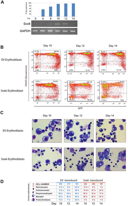
Because of the profound effect of Sox6 overexpression on K562 cells, we moved to an ex vivo model of human erythroid cell differentiation, starting from cord blood–derived CD34+ cells. In this culture, erythroid progenitors first expand to the erythroblast stage (day 0 to day 8) and then undergo terminal differentiation (day 9 to day 14). Real-time PCR analysis showed that Sox6 expression was absent in the most immature progenitors (day 0 to day 6), began to be detectable at the beginning of the erythroblast differentiation phase (day 8, which corresponded to approximately 40% of GpA+ cells), reached a peak at approximately day 12 (approximately 80% of GpA+ cells), and finally declined at the end of the culture (day 14; > 80% of GpA+ cells; Figure 2A). We therefore transduced the culture at day 6, immediately before the onset of endogenous Sox6 expression, and we subsequently performed analysis on samples taken at days 8, 10, 12, and 14. The percentage of GFP+ cells evaluated by FACS demonstrated similar transduction efficiencies with EV-GFP and Sox6-GFP vectors (approximately 85%). The result of a representative experiment (1 of 3) is shown in Figure 2B.
Sox6 enhances erythroid differentiation in cord blood–derived cell cultures. (A top panel) Percentage of GpA+ cells, estimated by FACS analysis on different days of the culture. (Bottom panel) Semiquantitative reverse-transcription PCR on the same days. Gels show Sox6 expression (top gel) and glyceraldehyde-3-phosphate dehydrogenase (GAPDH; bottom gel). (B) FACS analysis on erythroblasts transduced either with the EV (EV-erythroblasts) or the Sox6-overexpressing vector (Sox6-erythroblasts). The x-axis represents GFP expression; y-axis, GpA expression. (C) May- Grünwald-Giemsa staining on cytospin preparations of the same samples as above. (D) Differential counts on cells from the same cells as in panel C. More than 200 cells were scored for each sample.
Sox6 enhances erythroid differentiation in cord blood–derived cell cultures. (A top panel) Percentage of GpA+ cells, estimated by FACS analysis on different days of the culture. (Bottom panel) Semiquantitative reverse-transcription PCR on the same days. Gels show Sox6 expression (top gel) and glyceraldehyde-3-phosphate dehydrogenase (GAPDH; bottom gel). (B) FACS analysis on erythroblasts transduced either with the EV (EV-erythroblasts) or the Sox6-overexpressing vector (Sox6-erythroblasts). The x-axis represents GFP expression; y-axis, GpA expression. (C) May- Grünwald-Giemsa staining on cytospin preparations of the same samples as above. (D) Differential counts on cells from the same cells as in panel C. More than 200 cells were scored for each sample.
Erythroid maturation was evaluated by measuring the proportion of GFP+GpA+ cells by FACS. In control EV-erythroblasts, GFP+GpA+ cells were approximately 70% at day 10 and remained essentially stable until day 14 (Figure 2B top panels). By contrast, Sox6-erythroblasts reached a peak of GFP+GpA+ double positivity at day 10 (74.6%) and then declined to 56.1% at day 12 and to 40.6% at day 14, which suggests a progressive loss of transduced cells (Figure 2B bottom panels).
Cells from the same samples as above were spun onto a glass slide, stained with May Grünwald/Giemsa, and differentially counted to score the relative number of cells at the different stages of erythroid maturation (Figure 2C-D). At day 10, many polychromatic and orthochromatic erythroblasts, which usually appear later in control cultures, were already present in Sox6-transduced cells (Figure 2C-D). More strikingly, 3%-5% of reticulocytes were scored, whereas in the control culture, a maximum of 0.5%-1% were observed at the end of the culture (day 14). In parallel with the increased number of more mature cells (polychromatic and orthochromatic erythroblasts, as well as reticulocytes), a decrease in the number of more immature cells (proerythroblasts and basophilic erythroblasts) was observed in Sox6-overexpressing cells at day 10 (25% + 3% = 28% vs 43% + 14% = 57%). Finally, the distribution of the different cell types between the 2 cultures returned to equal levels at day 14, when GFP+ (Sox6-overexpressing) cells reached the minimum of their contribution to the culture (Figure 2B-D).
Analysis of the expression of globin genes (α, ϵ, γ, and β) demonstrated that Sox6 transduction greatly stimulated α, β, and γ expression but not ϵ expression, which likely reflects the accelerated cell maturation (supplemental Figure 3A). Interestingly, in these cells, the ϵ/α and γ/α ratios were decreased (supplemental Figure 3B), as in K562 cells, which supports the notion that besides the general induction of maturation, Sox6 specifically contributes to inhibit ϵ and γ genes transcription.
Sox6 induces early loss of CD34+ progenitor cells
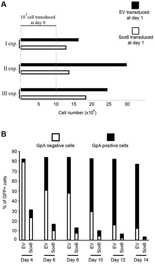
In a second set of experiments, we directly transduced CD34+ cells with either the control or the Sox6 vector as above, and then an aliquot of transduced cells was either placed in the unilineage erythroid culture or plated in methylcellulose medium for performance of a colony-forming unit assay (experiments n = 3). The proportion of GFP+ cells in the Sox6-transduced culture declined progressively from approximately 30% at day 4 to 3% by day 14 (Figure 3B). This was in strong contrast to the high level of GFP+ cells (80%-90%) transduced with the control vector, which remained constant during erythroid maturation (day 14). The low proportion of GFP+ cells after Sox6 transduction suggests an early loss of Sox6-overexpressing cells, in agreement with the slow proliferation of these cells in the first 24 hours after transduction (Figure 3A). Of interest, the proportion of Sox6-transduced cells that were GPA+ was already very high (34%) at day 4, in contrast to the very low GpA positivity (3.7%) of EV-transduced cells, which suggests that cells that survive Sox6 overexpression undergo accelerated erythroid differentiation (Figure 3B). Finally, the same transduced cells, seeded in methylcellulose, yielded no GFP+ colonies at day 14 (FACS and reverse-transcription PCR analyses), which indicates the loss of Sox6-transduced progenitors and/or their progeny.
Sox6 transduction of cord blood–derived CD34+ cells causes early loss of progenitor cells. (A) CD34+ cells transduced with Sox6 grew more slowly than EV-transduced cells. A total of 105 freshly purified CD34+ cells were transduced and counted 24 hours after transduction in 3 independent experiments. Black bars indicate EV-transduced cells; white bars: Sox6-transduced cells; and exp, experiment. (B) FACS analysis on CD34+ transduced cells at different time points after transduction. The x-axis represents days after transduction; y axis, percentage of GFP+ cells. Different colors in the histogram columns represent the proportion of GFP+ cells that were GpA− (white) or GpA+ (black), respectively.
Sox6 transduction of cord blood–derived CD34+ cells causes early loss of progenitor cells. (A) CD34+ cells transduced with Sox6 grew more slowly than EV-transduced cells. A total of 105 freshly purified CD34+ cells were transduced and counted 24 hours after transduction in 3 independent experiments. Black bars indicate EV-transduced cells; white bars: Sox6-transduced cells; and exp, experiment. (B) FACS analysis on CD34+ transduced cells at different time points after transduction. The x-axis represents days after transduction; y axis, percentage of GFP+ cells. Different colors in the histogram columns represent the proportion of GFP+ cells that were GpA− (white) or GpA+ (black), respectively.
SOCS3 is an early Sox6 target gene
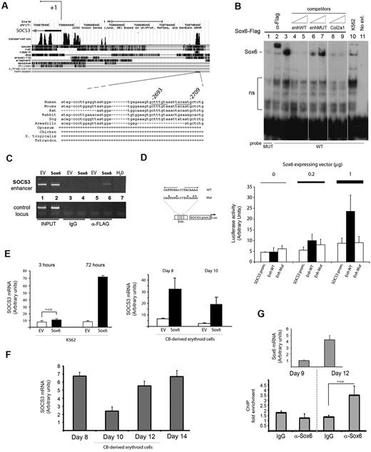
We then searched for direct Sox6 targets possibly responsible for the Sox6-induced erythroid differentiation. To this aim, we performed a genome-wide search for evolutionarily conserved potential Sox6 binding sites using TFBScluster software,27 taking the ϵ-globin Sox6 binding site as a model.28 The in silico experiment identified candidate targets (more than 800), which were filtered by selection of genes whose expression is known to be enriched in erythroid cells on the basis of literature and of DNA microarray data comparing the expression profile of 3 distinct cell populations, sorted by FACS from embryonic day 11.5 to embryonic day 13.5 mouse fetal liver: pluripotent hematopoietic progenitors cKit+/TER119−; erythroid-committed early progenitors cKit+/TER119+; and more differentiated erythroblast cKit−/TER119++ cells (Cantù et al28 and C.C., A.R., unpublished results, May 2006). Among the remaining genes,28 we focused on a double Sox6 consensus sequence lying 2700 nt upstream of the SOCS3 gene transcription start site (Figure 4A). SOCS3 is involved in the negative regulation of cytokine signaling, including EPO30,31 and insulinlike growth factor-1 (IGF-1),32,33 both of which regulate erythroid growth.34,35
SOCS3 gene is an early direct target of Sox6. (A) Mapping of the SOCS3 conserved region that contained the double Sox6 binding site. University of California Santa Cruz (UCSC) map indicates a block of conservation of approximately 100 nt centered on the Sox6 site. The double site was fully conserved in rat, mouse, and human. (B) Sox6 was bound in vitro to the SOCS3 enhancer in electrophoretic mobility shift assay. Nuclear extracts from Sox6-K562 (lanes 1-9) or from EV-K562 (lane 10) were incubated with either the WT (lanes 2-10) or the mutated (lane 1; MUT) SOCS3-labeled probes. The retarded band generated by the Sox6-FLAG protein was supershifted by the anti-FLAG antibody (lane 2) and competed for both by the SOCS3 oligonucleotide itself (lanes 4-5) and by a known Sox6 consensus (lanes 8-9; Col2a129 ). The SOCS3 probe mutated in the Sox6 consensus failed to give any binding (lane 1) or to compete for the Sox6 band (lanes 6-7). The WT probe alone (lane 11) yielded no bands. Ns indicates no specific binding. (C) ChIP on chromatins from transduced K562 cells. The SOCS3 enhancer element was immunoprecipitated by the anti-FLAG antibody (which recognizes the Sox6 transduced protein) but not by the corresponding preimmune serum, immunoglobulin G (IgG). (Top panel) SOCS3 region; (bottom panel) glyceraldehyde-3-phosphate dehydrogenase (GAPDH) locus, as negative control. Lanes 1 and 2 show input chromatins; lanes 3-4, IgG; lanes 5-6, anti-FLAG antibody; and lane 7, water. EV indicates empty vector; Sox6, Sox6 vector. (D) The SOCS3 enhancer containing the Sox6 double site was activated by Sox6 in cotransfection experiments in K562 cells. A 70-nt fragment containing either the WT (EnhWT) or the mutated Sox6 double site (EnhMut) was cloned upstream of the −393 + 12 region that corresponded to the SOCS3 minimal promoter (SOCS3prom). (Left panel) Mutations in the Sox6 consensus were the same as in panel B and were proven to abolish Sox6 binding in an electrophoretic mobility shift assay. (Right panel) All constructs were cotransfected in K562 cells together with increasing amounts (0.2 μg and 1 μg) of a Sox6-expressing plasmid. The EnhWT construct was activated in a dose-dependent manner by the addition of the cotransfected Sox6 plasmid (solid columns), whereas the corresponding mutated element, EnhMut, was insensitive to Sox6 cotransfection (open columns on right). (E) Real-time quantification of SOCS3 expression on Sox6 transduction in K562 (3 and 72 hours after transduction) and in primary cord blood–derived (CB) erythroblasts (48 and 96 hours after transduction). (F) SOCS3 expression during CD34+-derived erythroblast terminal maturation, determined by real-time PCR. (G top panel) Real-time quantification of Sox6 expression at day 9 and day 12. (Bottom panel) ChIP performed on chromatins from primary cells on the same days as above demonstrated that at day 12, Sox6 was bound to the SOCS3 enhancer. IgG antibodies were used as a control. Statistical significance is indicated above the chart.
SOCS3 gene is an early direct target of Sox6. (A) Mapping of the SOCS3 conserved region that contained the double Sox6 binding site. University of California Santa Cruz (UCSC) map indicates a block of conservation of approximately 100 nt centered on the Sox6 site. The double site was fully conserved in rat, mouse, and human. (B) Sox6 was bound in vitro to the SOCS3 enhancer in electrophoretic mobility shift assay. Nuclear extracts from Sox6-K562 (lanes 1-9) or from EV-K562 (lane 10) were incubated with either the WT (lanes 2-10) or the mutated (lane 1; MUT) SOCS3-labeled probes. The retarded band generated by the Sox6-FLAG protein was supershifted by the anti-FLAG antibody (lane 2) and competed for both by the SOCS3 oligonucleotide itself (lanes 4-5) and by a known Sox6 consensus (lanes 8-9; Col2a129 ). The SOCS3 probe mutated in the Sox6 consensus failed to give any binding (lane 1) or to compete for the Sox6 band (lanes 6-7). The WT probe alone (lane 11) yielded no bands. Ns indicates no specific binding. (C) ChIP on chromatins from transduced K562 cells. The SOCS3 enhancer element was immunoprecipitated by the anti-FLAG antibody (which recognizes the Sox6 transduced protein) but not by the corresponding preimmune serum, immunoglobulin G (IgG). (Top panel) SOCS3 region; (bottom panel) glyceraldehyde-3-phosphate dehydrogenase (GAPDH) locus, as negative control. Lanes 1 and 2 show input chromatins; lanes 3-4, IgG; lanes 5-6, anti-FLAG antibody; and lane 7, water. EV indicates empty vector; Sox6, Sox6 vector. (D) The SOCS3 enhancer containing the Sox6 double site was activated by Sox6 in cotransfection experiments in K562 cells. A 70-nt fragment containing either the WT (EnhWT) or the mutated Sox6 double site (EnhMut) was cloned upstream of the −393 + 12 region that corresponded to the SOCS3 minimal promoter (SOCS3prom). (Left panel) Mutations in the Sox6 consensus were the same as in panel B and were proven to abolish Sox6 binding in an electrophoretic mobility shift assay. (Right panel) All constructs were cotransfected in K562 cells together with increasing amounts (0.2 μg and 1 μg) of a Sox6-expressing plasmid. The EnhWT construct was activated in a dose-dependent manner by the addition of the cotransfected Sox6 plasmid (solid columns), whereas the corresponding mutated element, EnhMut, was insensitive to Sox6 cotransfection (open columns on right). (E) Real-time quantification of SOCS3 expression on Sox6 transduction in K562 (3 and 72 hours after transduction) and in primary cord blood–derived (CB) erythroblasts (48 and 96 hours after transduction). (F) SOCS3 expression during CD34+-derived erythroblast terminal maturation, determined by real-time PCR. (G top panel) Real-time quantification of Sox6 expression at day 9 and day 12. (Bottom panel) ChIP performed on chromatins from primary cells on the same days as above demonstrated that at day 12, Sox6 was bound to the SOCS3 enhancer. IgG antibodies were used as a control. Statistical significance is indicated above the chart.
To functionally validate the Sox6 site found upstream of the SOCS3 gene, we set up electrophoretic mobility shift assay experiments using as a probe an oligonucleotide that contained either the wild-type double Sox6 consensus sequence (WT) or a mutated version (Figure 4B). Nuclear extracts from Sox6-K562 and EV-K562 were used.
Sox6 bound to its consensus sequence in a specific manner (Figure 4B). The band generated by the Sox6-FLAG protein was correctly supershifted by the anti-FLAG antibody (lane 2). In competition experiments, increasing amounts of the WT oligonucleotide (lanes 4-5) and of a known Sox6 consensus sequence (Col2a1; lanes 8-9) efficiently competed for the Sox6 band, whereas the mutated oligonucleotide (lanes 6-7) did not. Finally, nuclear extracts from K562 cells transduced with the control vector generated a weak Sox6 band because of the endogenous Sox6 protein (lane 10), and the probe alone, in the absence of nuclear extracts, failed to yield any retarded band (lane 11).
ChIP experiments on K562 cells transduced with the Sox6-FLAG vector (Figure 4C) showed that the anti-FLAG antibody specifically immunoprecipitated the SOCS3 enhancer element (lane 6 top panel). No bands were detected when ChIP was performed with control immunoglobulin G (lanes 3-4).
We then cloned a region of 70 nt that surrounded the Sox6 binding sites (either WT or mutated within the Sox consensus [same mutation proven to abolish Sox6 binding in the electrophoretic mobility shift assay]) upstream of the SOCS3 promoter36 in a luciferase reporter plasmid, which we cotransfected in K562 cells together with a Sox6-expressing plasmid. Although the WT SOCS3 element was activated by Sox6, the mutated element was insensitive to Sox6 cotransfection (Figure 4D). Finally, real-time PCR performed on Sox6-K562 cells 3 and 72 hours after transduction revealed a strong early induction of SOCS3 mRNA on Sox6 overexpression (Figure 4E).
In untransduced CD34+-derived erythroid cultures, the profile of expression of SOCS3 showed a progressive increase in the last days of the culture (from day 10, when its expression was at a minimum, to day 14; Figure 4F). To correlate this pattern with the binding of Sox6 to the SOCS3 enhancer in vivo, we performed ChIP on chromatins from primary cells at day 9 and day 12. As shown in the bottom panel of Figure 5G, although no significant enrichment was observed at day 9, at day 12 (which corresponded to the peak of Sox6 expression; Figure 5G top panel), the anti-Sox6 antibody did immunoprecipitate the SOCS3 enhancer, which suggests that this element is recognized in vivo by Sox6 when Sox6 accumulates at late stages of erythroid differentiation. In agreement with this, when Sox6 was overexpressed in CD34+-derived erythroblasts at day 6, it induced SOCS3 mRNA overexpression (real-time PCR at day 8 and day 10; Figure 4E right panel). These data confirm the ability of Sox6 to bind in vitro and in vivo to the SOCS3 conserved element and to transactivate it in a dose-dependent manner in transfection experiments, which confirms that SOCS3 is a direct Sox6 target.
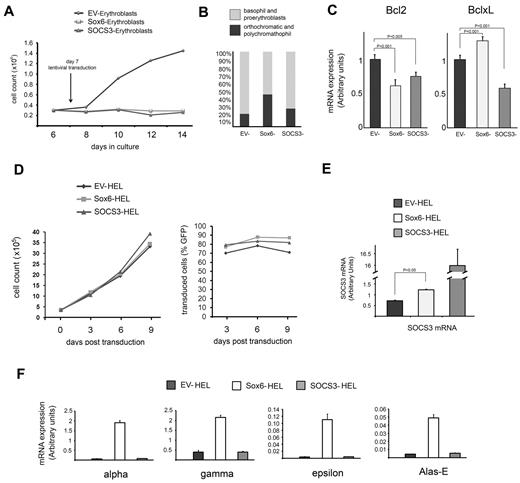
SOCS3 activation mediates inhibition of cell proliferation. (A-C) SOCS3 overexpression in CD34+-derived erythroblasts. (A) Growth curve of cultures infected with the empty vector (EV; ♦), the Sox6-overexpressing vector (■), or the SOCS3 overexpression vector (▴). The y-axis indicates the number of cells; x-axis, days in culture (all cultures were transduced at day 6 with similar efficiency). (B) Differential cell counts. The different colors in the columns represent the proportion of mature (orthochromatic plus polychromatophils; O+P) and immature (basophils plus proerythroblasts) cells, respectively. EV-transduced cells: O+P = 17%; Sox6-transduced cells: O+P = 43%; SOCS3-transduced cells: O+P = 24%. (C) Real-time PCR on Bcl-2 and Bcl-xL expression, 48 hours after transduction of day 6 erythroblasts with EV, Sox6, and SOCS3, respectively. Histograms show the relative levels of expression (mean ± SEM of at least 3 independent experiments) compared with glyceraldehyde-3-phosphate dehydrogenase (GAPDH) considered as 1. The differences shown were statistically significant, as indicated by P values. (D-F) Transduction experiments in HEL cells. (D) Neither Sox6 nor SOCS3 overexpression altered cell proliferation. Growth curve (left) and percentage of GFP+ cells (right) of HEL cells transduced with the empty control vector (EV-HEL), the Sox6-overexpressing vector (Sox6-HEL), and the SOCS3-overexpressing vector (SOCS3-HEL). (E) Quantitation by real-time PCR of SOCS3 expression in HEL cells transduced as above, relative to glyceraldehyde-3-phosphate dehydrogenase (GAPDH). (F) Real-time PCR. Only Sox6 activated erythroid gene transcription.
SOCS3 activation mediates inhibition of cell proliferation. (A-C) SOCS3 overexpression in CD34+-derived erythroblasts. (A) Growth curve of cultures infected with the empty vector (EV; ♦), the Sox6-overexpressing vector (■), or the SOCS3 overexpression vector (▴). The y-axis indicates the number of cells; x-axis, days in culture (all cultures were transduced at day 6 with similar efficiency). (B) Differential cell counts. The different colors in the columns represent the proportion of mature (orthochromatic plus polychromatophils; O+P) and immature (basophils plus proerythroblasts) cells, respectively. EV-transduced cells: O+P = 17%; Sox6-transduced cells: O+P = 43%; SOCS3-transduced cells: O+P = 24%. (C) Real-time PCR on Bcl-2 and Bcl-xL expression, 48 hours after transduction of day 6 erythroblasts with EV, Sox6, and SOCS3, respectively. Histograms show the relative levels of expression (mean ± SEM of at least 3 independent experiments) compared with glyceraldehyde-3-phosphate dehydrogenase (GAPDH) considered as 1. The differences shown were statistically significant, as indicated by P values. (D-F) Transduction experiments in HEL cells. (D) Neither Sox6 nor SOCS3 overexpression altered cell proliferation. Growth curve (left) and percentage of GFP+ cells (right) of HEL cells transduced with the empty control vector (EV-HEL), the Sox6-overexpressing vector (Sox6-HEL), and the SOCS3-overexpressing vector (SOCS3-HEL). (E) Quantitation by real-time PCR of SOCS3 expression in HEL cells transduced as above, relative to glyceraldehyde-3-phosphate dehydrogenase (GAPDH). (F) Real-time PCR. Only Sox6 activated erythroid gene transcription.
SOCS3 overexpression mediates growth arrest
To evaluate to what extent the Sox6-dependent SOCS3 induction recapitulates the phenotype induced by Sox6 overexpression, we transduced CD34+-derived erythroblasts (at day 6, as for Sox6) with a SOCS3-IRES-GFP vector. SOCS3 overexpression strongly decreased cell proliferation (Figure 5A) but, in contrast to Sox6, did not affect erythroid maturation. Differential cell counts at day 10 showed that whereas Sox6-transduced cultures had an increased number of more mature cells at the expense of more immature populations, in SOCS3-transduced cultures, the distribution of mature versus immature cells was similar to that of the control culture (Figure 5B). The Bcl-2 mRNA level was decreased in both Sox6- and SOCS3-overexpressing erythroblasts, which further confirmed the ability of SOCS3 (either induced by Sox6 or directly overexpressed) to induce a proapoptotic phenotype. Bcl-xL, which is known to increase in late erythroid differentiation, was down-regulated on SOCS3 overexpression but slightly stimulated on Sox6 transduction (Figure 5C).
In addition, SOCS3 overexpression in K562 (supplemental Figure 4) confirmed that SOCS3 was indeed able to inhibit cell growth, with kinetics similar to that observed on Sox6 overexpression, but it did not induce erythroid gene up-regulation. This reinforces the notion that Sox6-induced SOCS3 activation is responsible for reduced cell growth but not for erythroid differentiation and that additional Sox6 targets are required to fully recapitulate the Sox6-induced phenotype. This observation led us to predict that Sox6 overexpression, if achieved in a context insensitive to the SOCS3 functional contribution, would lead to increased erythroid gene expression, without any change in cell growth.
To verify this prediction, we took advantage of the HEL cell line. These cells carry the myeloproliferative disorder–associated JAK2V617F mutation,37 which makes them insensitive to SOCS3.38 Sox6 overexpression in these cells (at a level comparable with that attained in K562 and primary cells; supplemental Figure 1A) did not decrease cell growth (Figure 5D) but significantly increased erythroid specific gene expression, as demonstrated by real-time PCR of some prototypical erythroid genes (Figure 5F). In accordance with these data, SOCS3 overexpression in HEL cells did not induce a block in cell proliferation, as expected because of the JAK2V617F mutation, and did not induce erythroid-specific gene expression (Figure 5D-F). Bcl-2 and Bcl-xL mRNAs, assayed by real-time PCR, were not reduced (if anything, they were slightly increased; data not shown) by either Sox6 or SOCS3 overexpression. Together, these experiments confirmed the role of SOCS3 as a crucial Sox6 target for control of cell proliferation.
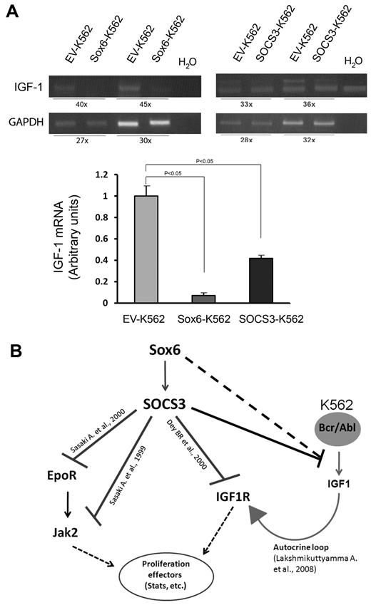
SOCS3 has inhibitory functions on signaling that are dependent on several cytokines, including the EPO/JAK/STAT and the IGF-1 receptor (IGF-1R) pathways, both of which are required for survival, proliferation, and differentiation of committed erythroid progenitor cells.38,39 To gain insight into the pathways perturbed by Sox6-dependent SOCS3 activation, we explored the effects of Sox6 overexpression on the aforementioned pathways in K562 cells, which are known to depend for their proliferation on autocrine IGF-1 signaling induced by Bcr/Abl.40
Whereas the EPO/JAK/STAT pathway was not altered by Sox6 overexpression (supplemental Figure 5), IGF-1 mRNA expression was reduced in K562 cells that overexpressed either Sox6 or SOCS3 (Figure 6A). Administration of recombinant IGF-1 (20, 100, and 200 ng/mL) to Sox6-K562 cells did not rescue Sox6-induced inhibition of cell proliferation (data not shown), as expected on the basis of the hypothesis that the decreased proliferation could be a consequence of Sox6-mediated activation of SOCS3 (which acts downstream of IGF-1R). These data suggest that inhibition of both IGF-1 expression and IGF-1 signaling pathway might contribute to the cell proliferation arrest elicited by Sox6 in K562.
The Sox6-SOCS3 axis negatively regulates IGF-1 gene transcription. (A) SOCS3 and Sox6 overexpression was accompanied by a strong repression of IGF-1 transcription. (Top panel) Reverse-transcription PCR; (bottom panel) real-time PCR. PCR cycles for IGF-1 and glyceraldehyde-3-phosphate dehydrogenase (GAPDH) amplifications are indicated below the figure. (B) Diagram showing the known relationships (references are indicated above the connecting lines38,40,41 ) between the genes/pathways studied in the present report. SOCS3 interferes with the EPO/JAK/STAT pathway by binding the EPO receptor (EpoR) and JAK,38 and it interferes with IGF-1 signaling by binding IGF-1R.41 K562 proliferation is sustained by an autocrine IGF-1 signaling loop activated by Bcr/Abl.40 Here, we propose that the SOCS3-mediated inhibition of IGF-1 transcription might be responsible for K562 decreased proliferation and increased apoptosis downstream of Sox6 (solid line). We cannot exclude that Sox6 could also act on IGF-1 through other unknown pathways (dashed line).
The Sox6-SOCS3 axis negatively regulates IGF-1 gene transcription. (A) SOCS3 and Sox6 overexpression was accompanied by a strong repression of IGF-1 transcription. (Top panel) Reverse-transcription PCR; (bottom panel) real-time PCR. PCR cycles for IGF-1 and glyceraldehyde-3-phosphate dehydrogenase (GAPDH) amplifications are indicated below the figure. (B) Diagram showing the known relationships (references are indicated above the connecting lines38,40,41 ) between the genes/pathways studied in the present report. SOCS3 interferes with the EPO/JAK/STAT pathway by binding the EPO receptor (EpoR) and JAK,38 and it interferes with IGF-1 signaling by binding IGF-1R.41 K562 proliferation is sustained by an autocrine IGF-1 signaling loop activated by Bcr/Abl.40 Here, we propose that the SOCS3-mediated inhibition of IGF-1 transcription might be responsible for K562 decreased proliferation and increased apoptosis downstream of Sox6 (solid line). We cannot exclude that Sox6 could also act on IGF-1 through other unknown pathways (dashed line).
Discussion
Sox6 is emerging as an important gene that controls different steps of erythropoiesis, ranging from cell differentiation to the control of globin gene expression.11,13,15,42,43 To gain insight into its role in human erythroid cells, we overexpressed Sox6 in the erythroleukemic cell line K562 and in primary erythroid cultures derived from cord blood purified CD34+ cells. In both cell types, Sox6-enforced expression resulted in significant erythroid terminal maturation.
K562 cells overexpressing Sox6 stop growing within 72 hours after transduction and undergo terminal erythroid differentiation, as shown by the strong induction of several erythroid-specific genes, including heme-synthesis enzymes and globin chains. Of note, K562 cells differentiate only on induction with a high concentration of chemical inducers, such as hemin.44 We obtained similar results in ex vivo erythroid cultures from CD34+ human cord blood cells (Figure 2). In these cultures, the early peak of lentivirus-mediated Sox6 expression caused a strong acceleration of differentiation, which led to the appearance of enucleated cells at day 10 of culture, which was absent in the control culture. This result confirms that Sox6 accelerates and boosts erythroid terminal differentiation of human progenitors.
Sox6 recently was reported to repress embryonic globin genes, possibly in cooperation with BCL11A,13,15 and its level of expression has been proposed to play a role in fetal hemoglobin repression in human erythroid progenitors.42 In these cells, knockdown of Sox6 leads to a modest induction of fetal hemoglobin, which is greatly increased by the combined knockdown of BCL11A, which suggests that although these 2 proteins cooperate to silence γ-genes, BCL11A has a preeminent role.15 We have shown here that Sox6 strongly stimulates the transcription (in absolute terms) of all globin genes normally expressed by K562 cells, including γ-globin and ϵ-globin (supplemental Figure 2). At first glance, this result might appear to be in conflict with the above reports. However, we suggest that Sox6 overexpression has 2 independent effects on globin synthesis: First, by inducing cell differentiation, it increases the overall expression of globin genes in general; on the other hand, it negatively modulates ϵ- and γ-globin transcription relative to other globin genes. In fact, the ϵ/α and γ/α ratios were reduced by 10- and 5-fold, respectively, in Sox6-K562 compared with EV-K562. This was supported by the observation that both promoters were occupied by Sox6 in ChIP experiments (supplemental Figure 2). The detailed molecular mechanism of Sox6 action remains to be elucidated in terms of identification of targets and interactors. Sox6 acts either as an activator or a repressor of transcription, exerting an architectural role on chromatin organization,3 and further studies are required to better study these aspects of Sox6 activity.
In the present report, among Sox6 direct target genes, we have identified SOCS3, the overexpression of which partially recapitulates the Sox6-induced phenotype by reducing cell proliferation. We have demonstrated that SOCS3 is an early direct Sox6 target and that SOCS3 overexpression partially recapitulates the Sox6 effect of cell growth arrest in primary cells and in K562, whereas it does not affect proliferation in HEL cells that are insensitive to SOCS347 (Figure 5; supplemental Figure 4).
Interestingly, in primary cells, although Sox6 overexpression was associated only with a decrease of Bcl-2 (with Bcl-xL expression being increased; Figure 5B), SOCS3 overexpression induced the down-regulation of both Bcl-2 and Bcl-xL. This might reflect both the accelerated erythroid differentiation induced by Sox6 (Bcl-xL is known to increase in late erythroid differentiation) and a possible direct transcriptional activation on Bcl-xL, in agreement with what happens in mouse erythropoiesis.12 In the latter case, the activation of Bcl-xL by Sox6, counteracting the parallel down-regulation imposed by SOCS3, might play a relevant role in ensuring the proper balance between proliferation and cell cycle withdrawal required for terminal maturation.
SOCS3 is involved in the down-regulation of different signaling pathways, and its deregulation in hematopoiesis has been implicated in the pathogenesis of myeloproliferative disorders.38,47-49 Among pathways controlled by SOCS3, there are those that involve EPO/JAK2/STAT5 and IGF-1/IGF-1R, both of which are required for erythroid progenitor cell survival.38,41 The present experiments in K562 cells show that the effects of Sox6 on proliferation are not mediated by alteration of the EPO/JAK2/STAT5 pathway (supplemental Figure 5). However, in K562 cells, in which Bcr/Abl promotes autocrine IGF-1 signaling, thus stimulating cell proliferation and protecting cells from apoptosis,40 IGF-1 mRNA was reduced when Sox6 or SOCS3 was overexpressed (Figure 6). This suggests that reduction of IGF-1 levels, together with SOCS3-dependent signaling inhibition, might contribute to the decreased cell proliferation downstream of Sox6 (and SOCS3).
Because autocrine or paracrine IGF-1 signaling is required in many stages of hematopoiesis and its inappropriate activation is an important event in leukemia,39,40 the SOCS3 activation elicited by Sox6 might be a relevant event in normal and pathologic erythroid differentiation. Further studies aimed at mapping in detail the molecular mechanisms downstream of the Sox6-SOCS3 induction will be crucial to understanding differentiation not only in erythropoiesis but also in other processes in which Sox6 plays a central role in cell commitment and differentiation.
The online version of this article contains a data supplement.
The publication costs of this article were defrayed in part by page charge payment. Therefore, and solely to indicate this fact, this article is hereby marked “advertisement” in accordance with 18 USC section 1734.
Acknowledgments
We thank Dr Fabio Gasparri for reagents and advice in signal transduction studies, Ileana Cantù and Maria Teresa Colzani for help with plasmid preparation and transfections, Dr Rebecca Favaro for suggestions and support, Dr Stefania Citterio and Dr Giuseppe Lamorte for FACS analyses, Dr Luisa Ronzoni for help with hemoglobin quantitation assays, and Dr Mario Colombo for scientific support.
This work was supported by the Italian Telethon Foundation (Core Grant TIGET; G.F.), My First AIRC grant 8726 (S.P.), Fondazione CARIPLO 2005 (S.O.), and PRIN 2006 (A.R.).
Authorship
Contribution: C.C. performed research and wrote the paper; R.I. performed ex vivo primary cell purification and erythroid cultures; I.A. assisted with electrophoretic mobility shift assay and transfection experiments; C.F., F.B., and L.C. assisted with lentiviral vectors and transduction; S.P. performed intracellular staining and FACS analyses on K562 cells; S.O. and G.F. supervised and interpreted the experiments; and A.R. assisted and supervised the research team, performed research, and wrote the paper.
Conflict-of-interest disclosure: The authors declare no competing financial interests.
Correspondence: Antonella Ronchi, Dipartimento di Biotecnologie e Bioscienze (BTBS), Università di Milano-Bicocca, Piazza della Scienza, 2, Ed.U3, 20126, Milano, Italy; e-mail: antonella.ronchi@unimib.it.
References
Author notes
C.C. and R.I. contributed equally to this study.

![Figure 1. Sox6 overexpression in K562 cells. (A) Schematic representation of the lentiviral vector used. LTR indicates Long Terminal Repeats; SFFV, Spleen Focus Forming Virus; WPRE, Woodchuck-Hepatitus-Virus-Posttranscriptoral-Regulatory Element. (B) Expression of the transduced Sox6 was assayed at the mRNA and protein level. (Top panel) Reverse-transcription PCR with primers detecting the exogenous, vector-derived Sox6 transcript. β-Actin primers were used as control. (Bottom panel) Western blot with the anti-FLAG antibody detected the exogenous Sox6 protein. The anti–β-actin antibody was used to normalize for protein loading. (C-E) Phenotypic changes of K562 cells on Sox6 overexpression. Sox6-K562 cells (right panels), compared with EV-K562 cells (left panels), showed (C) a reddish pellet that indicated the accumulation of hemoglobin chains, quantitated by enzyme-linked immunosorbent assay in the right panel; (D) an increased number of o-dianisidine–positive cells (brown staining), which indicated hemoglobin accumulation; and (E) increased GpA (CD235) positivity (FACS analysis; compare cells in R6 gate: 28.14%, multiplicity of infection [MFI] of 517.28 in Sox6-K562 vs 17.87%, multiplicity of infection of 384.47 in EV-K562). (F-H) Sox6-K562 cells stopped growing and underwent apoptosis: 1 × 10⁶ exponentially growing K562 cells were transduced at day 0 either with the Sox6 or the EV vector and were washed and replated in fresh medium 24 hours after transduction. (F) Sox6-transduced cells stopped growing 3 days after transduction, and the culture died by day 9. Error bars refer to 3 independent experiments. (G) Real-time PCR on Bcl-2 and Bcl-xL expression, 72 hours after transduction. Histograms show the relative levels of expression (mean ± SEM of at least 3 independent experiments) compared with glyceraldehyde-3-phosphate dehydrogenase (GAPDH) considered as 1. Statistical significance is indicated above the chart. (H) Seventy-two hours after transduction, FACS analysis was performed with anti–annexin V antibody and propidium iodide staining to evaluate apoptosis. In Sox6-K562 cells, 16.8% of cells were positive for both annexin V and propidium iodide, whereas only 7% of EV-K562 cells were double positive for the same markers.](https://ash.silverchair-cdn.com/ash/content_public/journal/blood/117/13/10.1182_blood-2010-04-282350/4/m_zh89991168260001.jpeg?Expires=1769895733&Signature=JI4GoeBKWgIuiPW8EFR4DTWbnDvNam5Hm2APpo97i-Xzws~vR77cEYQwBri3Oo5B6Jy0ug~MV5S4B265irhBMJ~6EeC71bh5NDaorWH~WlpJzSfOmB2w4RJQ3OirU7YGdh3LsoflaBSa4G19et3o-~OQN8LsLKXicEiIO51pSnZKbHd1Kjs5j3YflN5HSVPH-b~O1Y3CcRERht6rxRP9g5iZpjFLtoLCMqeVlp400FlEnSNOXlnXSWM6JcB6Fz0IVpzRIq5XlnVWoh8W~UubgL~udUbZYDcXB2Vcf3OTo19KYZhURkle9ZXjrdqd9RAx0~Nk4ClBH2Xb0BFVQA1ccQ__&Key-Pair-Id=APKAIE5G5CRDK6RD3PGA)
