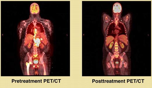
PET scans are almost always abnormal at diagnosis in the most common B-cell lymphomas (diffuse large B-cell lymphoma, follicular lymphoma, and Hodgkin lymphoma), and normalization after therapy is highly predictive of a good outcome. But how to apply PET scans to the management of these patients remains problematic. The major controversies include when (ie, for staging, after 2-4 cycles of therapy, after completion of therapy, surveillance in remission) and how (ie, we know they should always be combined with CT scanning, but the relative merits of visual interpretation and SUV [standard uptake value] calculation are debated).
The concept of interim (early) PET/CT scans to predict the eventual result of treatment is relatively new. In most but not all studies, patients with a positive scan after 2-3 cycles of treatment have a poorer outcome than those whose scans are negative.1-5 One hope of proponents of this approach is that patients with a positive interim scan might have their treatment changed and their chances for long survival increased. However, those patients whose interim scans are positive but turn negative by the end of treatment seem to do about as well as those with negative scans at both times.1,2,5-7 In fact, it appears that a negative scan at the end of therapy is the most powerful predictor of a long remission in the 3 common B-cell lymphomas.8-10 Thus the value of changing treatment on the basis of a positive interim scan depends on the new treatment still curing those patients destined to do well with the initial regimen, and curing some of the patients destined to do poorly. Of course, the results could vary from one disease to another and with different initial treatment regimens. These are questions for randomized trials, not standard therapy, and several such trials are in progress.
For studies such as PET/CT scans to be helpful in the clinic, they need to be reproducible (minimal intra- and interobserver variability) and need to be shown to correlate to the desired clinical end point. The report by Casasnovas et al in this issue of Blood addresses these issues for patients with diffuse large B-cell lymphoma.11 They used central review of all images and compared visual interpretation with the calculated maximal SUV at any site in the tumor (SUVMAX). They found the quantitative changes in the SUVMAX provided a much better prediction of treatment outcome than visual interpretation, and that later studies (after 4 cycles) were more predictive of outcome than those done after 2 cycles of therapy. The study did not address the issue of changing therapy based on early positive scans. Patients with positive scans after 2 cycles of therapy all received 2 more cycles of CHOP-R (cyclophosphamide, doxorubicin, vincristine, prednisone, and rituximab) and were then transplanted if the scan normalized after 4 cycles.
So where do we stand today in using PET/CT scans to benefit our patients? They seem to provide a useful staging study and can both upstage and downstage patients in comparison to CT scanning alone. The PET/CT scan at the end of treatment is the most powerful predictor of outcome. Currently both interim scanning and surveillance scanning in remission are topics for research, have not been shown to improve survival, and their use should be restricted to clinical trials. The study by Casasnovas et al makes a strong argument that interpretation using calculated SUVMAX is better than using visual interpretation.11 They identified cutoff points of reduction of the SUVMAX from pretreatment to scans after 2 and 4 cycles of therapy that provided the best discrimination between no relapse versus relapse and death versus survival. However, it seems likely that resolution of the SUVMAX to background at the end of therapy might provide the optimum chance for a good outcome. The percent reduction SUVMAX Casasnovas et al found optimal might not be the most advantageous in all situations (eg, interim versus end-of-therapy scanning, different treatment regimens) and with all types of lymphoma.
PET/CT scans provide a powerful tool to improve the care of our patients. Clinical trials aimed at defining the optimal use of these studies must be completed if we are ever to give our patients the maximum benefit the tests can provide. In the meantime, they represent a valuable staging study and a complete remission defined by PET/CT is the most powerful predictor of a durable remission (see figure).
Conflict-of-interest disclosure: The author declares no competing financial interests. ■

