Abstract
Abstract 1125
Race-related differences have been documented in the incidence of autoimmune diseases such as systemic lupus erythematosus and multiple sclerosis, in the clinical response to immunotherapies [such as IFNα (in HCV infections) and belimumab (in systemic lupus erythematosus)] and to hematopoietic stem cell transplantation. However, the basis for such race-associated differences remains poorly understood. Single Cell Network Profiling (SCNP) is a multiparametric flow cytometry based approach that simultaneously measures intracellular signaling activity in multiple cell subpopulations. Previously, SCNP analysis of peripheral blood mononuclear cells (PBMCs) from 60 healthy donors identified a race-associated difference in αIgD induced levels of pS6 and pAkt in B cells. The present study extended this analysis to a broader range of signaling pathway components downstream of the B cell receptor (BCR) in European Americans and African Americans using a subset of donors from the previously analyzed cohort of 60 healthy donors.
35 BCR signaling nodes (a node is defined as a paired modulator and intracellular readout) were measured by SCNP in PBMCs from 10 healthy donors [5 African Americans (36–51 yrs), 5 European Americans (36–56 yrs), all males]. Cryopreserved PBMCs were thawed, modulated at 37°C in 96-well plates, fixed and permeabilized. Permeabilized cells were stained with fluorochrome-conjugated antibodies that recognize extracellular surface markers and intracellular signaling molecules. The levels of 7 phospho-proteins [pLck (Y505), pSyk (Y352), pAkt (S473), pS6 (S235/S236), p-p38 (T180/Y182), pErk (T202/Y204), and pNFκB (S529)] were measured in CD20+ B cells at 0, 5, 15, 30, and 60 mins post αIgD exposure. CD20 and IgD surface markers were used to determine the frequency of IgD+ B cells.
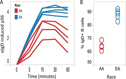
A) αIgD induced pS6 signaling (based on the log2fold increase in MFI in αIgD treated cells relative to the untreated control (0 min)) over time are shown for the African American (AA) and European American (EA) donors. The difference in pS6 signaling (averaged over time points) between racial groups is statistically significant. B) The percentage of CD20+ B cells that were IgD+ is shown for the AA and EA donors. The difference in IgD+ frequency between racial groups is statistically significant. In both (A) and (B), one of the ten donors was excluded due to an insufficient number (<200) of B cells collected for analysis.
A) αIgD induced pS6 signaling (based on the log2fold increase in MFI in αIgD treated cells relative to the untreated control (0 min)) over time are shown for the African American (AA) and European American (EA) donors. The difference in pS6 signaling (averaged over time points) between racial groups is statistically significant. B) The percentage of CD20+ B cells that were IgD+ is shown for the AA and EA donors. The difference in IgD+ frequency between racial groups is statistically significant. In both (A) and (B), one of the ten donors was excluded due to an insufficient number (<200) of B cells collected for analysis.
In conclusion, SCNP analysis allowed for the identification of statistically significant race-associated differences in BCR pathway activation within PBMC samples from healthy donors. Further characterization of racial functional differences in additional immune signaling pathways using this assay in samples from both healthy and diseased individuals may be critical for elucidating the basis for race-related differences in immune-mediated disease prevalence and treatment responses.
Longo:Nodality: Employment, Equity Ownership. Louie:Nodality: Employment, Equity Ownership. Mathi:Nodality: Employment. Hawtin:Nodality: Employment, Equity Ownership. Cesano:Nodality: Employment, Equity Ownership.
Author notes
Asterisk with author names denotes non-ASH members.


This feature is available to Subscribers Only
Sign In or Create an Account Close Modal