Abstract
Abstract 2228
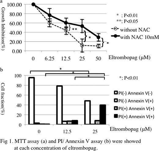
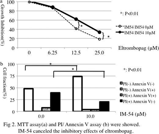
Eltrombopag is an oral thrombopoietin receptor agonist for the treatment of immune thrombocytopenic patients who are refractory to the medications including corticosteroid. In RAISE study (The Lancet 2011: 377; 393–402), 79% patients in the eltrombopag group responded to treatment at least once during the study. However, 7% of eltrombopag-treated patients had increase of alanine aminotransferase (ALT) concentration and 4% of total bilirubin. The mechanism of eltrombopag-induced hepatobiliary toxicity remains unknown. In this study, we evaluated the effects of eltrombopag on hepatocytes using HepG2, human hepatocellular carcinoma cell line. Method: Cell proliferation was analyzed by MTT assay. Analysis of apoptosis/necrosis was analyzed by flow cytometry assay using Annexin V and propium iodide (PI) (Annexin-/PI-, viable cells; Annexin+/PI-, early apoptosis cells; Annexin+/PI+, late apoptosis and necrosis cells; Annexin-/PI+, late necrosis cells). Reduced glutathione was measured by DTNB colorimetric method. Murine hepatoma cell line Hepa 1–6 and murine normal liver derived cell line NCTC clone 1469 were also used in this study. Results: HepG2 was incubated with eltrombopag for 72 hrs and then MTT assay was performed. Figure 1a showed the growth inhibition curve of HepG2. HepG2 growth was suppressed by eltrombopag in a dose dependent manner (Figure 1a: without NAC, 12.5 μM 44.3 ± 8.7 %). In the addition of N-acetylcysteine (NAC) canceled the inhibitory effect (Figure 1a: with NAC 10mM: 60.4 ± 22.3 % at 12.5μM eltrombopag, p<0.05). To investigate whether this suppression was due to apoptosis or necrosis, we used PI/ Annexin V assay using flow cytometry. Figure 1b showed that each cell fraction after 72 hrs incubation with eltrombopag. PI positive and PI negative fraction was markedly increased (0μM: 0.7 ± 0.4%, 12.5 μM: 8.0 ± 6.0% p<0.01, 25μM: 39.6 ± 12.2% p<0.001). To confirm that the inhibitory effects of eltrombopag might be due to necrosis, we used IM-54, which is inhibitory molecule against oxidative stress induced necrosis, in MTT assay and PI/ Annexin V assay. As shown in Figure 2 a and 2 b, IM-54 canceled the inhibitory effects of eltrombopag in MTT assay (IM-54 0μM: 18.3 ± 7.5 %, IM-54 10 μM: 33.9 ± 10.6 % at eltrombopag 25μM, p<0.01) and PI/Annexin V assay (IM-54 0μM: 48.3 ± 12.4 %, IM-54: 10 μM 74.2 ± 8.3 % in PI/Annexin V double negative fraction at eltrombopag 25μM, p<0.01). The addition of Caspase-Inhibitor III (Boc-D-FMK) did not cancel the growth inhibition by eltrombopag in MTT assey. We measured GSH concentration of HepG2 cells. After the treatment of eltrombopag for 72 hrs, GSH decreased at 12.5μM of eltrpmbopag, however IM-54 restored the GSH concentration (83 mM at IM-54 0μM and eltrombopag 0 μM, IM-54 0 μM: 42.0 mM and IM-54 10μM: 83.3 mM at eltrombopag 12.5μM). Similar data were obtained in the study using Hepa 1–6 and NCTC clone 1469. Conclusion: Basic structure of eltrombopag is similar to that of 3-bipheyl-carboxylic acid, which is a strong oxidizing agent. Taken together, eltrombopag have an oxidative stress on hepatocytes to result in hepatotoxicity.
No relevant conflicts of interest to declare.
Author notes
Asterisk with author names denotes non-ASH members.


This feature is available to Subscribers Only
Sign In or Create an Account Close Modal