Abstract 5079
IgD monoclonal proteins are rare. They are not seen as a MGUS and are present in 1% to 2% of patients with myeloma. In light chain amyloidosis (AL), IgD monoclonal proteins are rare. When an IgD protein is found, amyloidosis is often omitted from the differential diagnosis. An IgD protein in amyloidosis has been reported in single cases but never as a patient series. We report IgD AL in 53.
Clinical and demographic data for patients were retrieved from the patient records. Factors of interest were compared between patients who did and did not have an IgD protein.
Among 3,955 patients with AL amyloidosis seen, 53 patients (1.3%) had a serum IgD monoclonal protein. (Table 1) A serum monoclonal protein peak was visible on SPE in only 14, and only 5 had an M spike greater than 1 g/dL. On immunofixation of the serum, the IgD light chain was k in 11, λ in 35, and uncertain in 6; 1 patient had a biclonal D λ and G k protein. A urine monoclonal protein was detected in 43 of 51 patients; urinary immunofixation detected a λ light chain in 33 and a k light chain in 10. Biopsy of tissues showed amyloid deposits in the bone marrow in 47% and in the fat aspirate in 73%.
Patient and Clinical Characteristics (N=53)
| Characteristic . | Value . |
|---|---|
| Men | 30 (57) |
| Median (range) age, y | 60.5 (34.7–80.9) |
| Symptoms & signs | |
| Fatigue | 32 (60) |
| Lower extremity edema | 23 (43) |
| Paresthesias | 17 (32) |
| Weight loss | 17 (32) |
| Dyspnea on exertion | 15 (28) |
| Carpal tunnel syndrome | 14 (26) |
| Enlarged tongue | 11 (21) |
| Hepatomegaly | 9 (17) |
| Neuropathy | 8 (15) |
| Purpura | 7 (13) |
| Status at last follow-up | |
| Dead | 41 (77) |
| Cause of death (n=41) | |
| Unknown | 24 (59) |
| Cardiac failure or cardiac arrest | 10 (24) |
| Infection | 3 (7) |
| Myeloma | 1 (2) |
| Renal failure | 1 (2) |
| Cachexia | 1 (2) |
| Treatment-related leukemia | 1 (2) |
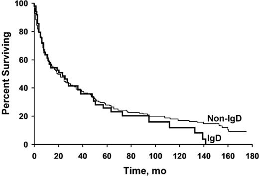
Six patients (age 47–63 years) underwent autologous SCT. Four had renal & two had cardiac AL. All 6 had hematologic PR, 4 CR, and 4 had organ response. One patient had relapse of disease and is now on dialysis, and another had relapse and is alive with salvage chemotherapy. One patient had disease relapse and died of progressive GI amyloidosis at 22 months. The other 5 are alive at a median of 68 months (range, 7.5–83.5 months). We compared the 53 patients with IgD amyloidosis with 144 patients with non-IgD amyloidosis. (Table 2) Findings that were significantly different between groups included a lower frequency of renal amyloidosis (P=.005) and a lower prevalence of cardiac amyloidosis (P=.047). There was a higher serum albumin level (P=.04) related to the lower level of proteinuria. No difference in survival was seen between the groups. (Figure). Variables that might affect survival—liver size, performance status, septal thickness, serum creatinine level, and β2-microglobulin level—were not different between groups.
Comparison of IgD and Non-IgD Amyloidosis
| Characteristic . | Patient Group . | P Value . | |
|---|---|---|---|
| IgD (n=53) IQR . | Non-IgD (n=144)IQR . | ||
| Age, y | 60 (50–67) | 61.5 (53–68) | .34 |
| Men | 30 (57) | 94 (65) | .31 |
| Hepatomegaly | 9 (17) | 39 (27) | .16 |
| Interventricular septal thickness, mm | 15 (11–17) | 13 (11–15) | .11 |
| Ejection fraction, % | 60 (44–67) | 60 (50–68) | .17 |
| Creatinine, mg/dL | 1.1 (.9–1.6) | 1.1 (.9–1.5) | .34 |
| Total cholesterol, mg/dL | 196 (146–299) | 242 (186–328) | .047 |
| β2-Microglobulin, mcg/mL | 2.77 (2.15–5.38) | 2.83 (2.07–3.99) | .39 |
| Albumin, g/dL | 2.97 (2.43–3.54) | 2.69 (2.07–3.37) | .04 |
| Urine protein, g/24h | 1.5 (.3–5.1) | 2.9 (.36–7.4) | .12 |
| Serum M spike | Immunofixation only (fixation only-0.6 g/dL) | Immunofixation only (fixation only-0.9g/dL) | .31 |
| % Marrow plasma cells | 10(3–30) | 8(4–16) | .09 |
| N=44 | N=137 | ||
| Renal amyloid | 19 36% | 84 58% | .005 |
| Cardiac amyloid | 24 45% | 81 56% | .047 |
| Neuropathy | 5 9% | 19 13% | .47 |
| Autonomic failure | 3 6% | 10 7% | .74 |
Patients With IgD and Non-IgD Amyloidosis Have Similar Survival. Kaplan-Meier analysis of survival in patients with IgD-associated (n=53) and non- IgD–associated (n=144) amyloidosis shows nearly overlapping survival curves. No difference between the groups was seen (P=.51).
IgD AL patients have a lower frequency of renal involvement and possibly also of cardiac involvement. The overall survival of these patients does not appear to be different from that of patients who have AL associated with another monoclonal protein. IgD monoclonal proteins are so closely linked to the diagnosis of multiple myeloma in the mind of a clinician that the possibility of amyloidosis may be overlooked.
No relevant conflicts of interest to declare.


This feature is available to Subscribers Only
Sign In or Create an Account Close Modal