Abstract
Abstract 5191
Extranodal natural killer T-cell lymphoma, nasal type (nasal ENKTL) is a distinct clinicopathologic entity of lymphoid tumors with variable size and differentiation of tumor cells. Nasal ENKTL is related to infection of the tumor cells with Epstein-Barr virus (EBV) and virtually all cases contain monoclonal episomal EBV DNA and detectable EBV encoded small nuclear RNAs (EBERs). Several clinical factors were known for their relation to the prognosis, but histopathologic prognostic factors of nasal ENKTL have not yet been well established. We evaluated prognostic value of the size and density of EBER-positive tumor cells along with immunohistochemistry (IHC) expression of CD30 and CD56.
Patients with newly diagnosed nasal ENKTL treated in a single institution (Gachon University Gil Hospital) were evaluated with respect to clinical characteristics, survival, and histopathologic characteristics. IHC were performed using anti-CD56 (DAKO), CD30 (DAKO). EBV RNA was detected by an ISH (in situ hybridization) technique. Paraffin sections were pretreated with xylene followed by treatment with proteinase K and hybridized with FITC-conjugated EBV oligonucleotides (Novocastra) complementary to the nRNA portion of the EBER-1 and EBER-2 genes. The numbers of EBER-positive tumor cells were counted and the nuclear length of tumor cells were measured using a computerized image analysis system (IMT i-Solution Inc., Vancouver, British Columbia, Canada) that included a DP70 Digital camera (Olympus, Tokyo, Japan) installed on an Olympus light microscope (Olympus BX51) and attached to a personal computer. Five independent microscopic fields (at x400 magnification), representing the densest tumor infiltration, were selected for each patient sample to ensure representativeness and homogeneity. Each independent microscopic field contained an approximately 80% tumor ratio. The results were expressed as the mean number of cells (± standard error) in 1 computerized x400 microscopic field (9941.38mm2/field). More than 50 tumor cells were selected for the measurement of nuclear length of tumor cells. The results were expressed as the mean diameter of cells (± standard error). The mean density and mean diameter were used for statistical analysis.
Twenty-two patients were analyzed. Median age were 48.5 (range 15–81) and 20 of them were male (91%). Seven (32%) patients were Ann Arbor stage I, and 6 (27%), 3 (14%), and 6 (27%) were stage II, III, IV, respectively. Sixteen patients (73%) received combined chemotherapy and radiotherapy whereas 5 patients (23%) received chemotherapy alone and 1 patient received only radiotherapy. Median of the mean EBER-positive tumor cell diameters of the patients were 7.32 mm (range 5.15–11.27). Median of the mean tumor cell counts of the patients were 71.7/field.
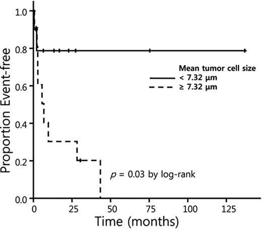
Patients with larger mean tumor cell diameters (≥ 7.32 mm) had a poorer event-free survival (EFS), which was defined as survival free from failure or death from any cause, than those with smaller (<7.32 mm) mean cell diameters (table 1 and figure 1). Density of tumor cells (≥71.7 vs. < 71.7/field) and IHC to CD56 or CD30 (≥50% vs. < 50% of expression) had no impact on EFS, whereas previously known clinical factors - local tumor invasion, type of therapy, performance status, and splenomegaly - maintained power enough to predict EFS (table 1).
univariate Cox analysis
| Factors . | Patient No. . | Hazard ratio (95% CI) . | p . |
|---|---|---|---|
| Age (>60) | 8 | 2.72 (0.76–9.79) | 0.125 |
| Local tumor invasion (+) | 6 | 4.26 (1.10–16.51) | 0.036 |
| Cervical nodes (+) | 9 | 1.36 (0.41–4.53) | 0.616 |
| Combined therapy (not doing) | 6 | 3.94 (1.1519–13.47) | 0.029 |
| ECOG PS (2,3,4) | 5 | 14.82 (2.57-85.36) | 0.003 |
| Serum LDH (elevated) | 9 | 1.72 (0.5219–5.70) | 0.378 |
| Ann Arbor stage (III and IV) | 9 | 1.74 (0.53–5.80) | 0.364 |
| Splenomegaly (present) | 9 | 4.52 (1.15-17.73) | 0.031 |
| Mean cell diameter (≥ 7.32 mm) | 12 | 4.73 (1.01–22.16) | 0.049 |
| Mean cell count (≥ 71.7/field) | 12 | 1.91 (0.51–7.23) | 0.340 |
| CD56 (≥ 50% expression) | 14 | 0.57 (0.17–1.91) | 0.572 |
| CD30 (≥ 50% expression) | 8 | 2.51 (0.67–9.38) | 0.173 |
Larger EBER-positive tumor cell size had an adverse impact on EFS in patients with nasal ENKTL. Further larger scale study to define the prognostic value of EBV infected tumor cell size is warranted.
No relevant conflicts of interest to declare.
Author notes
Asterisk with author names denotes non-ASH members.


This feature is available to Subscribers Only
Sign In or Create an Account Close Modal