Abstract
The transfer of membrane proteins between cells during contact, known as trogocytosis, can create novel cells with a unique phenotype and altered function. We demonstrate that trogocytosis is more common in multiple myeloma (MM) than chronic lymphocytic leukemia and Waldenstrom macroglobulinaemia; that T cells are more probable to be recipients than B or natural killer cells; that trogocytosis occurs independently of either the T-cell receptor or HLA compatibility; and that after trogocytosis, T cells with acquired antigens can become novel regulators of T-cell proliferation. We screened 168 patients with MM and found that CD86 and human leukocyte antigen G (HLA-G) were antigens commonly acquired by T cells from malignant plasma cells. CD3+CD86acq+ and CD3+ HLA-Gacq+ cells were more prevalent in bone marrow than peripheral blood samples. The presence of either CD86 or HLA-G on malignant plasma cells was associated with a poor prognosis. CD38++ side population cells expressed HLA-G, suggesting that these putative myeloma stem cells could generate immune tolerance. HLA-G+ T cells had a regulatory potency similar to natural Tregs, thus providing another novel mechanism for MM to avoid effective immune surveillance.
Introduction
The term trogocytosis was coined to describe the transfer of cell-surface membrane proteins and membrane patches from one cell to another at the time of contact.1 Although there are several reports of trogocytosis at the immunologic synapse, where molecules engage at a complex interface of functional ligands and receptors, our understanding of the extent and clinical significance of trogocytosis is still limited.2-7 The consequences of acquired expression of membrane proteins on the recipient cells needs to be determined as the inappropriate juxtaposition of acquired ectopic antigens after trogocytosis could result in a range of functional changes.7-9
There is evidence to suggest that trogocytosis is physiologically relevant in tumor immunology and several studies have demonstrated that tumor cells donate membrane proteins or patches to immune effectors.1,3,7,10 One example is the transfer of human leukocyte antigen G (HLA-G). This is a tolerogenic nonclassic HLA class I molecule, which has been associated with immune tolerance in 3 clinical settings: the feto-maternal interface, after allotransplantation, and in patients with a malignancy. There is evidence that the acquisition of HLA-G from tumor cells can change immune effector T cells into induced or acquired regulatory T cells (Tregacq), thus providing a possible mechanism of tumor escape.8,9 It has also been reported that components of the immune synapse, when acquired by trogocytosis from antigen presenting cells (APCs), alter the function of immune cells leading to the suggestion that the T cells, which acquire B7 costimulating molecules from malignant cells by trogocytosis may (such as, B7+ malignant cells) act as pseudo-APCs and cause ineffective T-cell signaling.11,12 Few studies have reported on the incidence of trogocytosis in patients with malignancies and the proteins involved and their significance has not been investigated. There are no published reports of trogocytosis in multiple myeloma (MM). Before the term trogocytosis was introduced, there were several reports, which demonstrated that CD80 or CD86 could be acquired by T cells,2,3 but the extent of trogocytosis involving other functionally significant molecules at the immunologic synapse has not been investigated. Machlenkin et al suggested that the T cells, which captured tumor antigens by trogocytosis were tumor-specific cytotoxic T cells; however, this has not been verified in another study.13
We investigated trogocytosis in patients with MM and other B-cell malignancies and have identified 2 molecules, HLA-G and also the B7 molecule CD86, which is from the molecules recruited to the immunologic synapse, as probable candidates for transfer and T cells as the most common recipient cells among the lymphocyte subpopulations. Phenotypic and functional changes in T cells, which acquired these antigens were evident. The results suggest that malignant plasma cells can donate antigens, which alter immune effector cells, especially those in the bone marrow (BM) environment, so that they acquire regulatory capacity, which inhibits the proliferation of other T cells. Our findings suggest a novel mechanism for suppression of the hosts's antitumor cytotoxic T-cell response.
Methods
Patient samples and cell lines
Peripheral blood (ethylenediaminetetraacetic acid [EDTA]) and BM biopsy (heparin) samples were obtained from patients and age-matched controls as part of their routine investigations at the Royal Prince Alfred Hospital. Informed consent was obtained in accordance with the Declaration of Helsinki and approval was given by the institutional ethics review committee (Protocol X07-201). The myeloma cell lines U266, RPMI 8226, KMS11, OPM2, and the T-cell receptor (TCR)–negative Jurkat cell line J.RT3-T3.5 were originally obtained from ATCC (Cryosite). Blood and BM samples were diluted 1:2 in phosphate-buffered saline (PBS) and separated on Ficoll-Paque Plus (GE Healthcare).
In vitro model of trogocytosis
Plasma cells (5 × 10⁵/mL) from patients or cells lines were biotinylated by incubating for 10 minutes at 25°C with 500 uL of 1 mg/mL EZ link sulfo-NHS-LC-biotin (Pierce Biotechnology). Fetal calf serum (FCS; 500 uL) was added to the cells and incubated for a further 10 minutes at 4°C. Cells were washed 3 times in RPMI-1640 (Invitrogen,) containing 10% heat-inactivated FCS, 1mM l-glutamine, 100 IU/mL penicillin, and 160 IU/mL gentamicin. Biotinylated plasma cells were cocultured with peripheral blood mononuclear cell (PBMCs; at a ratio of 5:1) in round bottom, 96-well microtiter plates (BD Bioscience) and placed at 37°C in a 5% CO2 incubator (NuAire) for 2 hours, a time, which had been determined in preliminary studies as the optimum (data not shown). After coincubation, cells were transferred to fluorescence-activated cell sorter (FACS) tubes (BD Bioscience) and washed at 360g for 5 minutes and the supernatant discarded. For confocal microscopy, cytospin slides were prepared with 100 μL cell suspensions (plasma cells, T cells, and plasma cells + T cells), fixed with methanol for 10 minutes, washed in H2O, stained with 10 μL anti-CD3 Alexa Fluor 488 and 10 μL streptavidin Alexa Fluor 594 (Invitrogen). Images were acquired on a Carl Zeiss LSM 510 Meta Confocal microscope with Argon 488 nm and 561 nm DPSS lasers and a Plan-Apochromat 63×/1.40 oil objective (Carl Zeiss MicroImaging). Flow cytometry was performed using a FACS ARIA II (BD Bioscience). The expression of strepavidin-phycoerythrin (PE) was determined on CD3+ (anti-CD3 PE-Cy7), CD19+ (anti-CD19 fluorescein isothiocyanate [FITC]), and CD16+ (anti-CD16 APC; all BD Bioscience) cells. Data were stored electronically for reanalysis (FlowJo Version 7.6.4 software; TreeStar).
Cell-surface staining for flow cytometry analysis
For screening the transfer of specific antigens, 100 μL of cells were incubated with 10 μL of a 1:10 dilution of anti–HLA-G FITC (Abcam; Clone MEM-G.9) or 10 μL of anti-CD80 FITC, anti-CD86 FITC, anti-CD273 FITC, anti-CD274 FITC, anti-CD275 FITC, anti-CD278 FITC, and anti-CD279 FITC for 15 minutes.
Flow sorting
Flow sorting was performed on a BD FACS ARIA II (BD Bioscience). Ficoll-separated BM cells were fluorescently labeled with anti-CD38 PerCP-Cy5.5 (BD Bioscience) and HLA-G FITC (Abcam). Stained cells were washed once with PBS and resuspended in 500 μL PBS. The cell suspension was transferred to a FACS tube with a cell strainer cap (BD Bioscience). Cells were then placed on ice and remained on ice until sorting. CD86+ or HLA-G+ plasma cells were sorted into FACS tubes containing culture media (RPMI-1640 containing 10% heat-inactivated FCS, 1mM l-glutamine, 100 IU/mL penicillin, and 160 IU/mL gentamicin).
CD86 and HLA-G acquisition from plasma cells
CD86+ plasma cells (RPMI 8226) were cocultured at a ratio of 5:1 with normal or myeloma flow sorted CD3+ T cells for 2 hours. Flow sorted HLA-G+ CD38++ plasma cells from BM samples (> 95% pure) were co-cultured at a ratio of 5:1 with flow sorted CD3+ cells for 2 hours. The expression of CD86 and HLA-G was determined on CD3+ (anti-CD3 PerCP-Cy5.5) cells either exposed or not exposed to the plasma cells during culture.
CD80 and CD86 mRNA expression
mRNA was extracted using the QuickPrep Micro mRNA purification kit (Amersham Pharmacia). Extraction buffer was added to pelleted cells, mRNA was bound to oligo(dT)–cellulose and washed 5 times in high-salt buffer followed by 5 times in low-salt buffer. mRNA was eluted at 65°C and stored at −80°C with 0.5 μL of RNasin ribonuclease inhibitor (Promega). cDNA was synthesized using the Ready-To-Go T-Primed First Strand kit (Amersham Pharmacia). Briefly, 33 μL of mRNA was heated to 65°C for 5 minutes, followed by incubation at 35°C for 60 minutes in First Strand kit reaction tubes. The resultant cDNA was heated to 94°C for 10 minutes, chilled on ice and diluted to 150 μL. cDNA was stored at −80°C. Oligo-nucleotide primers specific for CD86 were prepared by Genset Pacific. CD86 primers for a 361-bp fragment as described14 were 5′-CCAAAGCCTGAGTGAGCTAGT-3′ (sense) and 5′-ATTAGGTTCTGGGTAACCGTG-3′ (antisense). Polymerase chain reaction (PCR) was performed in 50 μL reaction volumes containing 200μM dNTP, 2.5mM MgCl2, 20pM of primers, and 2.5 U Amplitaq Gold (PerkinElmer) using a PTC DNA engine (MJ Research; 1 cycle of 95°C for 10 minutes followed by 39 cycles of 94°C for 30 seconds, 63°C for 30 seconds, 72°C for 30 seconds, and a final stage 72°C for 10 minutes). PCR product (10 μL) was run on 10% polyacrylamide gels in Tris borate EDTA buffer, at 200 V for 26 minutes (BioRadA). Gels were stained with 0.5 μg/mL ethidium bromide.
Real-time PCR was performed on a RotorGene (Corbett Research) with 45 cycles. Standards were obtained by running CD80, CD86, and β-actin PCR products in a 2% agarose gel. Bands were excised from the gel and then purified using the QIAEX II Gel Purification kit (Clifton Hill). Purified cDNA was quantitated on a Beckman Coulter DU 640 spectrophotometer. Standards were diluted to 108 and then in 10-fold dilutions down to 103 copies/μL. Copy number of CD86 were compared with the copy number of β-actin from each isolate.
Suppression assays
CD3+ HLA-G− or CD3+ CD86− sorted cells were washed with PBS and cells resuspended in 1 mL RPMI-10. Warm media (144 μL) was added to 6 μL of 5mM carboxyfluorescein succinimidyl ester (CFSE; Invitrogen). Diluted CFSE (25 μL) was added to the cell suspension. Cells were placed in a 37°C water bath for 10 minutes. Cold media (3 mL) was then added to the cell suspension, placed on ice for a further 10 minutes and centrifuged for 5 minutes at 300g. HLA-G− CD3+ CFSE+ T cells (0.5-2 × 10⁵) were stimulated with anti-CD2, anti-CD3, and anti-CD28 MACSiBeads (Miltenyi Biotec) at a ratio of 1 bead per T cell in the presence and absence of HLA-G+ or CD86+ T cells or HLA-G+ CD38++ plasma cells. Proliferation of CFSE+ CD3+ cells was measured on day 4 by flow cytometry. The Treg suppression assay was similar. Treg cells15 were identified as CD4+CD25++CD127− (BD Bioscience), and then sorted on a BD ARIA II. The proliferation of CFSE labeled non-Treg T cells with and without Treg cells was determined and the percentage inhibition of the Treg cells was calculated.
HLA-G on side population (SP) cells
BM cells from patients with MM were stained with 5 μg/mL Hoechst 33342 (Molecular Probes) with and without 100μM verapamil (Sigma-Aldrich) for 90 minutes, and then anti-CD38 PE, anti-CD138 APC, and anti–HLA-G FITC (Abcam). Cells were analyzed on a BD ARIA II (Hoechst at 440/30 and 670/40 nm). Data were reanalyzed using FlowJo Version 7.6.4 software (TreeStar).
Statistical analysis
Statistical analysis including the Student t test, Mann-Whitney test, Pearson correlation, and Kaplan-Meier survival curves were determined using Prism Version 5.01 (GraphPad Software).
Results
T cells from either patients with myeloma or age-matched controls acquire more myeloma cell membrane proteins than B cells or NK cells
Co-culture of biotinylated myeloma cell lines with blood mononuclear cells from either patients with myeloma (n = 6) or age-matched normal control cells (n = 4) resulted in a significant transfer of cell membrane proteins and/or patches from myeloma cells to T cells. Confocal microscopy provided a visual demonstration that T cells (Figure 1B) acquired biotinylated surface proteins or patches from U266 myeloma cells (Figure 1A) to a various degree after coculture (Figure 1C-E). This was confirmed by flow cytometry, which also established that CD3+ lymphocytes (Figure 1F) rather than CD3− lymphocytes (Figure 1G) preferentially acquired plasma cell membrane proteins.
Detection of trogocytosis by confocal microscopy and flow cytometry. (A) Biotinylated U266 plasma cells stained with streptavidin Alexa Fluor 594. Images were acquired on a Zeiss LSM 510 Meta Confocal microscope (Carl Zeiss). (B) Anti-CD3 Alexa Fluor 488 stained T cells. (C-E) Coculture demonstrating the variable (low, intermediate, and high) density of biotinylated U266 membrane proteins acquired by CD3+ cells. (F) Biotin-strepavidin PE transfer to CD3+ (35%) and (G) CD3− cells (2.0%). (H) Biotin transfer from U266 plasma cells to normal PBMC (n = 4) and myeloma PBMC (n = 6) was similar with greater transfer to T cells than to B or NK cells. Coculture with the other chronic B cell malignancies (CLL and Waldenstrom macroglobulinemia) resulted in less than 1% acquisition by T cells (n = 5). (I) Representative flow scatterplots of the biotin/strepavidin staining of U266 plasma cells and expression after coculture showing greater transfer of biotinylated proteins from U266 cells (bottom left panel) to CD3+ cells than CD19+ and CD16+ cells. (J) Summary of 5 experiments, which demonstrated that transfer was predominantly unidirectional from CD38++ plasma cells to CD3+ cells (t = 3.5; P < .007). (K) Both autologous and allogeneic CD3+ cells acquired a similar level of biotinylated proteins from CD38++ flow sorted primary plasma cells.
Detection of trogocytosis by confocal microscopy and flow cytometry. (A) Biotinylated U266 plasma cells stained with streptavidin Alexa Fluor 594. Images were acquired on a Zeiss LSM 510 Meta Confocal microscope (Carl Zeiss). (B) Anti-CD3 Alexa Fluor 488 stained T cells. (C-E) Coculture demonstrating the variable (low, intermediate, and high) density of biotinylated U266 membrane proteins acquired by CD3+ cells. (F) Biotin-strepavidin PE transfer to CD3+ (35%) and (G) CD3− cells (2.0%). (H) Biotin transfer from U266 plasma cells to normal PBMC (n = 4) and myeloma PBMC (n = 6) was similar with greater transfer to T cells than to B or NK cells. Coculture with the other chronic B cell malignancies (CLL and Waldenstrom macroglobulinemia) resulted in less than 1% acquisition by T cells (n = 5). (I) Representative flow scatterplots of the biotin/strepavidin staining of U266 plasma cells and expression after coculture showing greater transfer of biotinylated proteins from U266 cells (bottom left panel) to CD3+ cells than CD19+ and CD16+ cells. (J) Summary of 5 experiments, which demonstrated that transfer was predominantly unidirectional from CD38++ plasma cells to CD3+ cells (t = 3.5; P < .007). (K) Both autologous and allogeneic CD3+ cells acquired a similar level of biotinylated proteins from CD38++ flow sorted primary plasma cells.
The representative plots in Figure 1I show that 22.7% of CD3+ cells but only 2.3% of CD19+ cells and 1.4% of CD16+ cells acquired biotinylated membrane proteins from U266 cells. The myeloma cell lines U266, RPMI 8226, KMS11, and OPM2, as well as BM plasma cells from patients, all contributed to significant and similar levels of trogocytosis. The transfer of biotinylated BM plasma cell membrane to T cells is shown in Figure 1K.
Acquisition is greater from malignant plasma cells than from other malignant B cells
In contrast to myeloma plasma cells, when malignant B cells from patients with B-chronic lymphocytic leukemia (B-CLL) and Waldenstrom macroglobulinemia (WM) were biotinylated, there was very little acquisition of biotinylated proteins by the cocultured T cells (Figure 1H). Controls demonstrated comparable biotinylation of the CLL and WM B cells to the plasma cells but the acquisition of biotinylated protein by T cells from each of the 5 samples tested was less than the lowest value obtained with the myeloma plasma cells (Figure 1H).
Acquisition was predominantly unidirectional, did not require HLA compatiblility nor the presence of a TCR and involved both CD4 and CD8 cells
When CD3 cells were flow sorted and biotinylated before coculture with plasma cells, there was little transfer of CD3+ cell biotinylated proteins to the CD38++ plasma cells (t = 3.5; P < .007; n = 5; Figure 1J). Flow sorted primary plasma cells (> 98% CD38++) transferred similar levels of biotinylated protein to both autologous (n = 4) and allogeneic (n = 7) T cells (Figure 1K) suggesting that HLA compatibility was not a prerequisite for trogocytosis. There was no significant difference between the degree of trogocytosis involving CD4 or CD8 cells. Trogocytosis also occurred from U266 plasma cells to the TCR negative Jurkat cell line J.RT3-T3.5 suggesting that the TCR was not required (data not shown).
The presence of either CD86 or HLA-G expression on primary malignant plasma cells was associated with a poor prognosis
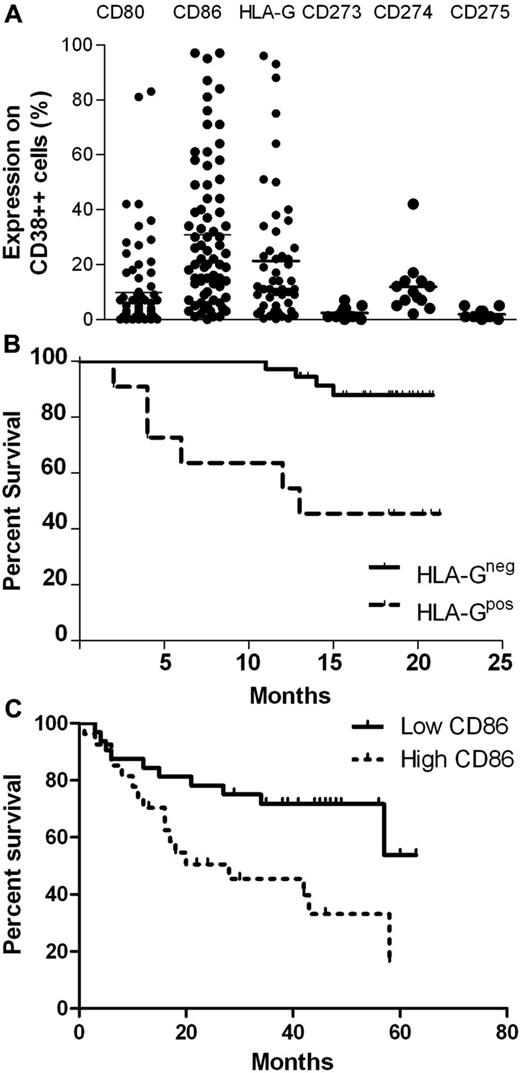
The expression of CD80 (mean 9.8%), CD86 (mean 47.5%) and HLA-G (mean = 10.6%) on CD38++ cells (plasma cells) in BM samples from patients with MM was quite variable (range = 0.2 to 96%; Figure 2A). The expression of other B7 family molecules (CD273, CD274, and CD275) on malignant plasma cells (Figure 2A) was low. As up to 60% of plasma cells in BM samples from a group of normal allogeneic donors had CD86 expression (data not shown), only those MM BM samples with > 60% CD38++ cells were considered to be CD86+.
Expression and functional significance of B7 antigens and HLA-G on plasma cells. (A) Expression of CD80, CD86, HLA-G, CD273, CD274, and CD275 on BM plasma cells (CD38++) of patients with myeloma. (B) Overall survival of patients according to HLA-G expression on BM plasma cells with HLA-G expression > 12% (HLA-G–positive) and < 12% (HLA-G–negative; χ2 = 12.4; P < .0004). (C) Overall survival of patients who had CD86 positive n = 33) and CD86 negative plasma cells (n = 28; χ2 = 6.5; P < .01) at diagnosis with CD86 positivity defined as > 60% (the upper limit of CD86 expression on normal plasma cells).
Expression and functional significance of B7 antigens and HLA-G on plasma cells. (A) Expression of CD80, CD86, HLA-G, CD273, CD274, and CD275 on BM plasma cells (CD38++) of patients with myeloma. (B) Overall survival of patients according to HLA-G expression on BM plasma cells with HLA-G expression > 12% (HLA-G–positive) and < 12% (HLA-G–negative; χ2 = 12.4; P < .0004). (C) Overall survival of patients who had CD86 positive n = 33) and CD86 negative plasma cells (n = 28; χ2 = 6.5; P < .01) at diagnosis with CD86 positivity defined as > 60% (the upper limit of CD86 expression on normal plasma cells).
The clinical relevance of HLA-G+ plasma cells was demonstrated by a significant reduction in overall survival (13 months versus median not achieved) for the 11 of 49 patients with HLA-G+ plasma cells defined as > 5% CD38++ cells and expressing > 12% HLA-G (χ2 = 12.4; P < .0004; Figure 2B). In a univariate analysis CD86 expression on plasma cells at diagnosis (n = 27 of 59) was also associated with a poor prognosis (Figure 2C; χ2 = 6.5; P < .01); however, there was no prognostic significance of the level of CD80 expression on plasma cells. For the current data there was no significant difference in β2-microglobulin level, age or M-protein isotype between either the HLA-G+ and HLA-G− or CD86+ and CD86− groups.
A significant number of myeloma patients have T cells with increased levels of B7 molecules and HLA-G expression
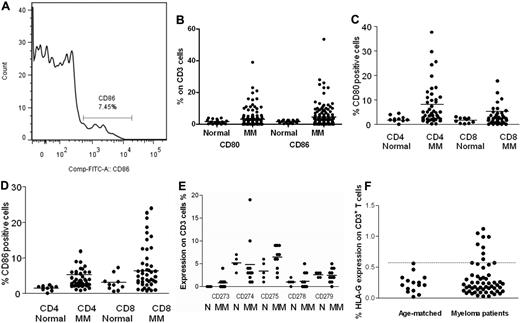
Although T cells normally express counter receptors for costimulatory molecules found at the immunologic synapse (eg, CD28), T cells do not normally express the B7 costimulatory molecules found on APCs (eg, CD80, CD86, CD273, CD274, and CD275). The expression of B7 receptors and ligands involved with the immunologic synapse as well as HLA-G expression on peripheral blood T cells from age-matched controls and myeloma patients was determined (Figure 3). A representative flow histogram of CD86 expression on T cells (Figure 3A), as well as data of CD80 and CD86 expression on CD3, CD4, and CD8 cells of patients with MM (Figure 3B-D) and the expression of B7 costimulatory receptors and their ligands on CD3 cells (Figure 3E) are shown. The expression of CD80 on T cells from MM patients (n = 143) ranged from 0% to 40% (mean = 2.8%), whereas CD86 expression ranged from 0% to 52% (mean = 3.7%). The increased expression of both CD80 (χ2 = 4.7; P < .02) and CD86 (χ2 = 15.3; P < .001) on peripheral blood CD3+ cells of patients with myeloma (n = 143) was significantly different from an aged-matched control group (n = 20). The data shows that CD80 is more probable to be present on CD4 cells than CD8 cells, whereas CD86 is more probable to be present on CD8 cells (Figure 3C-D). CD80 and CD86 expression on both CD4+ and CD8+ cells was almost exclusively found on CD45RO+ cells rather than CD45RA+ cells (data not shown).
B7 antigens, their ligands, and HLA-G on T cells. (A) Representative histogram of CD86 positive T cells. (B) Expression of increased CD80 (χ2 = 4.7; P < .02) and CD86 (χ2 = 15.3; P < .001) on peripheral blood CD3+ cells of patients with myeloma (n = 143) and an aged-matched control group (n = 20). (C) Expression of CD80 and (D) CD86 on peripheral blood CD4+ and CD8+ cells of patients with myeloma (n = 98) and an aged-matched control group (n = 10). (E) Expression of other B7 receptors and ligands involved at the immunologic synapse on CD3 cells of patients with myeloma and age-matched controls. (F) HLA-G expression on normal and myeloma patient T cells. Although expression is low, approximately 20% of myeloma patient's T cells express HLA-G above the age-matched range (dotted line, range 0.02%-0.56%; χ2 = 4.9; P < .03).
B7 antigens, their ligands, and HLA-G on T cells. (A) Representative histogram of CD86 positive T cells. (B) Expression of increased CD80 (χ2 = 4.7; P < .02) and CD86 (χ2 = 15.3; P < .001) on peripheral blood CD3+ cells of patients with myeloma (n = 143) and an aged-matched control group (n = 20). (C) Expression of CD80 and (D) CD86 on peripheral blood CD4+ and CD8+ cells of patients with myeloma (n = 98) and an aged-matched control group (n = 10). (E) Expression of other B7 receptors and ligands involved at the immunologic synapse on CD3 cells of patients with myeloma and age-matched controls. (F) HLA-G expression on normal and myeloma patient T cells. Although expression is low, approximately 20% of myeloma patient's T cells express HLA-G above the age-matched range (dotted line, range 0.02%-0.56%; χ2 = 4.9; P < .03).
Surface expression of CD273 (mean = 0.25%), CD274 (mean = 5.09%), CD275 (mean = 3.30%), CD278 (mean = 1.25%) and CD279 (mean = 2.25%) was measured on CD3+ T cells from normal individuals (n = 5; Figure 3E). Expression of CD273 (mean = 0.13%), CD274 (mean = 3.34%), CD275 (mean = 3.15%), CD278 (mean = 0.46%), and CD279 (mean = 1.39%) was also measured on T cells from MM patients (n = 11). There was no evidence of any significant variation from normal in the MM patients for these antigens.
HLA-G expression was found at low levels on normal T cells (n = 15; mean = 0.24%) and there was a trend toward higher expression on the T cells from patients with MM (n = 56; mean = 0.31%; Figure 3F). There was a significant number (20%) of MM patients with more HLA-G expressing CD3+ T cells than the age matched control range of 0.2% to 0.56% (χ2 = 4.9; P < .03; Figure 3F). There was no significant difference between HLA-G expression on CD4 and CD8 cells and the HLA-G+ T cells were CD25−, thus these cells were not natural Tregs (nTregs).
Both HLA-G and CD86 expression on BM T cells of patients with myeloma correlate with the expression on malignant plasma cells and but not with peripheral blood T cells
Both CD86 and HLA-G expression was significantly higher on the T cells in BM biopsy samples (10.5% and 1.25%, respectively) compared with T cells in peripheral blood samples (3.9% and 0.5%, respectively) tested on the same day (Figure 4A; for CD86: Mann Whitney U = 2973; P < .0001, and for HLA-G: U = 1216; P < .0001). CD86 and HLA-G expression was determined on both CD3+ as well as CD38++ cells in the same BM samples. There was a correlation in the expression of CD86 (r = 0.67; P < .001; n = 29; Figure 4B) and HLA-G (r = 0.82; P < .001; n = 29; Figure 4C) on plasma cells and T cells within the same BM sample for individual patients but there was no correlation between CD86 or HLA-G on T cells in the BM with the corresponding peripheral blood T cells (data not shown).
Correlation between CD86+ and HLA-G+ expression on BM T cells and plasma cells. (A) Increased expression of CD86 on BM T cells (mean = 10.5%) compared with peripheral blood T cells (mean = 3.9%) of patents with MM (U = 2873; P < .0001) and the increased expression of HLA-G on BM T cells (mean = 1.25%) compared with peripheral blood T cells (0.5%; U = 1216; P < .0001). (B) Correlation between expression of CD86 on BM T cells and BM CD38++ cells (r = 0.67; P < .001; n = 29). (C) Correlation between expression of HLA-G on BM T cells and BM CD38++ cells (r = 0.82; P < .001; n = 29).
Correlation between CD86+ and HLA-G+ expression on BM T cells and plasma cells. (A) Increased expression of CD86 on BM T cells (mean = 10.5%) compared with peripheral blood T cells (mean = 3.9%) of patents with MM (U = 2873; P < .0001) and the increased expression of HLA-G on BM T cells (mean = 1.25%) compared with peripheral blood T cells (0.5%; U = 1216; P < .0001). (B) Correlation between expression of CD86 on BM T cells and BM CD38++ cells (r = 0.67; P < .001; n = 29). (C) Correlation between expression of HLA-G on BM T cells and BM CD38++ cells (r = 0.82; P < .001; n = 29).
CD86 and HLA-G can be acquired by T cells from malignant plasma cells in vitro
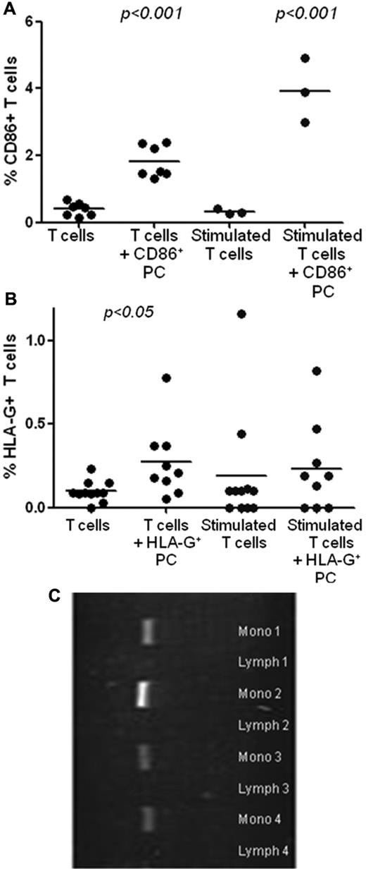
After coculture with CD86+ or HLA-G+ plasma cells, both flow sorted CD3+ cells of patients with myeloma and normal controls acquired significant levels of CD86 or HLA-G, respectively (P < .001; Figure 5A-B). Acquisition of CD86 was significant at 2 hours. T-cell activation (with anti-CD3/2/28 beads) up-regulated HLA-G (t = 6.06; P < .004) but not CD86 expression and activated T cells acquired additional CD86 but not HLA-G from malignant plasma cells.
CD86 on T cells is acquired not produced. (A) Significant acquisition of CD86 by stimulated and unstimulated T cells from CD86+ plasma cells after 2 hour coincubation. (B) Significant acquisition of HLA-G by unstimulated but not stimulated T cells after 2 hour coincubation. (C) CD86 mRNA expression in monocytes and T cells (> 90% pure) of 4 blood samples, which had a high CD86 protein expression on peripheral blood T cells.
CD86 on T cells is acquired not produced. (A) Significant acquisition of CD86 by stimulated and unstimulated T cells from CD86+ plasma cells after 2 hour coincubation. (B) Significant acquisition of HLA-G by unstimulated but not stimulated T cells after 2 hour coincubation. (C) CD86 mRNA expression in monocytes and T cells (> 90% pure) of 4 blood samples, which had a high CD86 protein expression on peripheral blood T cells.
CD86 protein but not mRNA expression is present in T cells
T cells (CD3+) and monocytes (CD14+) were purified from the peripheral blood of 4 patients whose T cells had 10% to 30% CD86 protein expression detected by flow cytometry. The T-cell preparations were > 99% CD3+ and the monocyte preparations were 67% to 83% CD14+. CD86 mRNA transcripts were detected in all the monocyte preparations but CD86 mRNA transcripts were either not detected or only trace amounts seen in CD3+ cell preparations (Figure 5C). Quantitation of CD86 mRNA by real-time PCR confirmed these findings with an average 24.4 times higher CD86 mRNA expression in CD14+ cells than CD3 cells.
HLA-G+ T cells are acquired Tregs with a similar inhibitory function to natural Tregs
Flow-sorted HLA-G+ T cells significantly reduced the proliferation of autologous CFSE-labeled, flow-sorted HLA-G− T cells in 4 day cocultures after stimulation with anti-CD2/3/28 beads (Figure 6A-B). CD86+ T cells had only a minor effect on the proliferation of CFSE labeled CD86− T cells (Figure 6B). The level of inhibition by HLA-G+ T cells was similar to that of CD4+ CD25++ CD127− (nTreg) cells (Figure 6B).
Functional studies of CD86 and HLA-G–positive T cells. (A) Flow sorted HLA-G–negative T cells were labeled with CFSE and cocultured with and without HLA-G–positive T cells. After 4 days of culture, cell proliferation was assessed and the inhibition of proliferation by the HLA-G–positive T cells was determined. (B) The inhibition of HLA-G+ T cells compared with inhibition by CD86+ T cells and CD4+CD25++CD127− Treg cells in cocultures
Functional studies of CD86 and HLA-G–positive T cells. (A) Flow sorted HLA-G–negative T cells were labeled with CFSE and cocultured with and without HLA-G–positive T cells. After 4 days of culture, cell proliferation was assessed and the inhibition of proliferation by the HLA-G–positive T cells was determined. (B) The inhibition of HLA-G+ T cells compared with inhibition by CD86+ T cells and CD4+CD25++CD127− Treg cells in cocultures
HLA-G is present on both SP and non-SP primary myeloma cells
HLA-G expression was determined on both SP-positive and non-SP plasma cells. BM samples (n = 26) from patients with MM and > 2% CD38++ cells were stained with Hoechst 33342, anti-CD38 PE, anti-CD138 APC, and anti–HLA-G FITC. The SP population ranged from 0.0% to 16.9% of the CD38++ cells. A representative example is shown in Figure 7A. Sixteen of 26 samples analyzed had insufficient cells to analyze minor subpopulations of the small number of HLA-G+ SP+ cells and were not included. Of the remaining 10 patients, HLA-G expression was present on 0.4% to 19.1% of the SP cells. HLA-G expression tended to be higher on the SP cells than non-SP cells and using a cut off of 10% there were more SP than non-SP cells with HLA-G expression (χ2 = 5.0; P < .025; Figure 7B).
HLA-G expression on SP myeloma cells. (A) HLA-G expression on the side population content of plasma cells (CD38++ CD138+) and the remaining non-SP plasma cells in a BM sample of a representative patient. (B) Ten patients with sufficient CD38++ SP+ cells for analysis and with > 1% HLA-G expression were analyzed for HLA-G expression on CD38++ SP cells and demonstrated a trend for higher HLA-G on SP cells (χ2 = 5.0; P < .025).
HLA-G expression on SP myeloma cells. (A) HLA-G expression on the side population content of plasma cells (CD38++ CD138+) and the remaining non-SP plasma cells in a BM sample of a representative patient. (B) Ten patients with sufficient CD38++ SP+ cells for analysis and with > 1% HLA-G expression were analyzed for HLA-G expression on CD38++ SP cells and demonstrated a trend for higher HLA-G on SP cells (χ2 = 5.0; P < .025).
Discussion
The importance of the acquisition of ectopic, tumor-derived antigens onto cytotoxic effector cells in patients with hematologic malignancies is poorly understood. This study adds further evidence to previous suggestions that this process, is an important cause of immune dysfunction and contributes to the inadequate antitumor response in these patients.7-10,13 Although it is probable that trogocytosis will lead to the transfer of many molecules that have little functional significance, our studies have identified 2 clinically significant molecules, CD86 and HLA-G, which are acquired from malignant plasma cells by T cells. We have shown high levels of these molecules on malignant plasma cells are associated with a poor prognosis and that when there is transfer of these molecules to T cells there is an impact on the function of effector T cells.
T cells acquired significantly more MM membrane proteins than natural killer (NK) and B cells, and as malignant cells acquired little membrane protein, trogocytosis was considered to be predominantly unidirectional. We found that the uptake by T cells of patients with MM was not different from T cells of age-matched normals and the malignant plasma cells exhibited a greater level of trogocytosis than other malignant B cells, thus suggesting that the acquisition of antigens is more related to the nature of the donor cells than the recipient cells. This is consistent with previous studies that showed that the direction of membrane exchange by trogocytosis is determined by the nature of the donor cell and not the recipient T cell.16 It also suggests that the original concept that trogocytosis is a “nibbling” of the donor cells by the recipient cells may not be quite correct.1
Using in vitro mixing studies we demonstrated that there was a greater transfer of CD86 than HLA-G from malignant plasma cells to T cells. It was then not surprising to find that when assaying blood samples, HLA-G expression on T cells of patients with MM was never as high as the levels of CD86. It was also shown that T cells that acquired HLA-G were far more potent suppressor cells in vitro than CD86+ T cells. Thus the main novel function of T cells that acquire CD86 may be as inefficient APCs rather than as inhibitors.11,12 Data are presented from functional studies, which demonstrated that HLA-G+ T cells have a similar level of inhibitory function to that of natural Tregs in CFSE suppression assays.17 As these HLA-G+ T cells were CD25− and FoxP3− they should be considered to be acquired Treg cells (Tregacq) rather than natural Treg cells. Tregacq cells warrant considerable more attention than has been previously given to them.
For both HLA-G and CD86, the expression on BM plasma cell correlated with the expression on BM T cells but not on peripheral blood T cells. This supports the concept that the process of trogocytosis occurs in the BM microenvironment and suggests that HLA-G+ and CD86+ T cells may constitute a subpopulation of tumor-infiltrating and perhaps even tumor-specific lymphocytes in patients with MM.13
The clinical significance of HLA-G in patients with MM has been highlighted in these studies by the demonstration that MM patients with more than 23% HLA-G–positive cells had a significantly shorter progression-free survival time than those patients who had less than 23% positive cells (median progression-free survival: 120 months vs 23 months; P = .0001). Much of our understanding of HLA-G has come from the expression of HLA-G on cytotrophoblasts at the feto-maternal interface where HLA-G inhibits the cytolytic function of maternal NK cells allowing immune tolerance of the semiallogeneic fetus.18 Both membrane-bound and soluble forms of HLA-G have been shown to prevent cytolytic T cells from carrying out antigen-specific cytolysis, inhibit the function of circulating NK cells, prevent CD4+ T cells alloproliferating,19 and inhibit maturation of dendritic cells.20 In addition, Fournel et al demonstrated induction of apoptosis of CD8+ T cells by soluble HLA-G (sHLA-G).21 High level HLA-G expression was found in an increasing number of pathologic conditions, including hematologic and nonhematologic malignancies, on monocytes in HIV patients, and in autoimmune and inflammatory diseases. It has been hypothesized that expression of “tolerogenic” HLA-G may aid in the escape of allotransplants from immune surveillance,22 and that trogocytosis of HLA-G from HLA-G expressing tumors to T and NK cells may generate tolerance and contribute to tumor evasion of immune surveillance.7-9,23
Carosella et al reported that HLA-G transcription and protein expression were up-regulated in tumor cells and suggested that this protects them from cytolysis.23 They demonstrated that in breast and ovarian carcinomas, as well as in melanocytic lesions, HLA-G expression protected tumors against NK cytolysis and correlated with the stage of malignancy. An unfavorable outcome was also observed in gastric and colorectal cancers as well as in CLL. Increased HLA-G expression was observed in various malignant hematopoietic diseases including B-CLL, acute myeloid leukemia, and acute lymphoblastic leukemia.23
Feger et al reported that a subset of CD4+ and CD8+ T cells expressing HLA-G was reported by under normal physiologic conditions in peripheral blood from healthy donors.24 We confirmed this in our studies. Using the monoclonal antibodies against HLA-G (MEM/G9 and 87G clones), to assess surface expression of HLA-G by flow cytometry we found an average of 1.6% (range 0.1%-8.3%) of CD4+ and 3.3% (range 0.6%-5.2%) of CD8+ T cells express HLA-G in age-matched controls. We also confirmed that secondary stimulation by anti–CD3/CD28 beads or IL-2 failed to overcome the hypoproliferation of HLA-G+ subsets.24 Whereas the lack of CD86 mRNA in MM T cells clearly demonstrated that CD86 protein expression on T cells is acquired, the low levels of endogenous HLA-G mRNA present in T cells prevented similar observations for HLA-G.
It has been proposed that HLA-G plays a role in tumor escape from the immune system in B-CLL. HLA-G expression on circulating B-CLL cells was found to vary from 1% to 54%.25,26 HLA-G transcription and expression in CLL patients demonstrated tumor cell protection against autologous NK lysis thus permitting tumor development by impairing antitumor immunity. Furthermore, higher levels correlated with increased immunosuppression as judged by immunoglobulin-G levels, CD4+ T cells, and total T-cell numbers.26,27 HLA-G expression has also been studied in patients with non-Hodgkin lymphoma (NHL). Although one study showed no expression,28 other studies reported 65%-100% HLA-G expression in B-NHL,29 and 58% expression in T-NHL.30 Similar results for MM are now reported in this study. In acute myeloid leukemia, no expression of HLA-G was reported by one group,28 and another study reported HLA-G expression in 20% of patients.31
It is considered that chemotherapy fails to remove residual drug-resistant cancer stem cells.32,33 The possibility that myeloma stem cells contribute to immune tolerance is an important question. Our observation that there is often a high level of HLA-G expression on myeloma SP cells compared with non-SP cells provides evidence to suggest that these putative myeloma stem cells33 are not only drug resistant but also that HLA-G expression may be protecting them from the host's immune defenses.
Feger et al performed suppression assays in which HLA-G+ T cells inhibited the proliferation of autologous CFSE-labeled HLA-G− T cells when cultured with allogeneic APCs.24 Inhibition was shown to be partially antagonized with the addition of anti–HLA-G antibodies. This suggested that HLA-G is not only a phenotypic marker but is also responsible for the impaired proliferation. Transwell experiments demonstrated that cell contact was required for suppression of proliferation.24
Previous studies demonstrated that an increase in CD86 expression on malignant cells is associated with a poor prognosis in a variety of malignancies including acute myeloid leukemia and thyroid carcinoma34,35 ; however, there has been a lack of consensus on the prognostic significance of CD86 in myeloma.36,37
In murine studies, Sabzevari et al analyzed CD80 levels on T cells after stimulation with APCs and demonstrated various levels of CD80 expression.11 It was previously thought that CD80 was endogenously up-regulated on T cells, but these studies showed that CD80 is physically acquired by T cells from APCs soon after T-cell activation. CD80 acquisition was mediated via the TCR and CD28 ligand and the level of CD80 acquisition by T cells related to both the expression of CD80 on APCs and the strength of signal 1. Our studies using samples from patients with myeloma have verified this observation for CD86, by demonstrating that the T cells do not produce CD86 mRNA transcripts. It has been suggested that the acquisition of CD80 might be of significance for the antigen-presenting aptitude of T cells.11 CD80+ and CD86+ T cells may thus act as inept APCs causing anergy or apoptosis.12,38 However, it has also been elegantly demonstrated that acquisition of B7 molecules also leads to deletion or fraticide.11,16 It has been reported that direct contact with B-CLL cells can cause impaired actin polymerization in T cells resulting in a defective immunologic synapse formation with APCs.39 However, in our studies the acquisition of MM membrane patches by T cells was independent of the TCR, suggesting that other mechanisms are also involved.
It was not surprising that both the CD4 and CD8 cells that acquired CD86 were almost exclusively memory T cells (CD45RO) rather than inexperienced naive T cells. However one unresolved issue is why CD4 cells tend to have higher CD80 expression and CD8 cells tend to have higher CD86 expression (Figure 3C-D). Perhaps there is preferential binding by CD86 to the tolerance inducing coreceptor CD152 in the BM microenvironment.
Trogocytosis may contribute to lymphocyte activation by prolonging the engagement of antigen receptors after APC and T-cell separation. Trogocytosis by cytotoxic lymphocytes might contribute to the selection of high-affinity T cells through the removal of antigenic complexes from the APCs and to the elimination of cytotoxic T cells through a process known as fraticide after the acquisition of pMHC molecules.9 However, this would only occur when there is an extremely high concentration of MHC class I–restricted antigenic peptide.1,11,40 Interestingly this situation occurs in patients with myeloma but not in CLL as patients with MM would at one stage have had overwhelming levels of tumor antigen causing T-cell exhaustion. Therefore, we should now consider that there is probably T-cell fracticide and deletion in these patients. There is strong support for this concept from the MOPC315 murine model in which it has been shown that idiotype-specifc T cells are deleted after the myeloma idiotype level in blood exceeds 50 ug/L.40
A better understanding of the mechanisms involved with tumor-induced suppression of the immune system is crucial for overcoming tolerance and introducing immune based therapies. The current studies highlight that it is a common phenomena for cytotoxic effector cells from patients to change their function after acquiring molecules from malignant cells. We need to acknowledge that trogocytosis is a common mechanism which enables tumors to disrupt the immune response.
There is an Inside Blood commentary on this article in this issue.
The publication costs of this article were defrayed in part by page charge payment. Therefore, and solely to indicate this fact, this article is hereby marked “advertisement” in accordance with 18 USC section 1734.
Acknowledgments
The work was supported by Sydney Foundation for Medical Research and the Cancer Institute of New South Wales.
Authorship
Contribution: R.B. designed the research and wrote the paper; K.K., J.F., S.Y., and H.S. performed research; J.G., D.H., P.J.H., P.F., and D.J. designed the research and contributed to writing the paper; and N.W. and N.N. contributed to writing the paper.
Conflict-of-interest disclosure: The authors declare no competing financial interests.
Correspondence: Ross Brown, Institute of Haematology, Royal Prince Alfred Hospital, Missenden Rd, Camperdown, NSW 2120, Australia; e-mail: ross.brown@sswahs.nsw.gov.au.


This feature is available to Subscribers Only
Sign In or Create an Account Close Modal