Human Vγ9Vδ2 T cells kill a broad range of solid tumor and leukemia/lymphoma cells. In this issue of Blood, Harly et al demonstrate a pivotal role of CD277/butyrophilin-3 for the activation of γδ T cells.1
Gamma delta (γδ) T cells expressing the Vγ9Vδ2 T-cell receptor account for approximately 5% of peripheral blood T cells. Many solid tumor and leukemia/lymphoma cells are susceptible to Vγ9Vδ2 T cell–mediated killing.2-4 Antitumor reactivity and the ability to intentionally increase this activity (see below) have raised great interest in exploring the immunotherapeutic potential of Vγ9Vδ2 T cells.5 In contrast to conventional αβ T cells, γδ T cells do not recognize antigenic peptides presented by MHC class I or class II molecules. It has been shown that human (and primate) γδ T cells expressing the Vγ9Vδ2 T-cell receptor recognize phosphorylated nonpeptide metabolites that can be secreted by many microbes. Such microbial “phosphoantigens” are extremely potent and specific activators of Vγ9Vδ2 T cells and cause their transient increase in the peripheral blood during the acute phase of many bacterial and parasitic infections.6 The microbial phosphoantigens are intermediates of the prokaryotic nonmevalonate pathway of cholesterol synthesis, and are active at pico- to nanomolar concentrations. Eukaryotic cells use the mevalonate pathway for cholesterol synthesis. Isopentenyl pyrophosphate (IPP), a naturally occurring endogenous phosphoantigen, is also seen by the Vγ9Vδ2 T-cell receptor but requires 3-log higher (micromolar) concentrations for γδ T-cell activation. Such concentrations do not accumulate in normal cells. On cellular transformation, however, much higher concentrations of IPP are produced, which then can be sensed by γδ T cells as tumor-associated antigen.
Interestingly, the levels of IPP production can be easily manipulated. Aminobisphosphonates (n-BP), which are in clinical use for the treatment of osteoporosis and bone metastasis in some cancer patients, inhibit an enzyme that degrades IPP, whereas statins reduce the endogenous production of IPP.3 In consequence, treatment of tumor cells with n-BP increases their susceptibility to γδ T cell–mediated lysis, because of enhanced IPP production.2,3 As a matter of fact, the therapeutic application of n-BP together with low-dose IL-2 results in increase of peripheral blood γδ T-cell numbers associated with some clinical benefit in patients with lymphoid malignancies7 or solid tumors.8
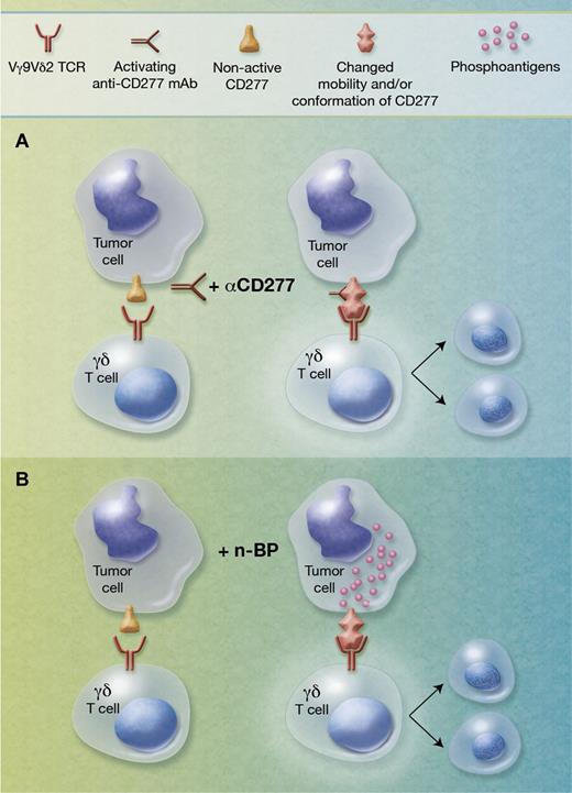
T-cell receptor gene transfer and mutagenesis studies have established that the T-cell receptor is crucially involved in the activation of Vγ9Vδ2 T cells by microbial or tumor-derived phosphoantigens. However, the mechanism of phosphoantigen recognition has remained undefined. Early studies indicated that such phosphoantigens do not require antigen processing nor do they require presentation by classic MHC molecules or MHC-related molecules such as CD1. Interestingly, however, a level of species restriction was noticed in that human but not murine cells were found capable of presenting such phosphoantigens to human γδ T cells. Previous work suggested that an ectopically surface-expressed ATPase might be involved in the cell-surface presentation of phosphoantigens.9 The work now reported by Harly et al represents a significant advancement in our understanding of how phosphoantigens activate human Vγ9Vδ2 T cells.1 These authors describe a central role for CD277, a member of the butyrophilin molecules, in this process. Butyrophylins are distantly related to known costimulatory molecules of the B7 superfamily. By generating a panel of activating or inhibitory anti-CD277 monoclonal antibodies (mAbs), Harly and coworkers uncovered that certain activating anti-CD277 mAbs induced a selective and exclusive activation and proliferation of Vγ9Vδ2 T cells when added to in vitro cultures of peripheral blood lymphocytes, similar to what is seen when phosphoantigens or n-BP are used for in vitro stimulation. On the other hand, they had developed other inhibitory anti-CD277 mAbs that specifically blocked the activation and proliferation of Vγ9Vδ2 T cells in response to phosphoantigens and n-BP. Moreover, the recognition of Vγ9Vδ2-susceptible lymphoma cells was also strongly inhibited by the blocking anti-CD277 mAbs. A series of elegant experiments then addressed the question of how the CD277 molecule contributes to the phosphoantigen-mediated γδ T-cell activation. To this end, they specifically knocked down the expression of different isoforms of the CD277 molecule and found out that the membrane mobility of CD277 was drastically influenced by phosphoantigens through the intracellular part of one particular isoform. Together with additional experiments, the results of this study suggest that activating anti-CD277 mAbs and phosphoantigens modify the CD277 molecule in such a way that the γδ T-cell receptor is triggered on coculture with CD277-expressing cells. It appears that agonistic anti-CD277 mAbs induce changes in the membrane mobility of the CD277 molecules that are somehow sensed by the Vγ9Vδ2 T-cell receptor (see figure panel A). In the case of phosphoantigens generated in tumor cells, particularly after tretament with n-BP, the presented data are in line with the assumption that the intracellularly generated phosphoantigens (ie, IPP) act through the intracytoplasmic domain of one CD277 isoform to induce a membrane reorganization of the CD277 molecule (see figure panel B).
Role of CD277/butyrophilin-3A in triggering human γδ T-cell activation. (A) The ubiquitously expressed CD277 molecule is not recognized by the Vγ9Vδ2 T-cell receptor (left). Upon incubation with agonistic anti-CD277 mAbs, changes (mobility and/or conformation) are induced in the CD277 molecule such that Vγ9Vδ2 T cells are activated in a T-cell receptor–dependent manner (right). (B) Some tumor cells are poorly recognized by Vγ9Vδ2 T cells (left). Upon treatment with aminobisphosphonates (n-BP), increased levels of phosphoantigens are produced that induce changes in the membrane mobility of CD277 molecules leading to activation of Vγ9Vδ2 T cells (right). Professional illustration by Alice Y. Chen.
Role of CD277/butyrophilin-3A in triggering human γδ T-cell activation. (A) The ubiquitously expressed CD277 molecule is not recognized by the Vγ9Vδ2 T-cell receptor (left). Upon incubation with agonistic anti-CD277 mAbs, changes (mobility and/or conformation) are induced in the CD277 molecule such that Vγ9Vδ2 T cells are activated in a T-cell receptor–dependent manner (right). (B) Some tumor cells are poorly recognized by Vγ9Vδ2 T cells (left). Upon treatment with aminobisphosphonates (n-BP), increased levels of phosphoantigens are produced that induce changes in the membrane mobility of CD277 molecules leading to activation of Vγ9Vδ2 T cells (right). Professional illustration by Alice Y. Chen.
The studies of Harly et al have undoubtedly discerned a pivotal role of the CD277 molecule for the activation of human Vγ9Vδ2 T cells. This notwithstanding, several issues require further analysis. Most importantly, it is presently unknown how the Vγ9Vδ2 T cells recognize the phosphoantigen (or anti-CD277 mAb)–induced changes of the CD277 surface molecule. The possibility of direct interaction of the Vγ9Vδ2 T-cell receptor with the modified CD277 molecule appears unlikely on the basis of preliminary experiments mentioned in their report.1 It is also possible that modified CD277 molecule might recruit additional molecules that are then sensed by the Vγ9Vδ2 T-cell receptor. In any case, the key role of CD277 for activation of Vγ9Vδ2 T cells described by Harly et al also provides an explanation as to why murine cells cannot act as presenting cells for phosphoantigen-reactive human Vγ9Vδ2 T cells: there is no CD277 ortholog in rodents.
Apart from providing novel insights into the mechanisms of phosphoantigen-dependent γδ T-cell activation, this study also opens new translational aspects. Agonistic anti-CD277 mAbs might be useful reagents to boost the antitumor and antilymphoma/leukemia efficacy of human Vγ9Vδ2 T cells. Considering that this population comprises up to 5% of all blood T cells, new strategies targeting the immunotherapeutic potential of Vγ9Vδ2 T cells might thus have major clinical implications.
Conflict-of-interest disclosure: The author declares no competing financial interests. ■


This feature is available to Subscribers Only
Sign In or Create an Account Close Modal