Abstract
Abstract 2637
Hodgkin lymphoma (HL) has traditionally been regarded as a disease primarily affecting the lymphatic organs. The introduction of dual modality [18]F-fluorodeoxyglucose positron emission tomography/computed tomography (PET/CT) may challenge this statement as PET/CT reveals more sites of extranodal disease than CT alone. The aim of this study was to characterize the clinico-pathological and prognostic features of newly diagnosed HL patients with PET/CT-ascertained skeletal involvement.
We performed a retrospective analysis of the pre-therapeutic PET/CT results from newly diagnosed HL patients at four Danish university hospitals. Criteria for inclusion in the study were (a) histologically verified HL, (b) age ≥15 years, and (c) PET/CT-based staging. Clinical information was obtained from the Danish Lymphoma Registry (LYFO) supplemented by review of medical records. Imaging reports were reviewed for extent of disease. Patients were divided into three groups according to their pattern of PET/CT-positivity: HL confined to lymphatic organs (lymph nodes, spleen, thymus, Waldeyer's ring) (group 1); HL with focal skeletal PET/CT lesions as the only extranodal involvement (group 2); HL with any extranodal involvement, including skeletal lesions if other extranodal sites were concurrently involved (group 3).
A total of 454 patients with newly diagnosed HL were included and the median follow-up time was 34 months (range 1–113). Focal skeletal PET/CT lesions were identified in 82 patients (18%). HL was confined to nodal areas in 336 (74%) patients (group 1). Among the 118 (26%) patients with extranodal disease, 39 had focal skeletal PET/CT lesions as their only extranodal manifestation (group 2), while 79 had involvement of other extranodal sites with or without co-existing skeletal lesions (groups 3). Group 1 patients exhibited the most favorable combination of clinico-pathological features, group 3 patients displayed most adverse combination of clinico-pathological features, whereas group 2 was intermediate (Table). In a 1:1 comparison between group 1 and 2 patients, the latter had significantly lower hemoglobin (Hgb) levels, higher erythrocyte sedimentation rates (ESR) and higher occurrence of B-symptoms.
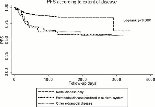
The PFS for group 2 patients was similar to that of group 3 patients (p=0.85), but significantly shorter than that of group 1 patients (p<0.001) (Figure). After adjusting for other risk factors in a multivariate Cox model, the presence of focal skeletal PET/CT lesions remained independently associated with shorter PFS (p=0.009).
PET/CT-based identification of focal skeletal lesions as the only extranodal involvement in patients with newly diagnosed HL is not uncommon. This group of patients frequently presents with systemic symptoms and elevated ESR. In addition, their PFS is inferior to that of patients with nodal HL, but similar to that of HL patients with other types of extranodal disease. Although rarely histologically verified, these focal skeletal lesions are thus likely to represent extranodal manifestations of HL.
| . | Group 1 (n=336) . | Group 2 (n=39) . | Group 3 (n=79) . | p-valueμ . |
|---|---|---|---|---|
| Median age, years | 37 | 46 | 50 | 0.002 |
| ECOG Performance, % | . | . | . | |
| .0 | 77 | 69 | 40 <0.001 | |
| .1 | 20 | 23 | 46 | |
| .2 | 3 | 3 | 6 | |
| .3 | 0 | 5 | 5 | |
| .4 | 0 | 0 | 3 | |
| Male:female ratio | 1.21 | 1.79 | 1.55 | 0.4 |
| Histological subtype, % | . | . | . | |
| . Nodular sclerosis | 56 | 59 | 60 | 0.09 |
| . Mixed cellularity | 21 | 21 | 15 | |
| . Lymphocyte rich | 5 | 3 | 3 | |
| . Lymphocyte depleted | 1 | 0 | 3 | |
| . Classical Hodgkin, unspecified | 10 | 15 | 19 | |
| . Nodular lymphocytic predominant | 7 | 2 | 0 | |
| Hemoglobin, g/dL | 13.2 | 12.4 | 11.6 | 0.0001* |
| Total leukocytes, 109/L | 8.7 | 9.3 | 9.6 | 0.06 |
| Thrombocytes, 109/L | 308 | 363 | 354 | 0.02 |
| Lymphocytes, 109/L | 1.60 | 1.70 | 1.34 | 0.01 |
| Erythrocyte sedimentation rate, mm/h | 26 | 46 | 63 | 0.0001* |
| Serum albumin, g/l | 42 | 40 | 35 | 0.0001 |
| Serum LDH, IU/L | 186 | 209 | 224 | 0.005 |
| B-symptoms, % | 38 | 56 | 78 | <0.001* |
Fisher's exact test for categorical variables; Kruskal-Wallis test for continuous values.
Group 2 differs significantly from group 1 (Wilcoxon rank-sum test).
Hutchings:Takeda/Millennium: Consultancy.
Author notes
Asterisk with author names denotes non-ASH members.


This feature is available to Subscribers Only
Sign In or Create an Account Close Modal