Abstract
Abstract 4302
Conventionally, renal involvement in pediatric acute leukemia have been diagnosed by ultrasonography according to the definition of unilateral or bilateral nephromegaly based on the length of major or minor axis >2SD, diffuse abnormal findings, or mass legions. However, if renal involvements are determined just only by the length of major or minor axis>2SD, overdiagnosis seems to be inevitable. Moreover, it might be difficult to detect renal mass legions by ultrasonography compared with other methods. To compensate for these shortcomings, in our hospital, we utilize contrast-enhanced CT to evaluate renal mass legions and to measure renal volume by utilizing 3D reconstruction models of CT imagings instead of measuring the length of major and minor axis of the kidney. Only when markedly decrease of renal volume before and after induction chemotherapy is observed, we define the nephromegaly at diagnosis as a true renal leukemic involvement. To assess whether our 3D-CT-based criteria for leukemic invasion to kidney could be superior to the conventional criteria, we retrospectively compared renal involvements in pediatric leukemia by conventional criteria with by 3D-CT-based criteria.
Between 2006 to 2012, 32 children were diagnosed with acute leukemia in our hospital. Enhanced CT scan was performed to the all patients before induction therapy, and a follow-up CT scan was performed after induction therapy or later stage on the patients who had abnormal CT findings at diagnosis. Mass legions, length of major and minor axis, and volume changes by utilizing enhanced CT and 3D reconstruction models calculated with the software SYNAPSE VINCENT (FUJIFILM MEDICAL), were evaluated.
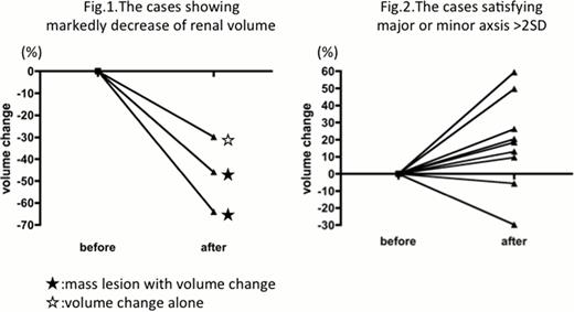
21 out of 32 patients (65.6%) presented with renal involvement when conventional criteria was used (major or minor axis >2SD, or diffuse abnormal findings or mass legions). However, when applied to our 3D-CT- based criteria (mass legions or marked decrease of renal volume before and after induction therapy), only 6 patients out of 29 patients (20.7%) showed renal involvement (volume change alone 1, mass lesions without volume change 3, mass lesion with volume change 2 as shown in fig.1). The main reasons for the discrepancy between criteria for renal invasions were as follows. In the cases diagnosed with nephromegaly by satisfying major or minor axsis >2SD, only one case showed markedly decrease of renal volume before and after induction chemotherapy, and other cases did not show any renal volume changes and even in some cases, contrary to our expectation, renal volume increased as shown in fig.2. It suggested that the nephromegaly defined by only renal length include many false positive cases. Most importantly, renal mass legions could be detected only by enhanced CT, not by ultrasonography in some cases.
Our 3D-CT-based criteria for detection of leukemia invasion to kidney is much more reliable than the conventional criteria based on nephromegaly measured by major or minor axis.
No relevant conflicts of interest to declare.
Author notes
Asterisk with author names denotes non-ASH members.


This feature is available to Subscribers Only
Sign In or Create an Account Close Modal