Abstract
After treatment with glucocorticoids (e.g. dexamethasone) or catecholamines (e.g. epinephrine), the white blood cell (WBC) count substantially increases. This is primarily due to WBCs shifting from the marginated to circulating pools (Nakagawa et al., Circulation, 2008) and is traditionally attributed to down-regulation of adhesion molecule expression (Weber et al., J Leukoc Biol, 2004).Recent research has described how mechanical properties determine the radial position of blood cells within the intravascular space (Reasor et. al, Ann Biomed Eng., 2013). In addition, because WBC demargination occurs rapidly (e.g.,<15 min after IV epinephrine infusion (Dimitrov et al., J Immunol. 2010)) on a timescale that may be shorter than that expected for alterations in gene expression, we hypothesized that alterations in WBC mechanical properties upon exposure to glucocorticoids or catecholamines mediate demargination.
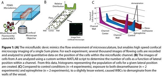
To that end, we developed an in vitro microfluidic system as a simplified microvasculature model (Fig 1A), which our laboratory has expertise in (Tsai et al., J Clin Invest., 2008 and Rosenbluth et al., Biophys J. , 2006). In the absence of confounding factors such as WBC release from bone marrow or endothelial interactions, this type of assay is ideally suited to determine the role of glucocorticoid and catecholamine treatment on the demargination of WBCs. By flowing whole blood into similar non-functionalized microfluidic devices, other groups have demonstrated that non-activated WBCs marginate to the microfluidic channel wall, which is likely due to their mechanical properties (Jain et al., PLoS One, 2009). Human whole blood was incubated at 37° C with acridine orange (WBC stain) and either dexamethasone or epinephrine at physiologically relevant concentrations. The blood was then flowed through our microfluidics at physiologic shear rates while confocal videomicroscopy was used to image the center plane of the channel. We developed custom analysis software that extracts the position of individual WBCs from a series of confocal images and plots histograms of their locations, tracking over 10,000 WBCs per experiment (Fig 1B). Overall, we found that both dexamethasone and epinephrine (to a slightly lesser extent) cause WBCs to demarginate from the walls of the vessel compared to control conditions (Fig 1C). This glucocorticoid and catecholamine-induced movement of WBCs toward the microchannel center mimics in vivo demargination and our reductionist microfluidic approach strongly suggests that alterations in WBC mechanics play a key role in this process.
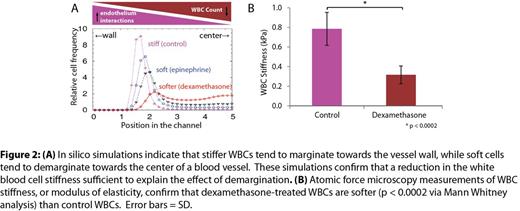
Indeed, using computational modeling, we confirmed that a reduction in the mechanical stiffness of WBCs is sufficient by itself to explain the observed demargination (Fig 2A) (Kumar et al., Phys Rev Lett., 2012). Using a range of WBC stiffnesses, our simulations revealed that decreases in WBC stiffness correlated with the degree of demargination. To corroborate our microfluidic data, we also directly measured WBC stiffness using atomic force microscopy. WBCs treated with dexamethasone were significantly softer (p< 0.0002) than control WBCs (Fig 2B), supporting our hypothesis that the demargination phenomenon is related to the biophysical changes in WBCs. Experiments measuring the stiffness of epinephrine-treated cells as well as experiments evaluating how these drugs affect the actin cytoskeleton are currently underway.
Overall, our data suggest that WBC mechanics play a major role in glucocorticoid- and catecholamine-induced demargination and that the underlying mechanisms may, at least in part, be biophysical in nature. This novel finding may have important implications in other hematologic processes such as WBC margination and recruitment during inflammatory responses or hematopoietic stem cell mobilization and homing.
No relevant conflicts of interest to declare.
Author notes
Asterisk with author names denotes non-ASH members.


This feature is available to Subscribers Only
Sign In or Create an Account Close Modal