Abstract
Next to Sub-Saharan Africa, the Caribbean has the second highest prevalence of human immunodeficiency virus type 1 (HIV-1) infection; and human T-cell lymphotropic virus type 1 (HTLV-1), another human retrovirus, is also endemic. Fewer than five percent of persons with HTLV-1, predominantly transmitted through breast feeding, progress to any of the myriad associated diseases, including adult T-cell leukemia, HTLV-1 associated myelopathy/Tropical Spastic Paraparesis (HAM/TSP), or polymyositis. HTLV-1, through its oncoprotein-like transactivating (Tax) protein, results in aberrant proliferation of CD4 cells through interaction with cell cycle regulators, and activation of nuclear factor kappa B and the interleukin-2 pathway. Elevated CD4 counts, often above normal range, observed in patients with HTLV-1/HIV-1 co-infection, may be spurious, posing a challenge for laboratory monitoring of lymphoproliferative disorders or HIV-1 progression.
We compared CD4 counts, HIV-1 viral load, and clinical parameters in a cohort of patients co-infected with HTLV-1 and HIV-1 (syphilis, hepatitis B and C non-reactive), with controls with HIV-1 infection only matched for age, sex, and duration of antiretroviral therapy, as well as donors without HTLV-1 or HIV-1 infection. We used flow cytometry to characterize CD4 and CD8 naïve and memory T-cell subsets, expression of T-cell survival and homeostatic cytokine interleukin-7 alpha receptor (CD127), and examined the role of co-inhibitory programmed death-1 in HTLV-1 infected (intracellular HTLV-1 Tax protein-expressing) and HIV-1 infected (intracellular HIV-1 Gag protein-expressing) CD4 cells in immune evasion. Additionally, we assessed the effects of exogenous interleukin-7 (IL-7) and programmed death-1 pathway blockade on the function of responding CD8 cytotoxic T-lymphocyte (CTL) subsets ex vivo.
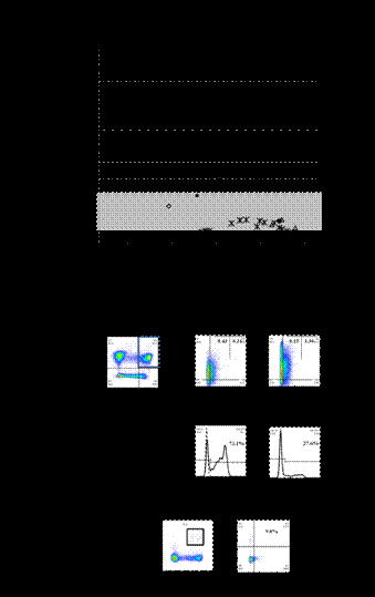
All patients with HTLV-1 and HIV-1 co-infection (n = 5) were female and of median age 42 years (range, 22 to 49 years), with normal or above normal CD4 counts. Four of five patients with HTLV-1/HIV-1 co-infection were WHO Stage 4 HIV/AIDS at diagnosis (e.g., oesophageal candidiasis, HIV nephropathy). Median duration since diagnosis with HIV-1 infection was six years (range, 1 to 18 years), and unexpectedly high CD4 count was the reason for HTLV-1/2 testing in all cases. Nadir CD4 count among patients with HTLV-1/HIV-1 co-infection was significantly higher than controls with HIV-1 infection only (n = 12) matched for age, sex, and duration of antiretroviral therapy (median 684, range 467 to 1474; and median 36, range, 8 to 352 cells per microliter, respectively; p = 0.007, Mann-Whitney U test). Flow cytometric analyses of CD8 and CD4 T-cell memory subsets revealed more profound loss of CD127 in the CD8 compared to the CD4 compartment; and CD8(+) effector memory T-cells showed the most significant downregulation of CD127 when patients with HTLV-1/HIV-1 co-infection were compared with seronegative donors (40.3±17.0%, n =5; and 73.1±7.48%, n = 10, respectively; p = 0.032, Mann-Whitney U). In ex vivo experiments in the context of peripheral blood mononuclear cells, preliminary data suggest that patients with HTLV-1/HIV-1 co-infection have impaired CD8(+) CTL function in response to HLA-restricted HTLV-1 Tax11-19 and cytomegalovirus (CMV) pp65 peptide stimulation. Further, we sought to describe expression of co-inhibitory marker programmed death-1 ligand (PDL1) on intracellular HTLV-1 Tax protein- as well as intracellular HIV-1 Gag protein-expressing CD4 cells. Blockade of PDL1 resulted in partial recovery of CTL function in patients with HTLV-1/HIV co-infection, and enhanced killing of target cells infected with either HTLV-1 or HIV-1.
Patients with HTLV-1/HIV-1 co-infection show profound CTL exhaustion; and CD4 cells, although within or above normal CD4 count range, show aberrant naïve and memory T-cell distribution and possible immune evasion by upregulation of the programmed death-1 pathway, blockade of which showed a tendency towards enhanced virus-specific CTL function and killing of target cells infected with HTLV-1 or HIV-1.
(A) CD4 T-cell counts for Jamaican patients with HTLV-1/HIV-1 co-infection. (B) Expression of CD127 in CD8(+) T-cell memory compartments. (C) Flow cytometry dot plot showing CMV-specific CD8(+) cells (left), and CD107a expression in CMV-specific CTLs.
(A) CD4 T-cell counts for Jamaican patients with HTLV-1/HIV-1 co-infection. (B) Expression of CD127 in CD8(+) T-cell memory compartments. (C) Flow cytometry dot plot showing CMV-specific CD8(+) cells (left), and CD107a expression in CMV-specific CTLs.
No relevant conflicts of interest to declare.
Author notes
Asterisk with author names denotes non-ASH members.


This feature is available to Subscribers Only
Sign In or Create an Account Close Modal