The ability of adoptively infused NK cells to home and traffic to the microenvironment where the tumor resides may be a critical determinant of their ability to mediate clinically meaningful anti-tumor effects. The initial step in leukocyte emigration from post-capillary venules, referred to as “tethering”, is a low-affinity interaction between leukocyte ligands with selectins expressed on endothelial cells. Since E-selectin is constitutively expressed on endothelium of skin and bone marrow in humans, leukocyte recruitment to bone marrow is thought to be largely dependent on E-selectin-binding. Among E-selectin ligands, only ligands bearing sialyl Lewis X with a terminal fucose (fucosylated) are functional forms that actively bind to E-selectin. One of the pitfalls of ex vivo NK cell expansion for adoptive infusion in humans is that expanded NK cells express predominantly non-fucosylated E-selectin ligands. We hypothesized that ex vivo fucosylation could enhance the binding capacity of E-selectin ligands on NK cells improving their homing into bone marrow where hematological malignancies reside.
CD56+/CD3- NK cells were isolated from normal human subjects by immuno-magnetic bead selection and were expanded ex vivo over 7-21 days by co-culturing with irradiated EBV-LCL feeder cells in IL-2 containing medium. Expanded NK cells were incubated for 30 minutes at room temperature with GDP-fucose and alpha1,3 fucosyltransferase-VI (ASC-101). The levels of fucosylation were determined by CLA surface expression measured by flow cytometry using the antibody HECA-452. After fucosylation, NK cells were analyzed by flow cytometry to assess for phenotype changes, viability and stability of fucosylation. Chromium release assays were performed to assess NK cell cytotoxicity against tumor cells. To determine whether fucosylated NK cells had enhanced binding to E-selectin, the binding capacity of NK cells to human recombinant E-selectin/Fc Chimera protein was evaluated by flow cytometry.
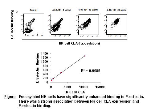
Expanded human NK cells had low levels of baseline fucosylation, ranging from only 10-25%. Expanded NK cells were successfully fucosylated with ASC-101 in a dose-dependent manner (figure); the MFI of CLA on NK cells peaked at 25 ug/ml of ASC-101, with nearly 100% of NK cells being fucosylated (CLA positive). Fucosylation did not affect NK cell viability nor was it associated with changes in NK cell phenotype including surface expression of CD16, CD56, KIR2DL1, KIR2DL2/3, KIR3DL1, NKG2A, NKG2D, TRAIL, perforin, or granzymes A/B. Ex vivo cell culture showed fucosylation was sustained at nearly 100% for 48 hours, but then rapidly declined returning to baseline levels by 96 hours. NK cell cytotoxicity against tumor targets including K562 cells and myeloma cells was preserved and unaffected by fucosylation. Fucosylation significantly enhanced the binding capacity of NK cells to human E-selectin. Further, NK cell binding to recombinant human E-selectin/Fc chimera protein directly correlated with the degree of NK cell fucosylation (figure), which was dose-dependent on the ASC-101 concentration. The effects of forced fucosylation on the ability of human NK cells to home to the bone marrow following adoptive transfer into immuno-deficient mice is currently being explored.
Expanded NK cells primarily express non-glycosylated ligands for E-selectin, potentially limiting their ability to home to the bone marrow following adoptive transfer in humans with hematological malignancies. Ligands for E-selectin on the surface of expanded NK cells can be glycosylated ex vivo rapidly and to high degrees using ASC-101, significantly enhancing their ability to bind E-selectin. These data suggest forced fucosylation of NK cells could be used as a novel approach to improve the antitumor effects of adoptive NK cell infusions in patients with hematological malignancies.
Miller:America Stem Cell Inc: Employment. Wolpe:American Stem Cell, Inc: Employment. Koh:America Stem Cell Inc: Employment.


This feature is available to Subscribers Only
Sign In or Create an Account Close Modal