Abstract
Paroxysmal nocturnal hemoglobinuria (PNH) is an acquired disorder, characterized by the clonal expansion of hematopoietic stem cells lacking glycosylphosphatidylinositol-anchored proteins. PNH clones are frequently found in patients with aplastic anemia (AA), myelodysplastic syndrome (MDS), unusual site thrombosis and non-inmume hemolysis. In Colombia, the DECF laboratory analyses blood samples from all over the country, summited by the APEC (association of patients with complement diseases) for PNH screening.
We reviewed the results of flow cytometry (FC) analyses from 1448 patients screened for PNH in DECF laboratory, between 2010 and 2013, and evaluated the association between clinical characteristics and distribution of PNH clone sizes. Clinical characteristics were considered as referred by treating physicians in the FC analysis request forms. All patients included gave written informed consent.
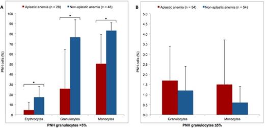
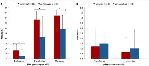
Mean age of the study population was 44.6±18.5 years and 60.5% were female patients. The most frequent indications for screening were thrombosis (23.5%) and unexplained cytopenias (21.8%). Only 14% of the samples were PNH positive. Table 1 shows the results of FC analysis according to the indications for screening. Median clone sizes were 0.1% (interquartile range (IQR): 0-5.8%) in erythrocytes, 4.2% (IQR: 1.1-53.9%) in granulocytes and 3.8% (IQR: 0.7-64.1%) in monocytes. PNH clone size in granulocytes showed strong correlation with clone size in monocytes (r=0.86, p<0.001) and moderate correlation with clone size in erythrocytes (r=0.65, p=0.001). Figure 1 depicts the distribution of PNH granulocyte clone sizes among indications for screening. In the group of patients with PNH granulocyte clone sizes >5%, those with AA had smaller clone sizes than the rest (Figure 2), while patients with hemolysis showed larger clone sizes than the rest (Figure 3). No significant differences in clone size distribution were found within patients with PNH granulocyte clone sizes ≤5%.
Indications for screening and results of flow cytometry analysis
| Indication for screening . | Screened* . | PNH positive . | ||
|---|---|---|---|---|
| n (%) . | Mean age (years) . | Female sex (%) . | ||
| Thrombosis | 340 | 13 (3.8) | 44.0±20.3 | 61.5 |
| Unexplained cytopenias | 316 | 44 (13.9) | 43.5±17.1 | 67 |
| Hemolysis | 284 | 67 (23.6) | 42.6±19.4 | 63.4 |
| Aplastic anemia | 235 | 82 (34.9) | 40.4±18.8 | 61 |
| Myelodysplastic syndrome | 86 | 13 (15.1) | 60.2±19.2 | 61.5 |
| Not provided | 410 | 27 (6.6) | 44.8±15.1 | 50 |
Some patients had more than one indication for screening
PNH granulocyte clone sizes distribution among indications for screening Dotted horizontal lines mark quartiles of the whole population. Solid horizontal lines mark medians of each group.
PNH granulocyte clone sizes distribution among indications for screening Dotted horizontal lines mark quartiles of the whole population. Solid horizontal lines mark medians of each group.
PNH clone sizes in patients with and without aplastic anemia (A) Group with PNH granulocytes >5%. (B) Group with PNH granulocytes ≤5%. Data presented as medians and 75th percentile. * p < 0.001.
PNH clone sizes in patients with and without aplastic anemia (A) Group with PNH granulocytes >5%. (B) Group with PNH granulocytes ≤5%. Data presented as medians and 75th percentile. * p < 0.001.
PNH clone sizes in patients with and without hemolysis (A) Group with PNH granulocytes >5%. (B) Group with PNH granulocytes ≤5%. Data presented as medians and 75th percentile. * p < 0.001.
PNH clone sizes in patients with and without hemolysis (A) Group with PNH granulocytes >5%. (B) Group with PNH granulocytes ≤5%. Data presented as medians and 75th percentile. * p < 0.001.
Our findings are comparable to those reported in other countries. PNH granulocyte clone sizes had stronger correlation with PNH monocyte clones than with PNH erythrocyte clones. About one third of the patients with AA and one fourth of those with hemolysis were PNH positive. Over 50% of patients with AA showed PNH clones sizes <5%. Patients with hemolysis have larger clones than the rest. Further studies are needed to establish the association between PNH clone sizes and clinical outcomes of related disorders.
No relevant conflicts of interest to declare.
Author notes
Asterisk with author names denotes non-ASH members.


This feature is available to Subscribers Only
Sign In or Create an Account Close Modal