Abstract
Allogeneic hematopoietic stem cell transplantation is an effective method for treatment of hematological malignancies, while GVHD and graft rejection are main complications, which seriously affect patients' survival rates and quality of life.
Establishing allo-transplantation mice model with mRFP and GFP transgenic mice, to simulate clinical hematopoietic stem cell transplantation and explore the mechanism of stem cell homing and GVHD.
1) Thirteen C57BL/6 GFP transgenic mice, used as recipients, were irradiated with 7 Gy. Each mouse was injected through caudal vein with 2*106 bone marrow cells isolated from FVB mRFP transgenic mice. 2) Symptoms like weight loss, depilation, diarrhea were observed as GVHD manifestation while survival rates were evaluated. Routine blood test and FACS were performed at different time points to confirm hematopoiesis reconstitution. 3) Mice were perfused with paraformaldehyde under anesthesia to fix the tissue, while pathological examination and real-time PCR were performed for studying donor and recipient cells interactions in different organs. 4) Semi-solid decalcification was used to treat the femora before observing under confocal microscope directly or after making frozen section, three-dimensional reconstruction were made to observe the cellular interaction, especially for cells within the bone marrow.
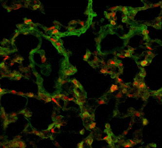
1) Depilation, wrinkled skin, hunchback and sharp decline of weight were observed in 8/13 mice. Routine blood test implicated hematopoietic reconstitution. FACS showed 86.1%±7.8% mRFP+ cells in peripheral blood of recipients. 2) mRFP+ cells were found distributing throughout the body's organs. mRFP+ Lymphocyte infiltration and inflammatory exudate were seen especially in the small intestine, lung, liver and skin (Fig.1). GFP+ cells were found surrounding mRFP+ cells in the bone marrow of the femora decalcified with semi-solid decalcification. Their interactions can be further observed clearly in bone marrow microenvironment in three-dimensional reconstruction by confocal microscope (Fig.2).
The donor and recipient cells' morphous, location, ratio and cellular interaction can be visually observed in recipient mice's lung under confocal microscope (250x).
The donor and recipient cells' morphous, location, ratio and cellular interaction can be visually observed in recipient mice's lung under confocal microscope (250x).
By semi-solid decalcification and three-dimensional reconstruction under confocal microscopy, the relationships of GFP and RFP marked cells in the bone marrow microenvironment are clearly demonstrated, with their original position, fluorescence and morphous. Several RFP donor's cells were found surrounded by the GFP recipient's cells, both cells cross-talking actively (200x).
By semi-solid decalcification and three-dimensional reconstruction under confocal microscopy, the relationships of GFP and RFP marked cells in the bone marrow microenvironment are clearly demonstrated, with their original position, fluorescence and morphous. Several RFP donor's cells were found surrounded by the GFP recipient's cells, both cells cross-talking actively (200x).
Owing to RFP on donors' cells and GFP on recipients' cells, together with our novel protocol named semi-solid decalcification, we can visually observe the donor and recipient cells' location, ratio and cellular interaction, as well as morphological changes. Within various tissues especially for such tissues as bone marrow and lung, the details between cells can be studied lively by fluorescence microscope and confocal microscope. In recipient mice with GVHD, donor cells can be found in various target tissues such as intestine, lung, liver and skin. Gene marked cells with fluorescence protein can benefit morphological, immunological, cytogenetic and molecular studies in recipients after HSCT.
The allo-transplantation model with mRFP and GFP transgenic mice is powerful in study of Stem Cell Homing and Donor-Recipient Cellular Interaction. The cellular interaction can be easily observed by three-dimensional reconstruction after semi-solid decalcification, especially for bone marrow and lung.
No relevant conflicts of interest to declare.
Author notes
Asterisk with author names denotes non-ASH members.


This feature is available to Subscribers Only
Sign In or Create an Account Close Modal