Abstract
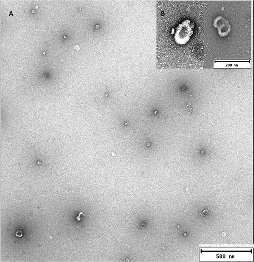
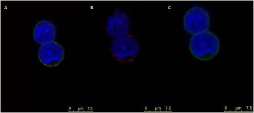
A new mechanism of intercellular communication has been proposed consisting in the secretion of exosomes/ microvesicles (MVs). Such mechanism has been shown to modify the functional properties of recipient cells by the transfer of proteins, mRNA, or micro-RNAs. The hypothesis of the present work was that MSC from MDS patients could differentially modify the HPC properties throughout the shedding of MVs when compared with those from controls due to their different content. Material and methods: MVs were isolated from MSC from bone marrow (BM) samples 18 patients diagnosed with ‘de novo’ and untreated low risk MDS and from MSC from 12 healthy BM. BM-MSC at third passage were cultured in DMEM deprived of FCS, and supernatants were collected after 6 or 24 hours. MVs purification was performed in the majority of the experiments (16 MDS/ 9 Controls) using the ExoQuick-TC exosome precipitation solution (ExoQuick; System Biosiences). To confirm the isolation of MVs by exosome precipitation solution, in some cases (2 MDS/3 Controls) the MVs were obtained by ultracentrifugation; MVs identification was done by transmission electron microscopy (TEM) as well as by flow cytometry (FC). To evaluate if the micro-RNA content into MSC-MVs from patients and controls was different, expression analysis of miRNAs was done using Megaplex™ RT Primers pool (Applied Biosystems) and 384-well microfluidic cards (TaqMan® MicroRNA Array A) were loaded with retro-transcription product and PCR runs were performed on a 7900HT Fast Real-time PCR system (eight MVs from MDS and 4 from HD).To demonstrate the incorporation of MVs from MSC into human hematopoietic progenitors (HPC: CD34+ cells obtained by immunomagnetic selection) HPC were co-cultured with MVs from MSC. Incorporation of Vybrant Dil-labelled MVs into HPC was evaluated at 1, 3, 6, and 24 h. by FC. To detect the incorporation of MVs by confocal microscopy (CM) an intracellular primary Ab for CD90 (Santa Cruz, Biotechnology) was used as MVs marker and anti-CD45 to detect HPC. A Zeiss LSM 510 CM connected to a digital camera (Leica DC 100) were used to obtain confocal images. Apoptotic rate of CD34+ cells that had the MVs-MSC from MDS and controls were evaluated by FC by using APC H7 Annexin V DY634 (Immunostep) and 7AAD (BD Biosciences). Results: More than 95% of MVs isolated by ExoKit system from supernatants of cultured MSC from 6 HD and 6 MDS patients showed scatter intensities lower than of 6µm beads. We observed, in all cases, the same FC pattern. Also, MVs/exosomes isolated by ultracentrifugation (3 MDS/ 5 HD) showed the same FC pattern. MVs from MDS and controls isolated by ultracentrifugation were identified by TEM (fig1). When co-cultures of CD34+ HPC and MVs were studied in both HD and MDS, MVs were incorporated into HPC in all cases (fig2). When the content of miRNAS in the MVs from MDS and HD were compared significant differences were observed between both groups. Twenty-one out of 384 evaluated miRNAs were over-expressed in the MVs from patients compared with the controls. To confirm these results, the expression of miR10a and miR-132 was analyzed by RT-PCR. In both cases their expression was significantly increased in MVs from patients. Recently, it has been suggested that the cargo of these structures are bioactive molecules, therefore we explored the possibility that MVs could modify the behavior of the target cell. For this purpose we searched in which pathways the overexpressed miRNAs could be involved and apoptosis was among them. Since it is considered a very important process in MDS pathophysiology we compared apoptosis by FC, after co-culturing CD34+ cells with MVs from MSC of MDS and HD. Interestingly, preliminary results show that the MVs from MDS protected better from apoptosis CD34+ cells than MVs obtained from controls. In summary, in the present study we show that BM-MSC produce MVs/exosomes with different microRNAs content according to their origin, MDS or HD. These structures can be incorporated into HPC and can modify their properties.
Funding: Instituto de Salud Carlos III. PI12/01775. Junta de Castilla y León.GRS 873/A/13. Portuguese FST Grant. SFRH/BD/86451/2012
Diez-Campelo:Celgene: Honoraria, Research Funding; Novartis: Honoraria, Research Funding. San Miguel:Jansen, Celgene, Onyx, Novartis, Millenium: Membership on an entity’s Board of Directors or advisory committees. del Cañizo:Celgene: Membership on an entity’s Board of Directors or advisory committees, Research Funding; Jansen-Cilag: Membership on an entity’s Board of Directors or advisory committees, Research Funding; Arry: Membership on an entity’s Board of Directors or advisory committees, Research Funding; Novartis: Membership on an entity’s Board of Directors or advisory committees, Research Funding.
MVs from MSC -MDS analysed by TEM. A. Scale bar 500nm. B. Scale bar 200nm
MVs from MSC -MDS analysed by TEM. A. Scale bar 500nm. B. Scale bar 200nm
CM images A- C Sequential images of CD34+ cells with MVs from MSC-MDS. A. overlay (CD90 in red and CD45 in green), B CD34+ with anti CD 90 (red) and C with anti-CD45 (green). DAPI (blue)
CM images A- C Sequential images of CD34+ cells with MVs from MSC-MDS. A. overlay (CD90 in red and CD45 in green), B CD34+ with anti CD 90 (red) and C with anti-CD45 (green). DAPI (blue)
Author notes
Asterisk with author names denotes non-ASH members.


This feature is available to Subscribers Only
Sign In or Create an Account Close Modal