Abstract
Background:
Extranodal disease is common in diffuse large B-cell lymphoma (DLBCL), and involvement of more than one extranodal site is associated with a worse outcome. 18F-fluorodeoxyglucose PET/CT (PET/CT) is the current state-of-the-art for staging of DLBCL, and has shown to be much more sensitive for the detection of extranodal involvement than a stand-alone CT scan. Therefore, a re-evaluation of the clinical significance of extranodal disease among PET/CT staged DLBCL patients is warranted.
Patients and Methods:
We retrospectively included patients from Aalborg (2007-2012), Copenhagen (2009-2012), and British Columbia (2011-2012) in the present study. The inclusion criteria were, i) newly diagnosed DLBCL, ii) R-CHOP or R-CHOP like first-line treatment, and iii) PET/CT staging. The written PET/CT files were reviewed for disease stage and extranodal sites of involvement. The relationship between number of involved sites, extranodal locations and outcome were assessed with simple Cox regression analyses. Extranodal locations with p<0.1 in univariate analysis were entered in a multivariable Cox regression analysis together with the following International Prognostic Index (IPI) factors: age > 60 years, elevated LDH, ECOG performance score >1.
Results:
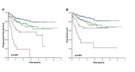
A total of 444 patients with a median age of 65 years (range 16-90) and a male:female ratio of 1.3 were included in the study. Of these patients 28% (n=98) had Ann Arbor stage I disease, 16% (n=72) stage II disease, 16% (n=71) stage III disease, and 46% stage IV disease (n= 203). LDH was elevated in 51% (n=224), and 17% (n=74) had ECOG performance status >1. B-symptoms were present in 37% (n=164) and 26% (n=114) had a bulky mass =/> 10 cm. With a median follow-up of 2.4 years (range 0.5-6.5) in patients still alive at the time of analysis, the 3-year OS and PFS were 73% and 69%, respectively. Extranodal disease was diagnosed in 286 (64%) of the patients. The anatomic locations of extranodal disease and their relations to outcome are shown in Table I. Figure 1A and B show the PFS and OS curves when patients are grouped according to the number of involved extranodal sites. Patients with one or two extranodal sites of involvement had similar outcome (3-year PFS 68% vs. 70%), whereas all patients with involvement of more than three extranodal sites progressed.
Conclusions:
Extranodal involvement is diagnosed in more than half of all newly diagnosed DLBCL patients staged with PET/CT. Bone/bone marrow involvement was the most common site and associated with a worse outcome. Thus, detection of these lesions with PET/CT is clinically important. The presence of extranodal disease is generally associated with a worse outcome, but our data suggest that the optimal cut-off for prognostication in PET/CT staged patients may be more than two sites rather than more than one site, as according to the IPI.
Extranodal DLBCL and their relationship with outcome in PET/CT staged patients treated with R-CHOP. Empty boxes represent variables not included in multivariate models.
| Site . | Frequency, n (%) . | HR, univariate . | HR, multivariate . | ||
|---|---|---|---|---|---|
| PFS | OS | PFS | OS | ||
| Lung | 33 (7%) | 1.56, p=0.002 | 1.46, p=0.26 | Not significant | |
| Liver | 34 (8%) | 2.39, p=0.001 | 2.43, p=0.002 | Not significant | Not significant |
| Bone/bone marrow (PET/CT) | 127 (29%) | 2.49, p<0.001 | 2.53, p<0.001 | 1.77, p=0.007 | 1.66, p=0.03 |
| Bone marrow indolent NHL (biopsy) | 28 (6%) | 0.86, p=0.70 | 0.94, p=0.87 | ||
| Bone marrow DLBCL (biopsy) | 43 (10%) | 2.55, p<0.001 | 2.66, p<0.001 | Not significant | Not significant |
| Gastrointestinal | 35 (8%) | 1.27, p=0.43 | 1.02, p=0.96 | ||
| Kidney | 13 (3%) | 2.10, p=0.06 | 1.63, p=0.29 | Not significant | |
| Soft tissue and muscle | 46 (10%) | 1.18, p=0.58 | 1.17, p=0.64 | ||
| Paranasal sinus | 15 (3%) | 1.57, p=0.28 | 1.69, p=0.25 | ||
| Pleural fluid | 16 (4%) | 2.82, p=0.005 | 3.23, p=0.003 | 2.43, p=0.02 | 2.53, p=0.02 |
| Testicular | 13/252 (5%) | 2.42, p=0.22 | 1.81, p=0.41 | ||
| Female genitals | 10/192 (5%) | 3.38, p=0.006 | 3.76, p=0.003 | ||
PFS (Figure 1A) and OS (Figure 1B) in patients grouped according to the number of extranodal sites involved: zero (blue), 1 (green), 2 (grey), 3 (purple), >4 (red).
PFS (Figure 1A) and OS (Figure 1B) in patients grouped according to the number of extranodal sites involved: zero (blue), 1 (green), 2 (grey), 3 (purple), >4 (red).
No relevant conflicts of interest to declare.
Author notes
Asterisk with author names denotes non-ASH members.


This feature is available to Subscribers Only
Sign In or Create an Account Close Modal