Abstract
Lytic bone involvement in primary systemic (AL) amyloidosis is uncommon. To-date, there is no systematic study of bone metabolism in patients with AL amyloidosis. For this reason, we evaluated prospectively 102 patients with previously untreated AL amyloidosis who were diagnosed between January 2000 and June 2008 in the Plasma Cell Dyscrasias Unit of the Department of Clinical Therapeutics (Athens, Greece). All patients had histologically confirmed AL amyloidosis, while the definition of organ involvement, hematological and organ response and progression were based on consensus criteria. The levels of the following bone remodeling indices were measured before the administration of any kind of therapy: i) osteoclast regulators: soluble receptor activator of nuclear factor-kappaB ligand (sRANKL), osteoprotegerin (OPG), chemokine C-C motif ligand 3 (CCL-3, previously known as MIP-1alpha) and osteopontin; ii) bone resorption markers: C- and N- telopeptide of type-1 collagen (CTX and NTX, respectively) and tartrate resistant acid phosphatase type-5b; iii) bone formation markers: bone-alkaline phosphatase and osteocalcin. The results were compared with those of 35 age- and gender-matched healthy controls, 40 patients with monoclonal gammopathy of undetermined significance (MGUS) and 35 newly diagnosed, untreated patients with symptomatic multiple myeloma (MM).
The median age of AL patient was 65 years (range: 39-80 years); 43% were males; 60% had heart involvement, 73% renal involvement, 11% liver involvement and 40% had peripheral/autonomous nerve system involvement; 67% of patients had two or more organs involved; 16% had a baseline serum creatinine >2 mg/dl. The median serum albumin was 3.2 g/dl, the median serum β2-microglobulin was 1.8 mg/L and the median 24h urine protein was 3350 mg/24h. Median NT-proBNP was 1954 pg/ml; 26%, 41% and 33% of patients were Mayo stage -1, -2 and -3, respectively.
None of AL patients had lytic bone lesions in plain radiography or other features suggestive of MM, such as hypercalcemia, significant anemia unrelated to renal impairment or predominant Bence-Jones proteinuria. Bone resorption (assessed by CTX or NTX) in AL-patients was increased (p<0.001) compared to healthy controls but bone formation had no difference between AL patients and controls. CCL-3, sRANKL and OPG were increased in AL patients compared to controls (p<0.01 for all comparisons), but the ratio of sRANKL/OPG was similar in AL patients vs. controls (p=0.854). MM patients had increased bone resorption and decreased bone formation markers compared to AL patients, while sRANKL/OPG ratio was markedly decreased in AL vs. MM, due to high levels of OPG in AL vs MM patients (>4 fold higher; p<0.001). Compared to MGUS, AL patients had decreased sRANKL/OPG ratio due to increased OPG. OPG levels correlated with NT-proBNP levels (p<0.001) and were slightly higher in patients with cardiac involvement (p=0.028).
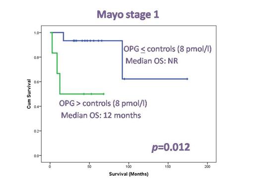
Patients with OPG levels above the upper value of healthy controls had a shorter survival (p=0.026), while AL patients with OPG levels in the top quartile had short survival (12 months vs. 58 months, p=0.024). In the multivariate analysis cardiac biomarkers outperformed OPG; however, in patients with Mayo stage-1 disease, OPG levels remained significant and identified patients with short survival within the favorable risk group (12 vs. >60 months, p=0.012; see the Figure). Although renal involvement did not affect bone markers, in AL patients with serum creatinine >1.5 mg/dl, CTX and NTX were higher than in patients with <1.5 mg/dl (p<0.001 and p=0.016, respectively). Neither the involvement of other organs nor the number of involved organs appeared to influence bone remodeling in any way.
We conclude that elevated bone resorption and high bone turnover is present in AL patients possibly due to direct and indirect activity of amyloid fibrils or light chains in bone cells. Increased OPG in AL is not only a compensation factor to osteoclast activation but it may also reflect tissue damage in the heart. There is published evidence that OPG is produced by cardiac cells and has been associated with poor prognosis in heart disorders, including heart failure and acute coronary disease. In AL amyloidosis, high OPG is able to identify patients at increased risk of death, mainly within the more favorable prognosis group by Mayo staging.
No relevant conflicts of interest to declare.
Author notes
Asterisk with author names denotes non-ASH members.


This feature is available to Subscribers Only
Sign In or Create an Account Close Modal