Abstract
Introduction
Ultra-large von Willebrand factor (ULVWF) strings are synthesized in ECs, packaged in Weibel-Palade Bodies (WPBs), and secreted by stimulated ECs. Complement components studied to date [C3, factor (F) B, FD, FP, FH, FI, C5] are released slowly and continuously from human umbilical vein endothelial cell (HUVEC) cytoplasm and are not packaged in WPBs (PLoS One. 2013;8(3):e59372). In contrast, a recent report (Blood. 2014;123(1):121-5) contended that FH co-localizes with VWF in the WPBs. If this were so, it could have therapeutic importance for the treatment of atypical hemolytic uremic syndrome (aHUS) resulting from deficiency of FH because it might be possible to increase circulating FH levels transiently by administration of the WPB secretagogue, des-amino-D-arginine vasopressin (DDAVP).
Hypothesis
FH is not co-localized with VWF in WBPs, but rather is released slowly and continuously from EC cytoplasm regardless of cell stimulation.
Methods
Immunofluorescent Microscopy
HUVECs were stimulated with histamine and stained with rabbit anti-VWF plus secondary donkey anti-rabbit Alexa Fluor IgG-488. The cells were then fixed and stained with goat-anti FH plus secondary chicken anti-goat Alexa Fluor IgG-647. The nuclei were detected with DAPI.
In vitro VWF and FH from HUVECs
HUVECs either were, or were not, stimulated with histamine. Supernatant was collected a variety of times over 7 hrs and assayed for VWF and FH antigen levels by ELISA. VWF assay antibodies (polyclonal): (1) capture, rabbit anti-human VWF (Ramco); (2) detection, goat anti-human VWF (Bethyl) and rabbit anti-goat IgG-HRP (Invitrogen). FH assay antibodies: (1) capture, polyclonal goat anti-human FH (Advanced Research Technologies); (2) detection, monoclonal mouse anti-human FH (Pierce, Thermo Scientific) and polyclonal goat anti-mouse IgG-HRP (Invitrogen).
In vivo VWF and FH
Plasma samples were obtained from 6 pediatric patients with von Willebrand disease (VWD) being tested for EC release of WPB VWF in response to DDAVP. For each patient, 1 sample was obtained prior to DDAVP administration, and 2 other samples were obtained 1 and 4 hours later. VWF levels were measured in each sample using standard clinical laboratory procedure at an affiliated hospital. FH antigen levels were quantified by ELISA, as above.
Results
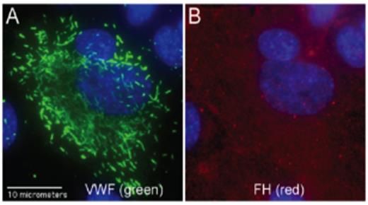
Using non-overlapping spectral secondary detection antibody pairs, VWF was seen in clusters in HUVEC WPBs (Fig. 1A). In contrast, FH was distributed throughout the HUVEC cytoplasm (Fig. 1B). The VWF and FH images did not overlap, indicating that VWF and FH did not co-localize in the WPBs.
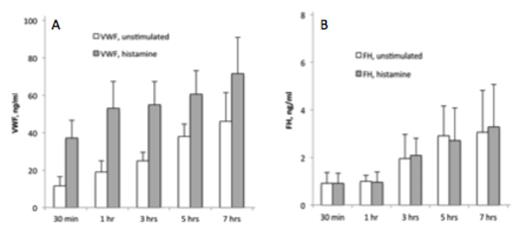
Histamine addition to HUVECs in vitro resulted in ~ 4-fold increases in VWF secreted from HUVEC WPBs at 30 min and 1 hour, and 2-fold increases at 3 hours (Fig 2A). In contrast, FH release was slow and continuous, regardless of histamine stimulation, suggesting that FH is located in EC cytoplasm and is not stored in WPBs (Fig. 2B).
In vitro VWF and FH release from ECs under non-stimulated and histamine-stimulated conditions.
In vitro VWF and FH release from ECs under non-stimulated and histamine-stimulated conditions.
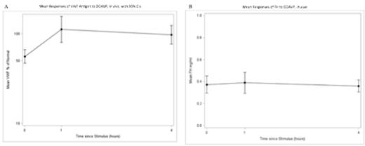
In all 6 patient samples, VWF antigen increased significantly at 1-hour post-DDAVP administration (Fig. 3A). In contrast, FH antigen levels did not change significantly at hour 1 or hour 4, compared to hour 0, indicating that FH is not co-localized and secreted along with VWF from the WPBs of stimulated ECs in vivo (Fig. 3B).
(A) Mean responses of VWF antigen to DDAVP, in vivo, with 95% CIs.The mean response was significantly greater 1-hour and 4-hours post-DDAVP compared with baseline (P=0.0085 and 0.0079, respectively). After 1-hour post-DDAVP, the mean response was 201% greater (95% CI: 129%, 314%) than baseline. After 4-hours, the mean response was 174% greater (95% CI: 123%, 247%) than baseline. (B) Responses of FH to DDAVP, in vivo. There was no statistically significant difference in FH response between time points (P=0.77).
(A) Mean responses of VWF antigen to DDAVP, in vivo, with 95% CIs.The mean response was significantly greater 1-hour and 4-hours post-DDAVP compared with baseline (P=0.0085 and 0.0079, respectively). After 1-hour post-DDAVP, the mean response was 201% greater (95% CI: 129%, 314%) than baseline. After 4-hours, the mean response was 174% greater (95% CI: 123%, 247%) than baseline. (B) Responses of FH to DDAVP, in vivo. There was no statistically significant difference in FH response between time points (P=0.77).
Conclusions
We used immunofluorescent microscopy and ELISA assays on samples obtained in vitro and in vivo to demonstrate that FH is not packaged in, or secreted from, the WPBs of stimulated human ECs. FH is, therefore, similar to all other complement components studied to date in that it is released slowly and continuously from ECs and is not influenced by cell stimulation. DDAVP is unlikely to be a viable treatment option for patients with aHUS secondary to deficiency or inhibition of FH.
Sartain:Hemostasis and Thrombosis Research Society: Research Funding. Turner:Mary R Gibson Foundation: Research Funding; Hinkson Memorial Fund : Research Funding. Moake:Mary R Gibson Foundation: Research Funding; Hinkson Memorial Fund: Research Funding.
Author notes
Asterisk with author names denotes non-ASH members.


This feature is available to Subscribers Only
Sign In or Create an Account Close Modal