Abstract
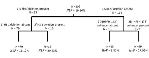
Hereditary persistence of fetal hemoglobin (HPFH) and (δβ)0 thalassemia are caused by deletions within the β-globin gene (HBB) cluster that remove elements that affect the expression of the γ-globin genes (HBG2 and HBG1, or HBG). These deletions are of different lengths and have different 5’ and 3’ breakpoints. The phenotypes associated with heterozygous carriers of (δβ)0 thalassemia and HPFH deletions are differentiated by levels of 5-15% HbF distributed heterocellularly in the former and 15-30% HbF distributed pancellularly in the latter. We found a novel 588.6 kb deletion that removed both the 3.5 kb fragment 5’ to HBD that is deleted in Corfu β thalassemia and contains a BCL11A binding site, and the known cis-acting elements downstream of HBB. The proband with this deletion had a HbF of 5.4% (Morrison et al, Blood, 2014 abstract 3452). To study the relative importance of 5’ and 3’ regulatory elements in HBG expression we studied 209 cases culled from the literature and from our laboratory where the 3.5 kb element 5’ to HBD and enhancers 3’ to HBB were deleted and HBG remained intact. We used a backwards stepwise regression statistical analysis to determine which deleted elements had the greatest effect on HbF levels. The combination of the deletion of 3.5 kb intergenic region 5’ to HBD, the presence of the HPFH-1 “3D” enhancer juxtaposed to HBG, and the deletion of the 3’ HS1 region accounted for 66.7% of the HbF variation in heterozygotes for HPFH and (δβ)0-thalassemia deletions. The HPFH-1 “3D” enhancer juxtaposed to HBG— the main difference between HPFH-1 and 2 compared with Spanish (δβ)0-thalassemia—was associated with an increase in HbF of 20.78% (p<2e-16) after adjusting for the effects of the other 5’ and 3’ cis-acting elements. The next most significant factor was the deletion of the 3.5 kb fragment 5’ to HBD which resulted in an increase of 10.62% HbF after similar adjustments (p<2e-16); deletion of the 3’ HS1 region accounted for an increase in HbF of 5.25% (p<1.05e-5). The HPFH-3 and HPFH-6 enhancer regions each accounted for a less than 1% increase in HbF and were not significantly associated with HbF in this model. Among 194 individuals where both 5’ and some 3’ elements affecting γ-globin gene expression—excluding the “3D” enhancer—were deleted, HbF was 20±9.3%; in 13 cases where all 3’ enhancers—including the “3D” enhancer—were deleted, HbF was 6.8±3.7% (p=8.9e-07). To determine which combinations of cis-acting elements were associated with high and low HbF levels we performed a classification and regression tree (cART) analysis on HbF. The results of the regression tree (Figure) only included the deletion of the 5’ 3.5 kb fragment region, the presence of the HPFH-1 “3D” enhancer and the deletion of the 3’ HS1 region and were consistent with the results of the backwards selection model. The absence of the 5’ 3.5 kb fragment 5’ to HBD combined with the presence of the HPFH-1 “3D” enhancer was associated with the highest average HbF of 27.02%. The absence of the 3.5 kb fragment 5’ to HBD combined with the absence of the HPFH-1 “3D” enhancer was associated with the lowest average HbF of 6.82%.The 588.6 kb deletion is the largest deletion reported in the HBB cluster that leaves the γ-globin genes intact, and the second to remove both the BCL11A binding site and all known 3’ enhancer elements. By studying deletions in the HBBgene cluster we have further defined the hierarchy of cis-acting elements that modulate HbF levels in adults and suggest a paramount role of the distal “3D” enhancer.
No relevant conflicts of interest to declare.
Author notes
Asterisk with author names denotes non-ASH members.


This feature is available to Subscribers Only
Sign In or Create an Account Close Modal