Key Points
Lack of KLRG1 and T-bet expression is a unique feature of DNT and subsets of single positive T cells in ALPS patients.
Genetic, phenotypic, and transcriptional evidence indicates that DNT in ALPS patients derive from both CD4+ and CD8+ T cells.
Abstract
Accumulation of CD3+ T-cell receptor (TCR)αβ+CD4−CD8− double-negative T cells (DNT) is a hallmark of autoimmune lymphoproliferative syndrome (ALPS). DNT origin and differentiation pathways remain controversial. Here we show that human ALPS DNT have features of terminally differentiated effector memory T cells reexpressing CD45RA+ (TEMRA), but are CD27+CD28+KLRG1− and do not express the transcription factor T-bet. This unique phenotype was also detected among CD4+ or CD8+ ALPS TEMRA cells. T-cell receptor β deep sequencing revealed a significant fraction of shared CDR3 sequences between ALPS DNT and both CD4+ and CD8+TEMRA cells. Moreover, in ALPS patients with a germ line FAS mutation and somatic loss of heterozygosity, in whom biallelic mutant cells can be tracked by absent Fas expression, Fas-negative T cells accumulated not only among DNT, but also among CD4+ and CD8+TEMRA cells. These data indicate that in human Fas deficiency DNT cannot only derive from CD8+, but also from CD4+ T cells. Furthermore, defective Fas signaling leads to aberrant transcriptional programs and differentiation of subsets of CD4+ and CD8+ T cells. Accumulation of these cells before their double-negative state appears to be an important early event in the pathogenesis of lymphoproliferation in ALPS patients.
Introduction
Autoimmune lymphoproliferative syndrome (ALPS) is a disorder of lymphocyte homeostasis associated with mutations in genes involved in the Fas apoptosis pathway. It is characterized by chronic benign lymphoproliferation, autoimmunity often manifesting as multilineage cytopenias and an increased risk of lymphoma.1,2 Most patients carry heterozygous germ line (ALPS-FAS) or somatic mutations of the FAS gene (ALPS-sFAS) leading either to decreased Fas expression and thus haploinsufficiency or to expression of dominant, negative Fas receptors.3,4 In some patients, particularly those with haploinsufficiency, a second somatic FAS mutation or somatic loss of heterozygosity (LOH) enhances clinical penetrance (ALPS-FAS-sLOH).5,6
The most prominent feature of disturbed T-cell differentiation and homeostasis in Fas deficiency is the accumulation of CD3+ T-cell receptor (TCR)αβ+CD4−CD8− double-negative T cells (DNT). Multiple lines of evidence suggest that DNT in Fas-deficient mice and humans arise from chronically activated CD8+ T cells. These include a demethylated CD8α gene in DNT from Fas lpr/lpr mice,7 reduced DNT in major histocompatibility complex (MHC) class I-deficient or β2 microglobulin-deficient Fas lpr/lpr mice8-10 and shared CDR3 sequences among TCR Vβ transcripts of CD8+ T cells and DNT of FAS mutant patients.11 The fact that DNT are not completely absent in β2 microglobulin-deficient and MHC class I-deficient mice and the finding that lpr/lpr CD4+ T cells can differentiate into B220+ DNT in adoptive nu/nu hosts12 may argue for an additional contribution of CD4+ T cells to DNT. However, there are no data supporting a CD4+ origin of DNT in humans.
Irrespective of their origin, the differentiation pathway of DNT and the role of Fas in this process are not clear. DNT could represent direct descendants of chronically activated single positive T cells (SPT), which downregulate coreceptors at a differentiation state when they are destined to undergo Fas-mediated apoptosis. In this case, DNT should phenotypically resemble chronically activated cells and the major role of Fas in T-cell homeostasis would be the regulation of the size of the pool of such T cells. The observation that murine and human DNT express markers of terminally differentiated T cells supports this view.11,13,14 However, it is also conceivable that defective Fas signaling alters T-cell differentiation already at an earlier stage. Such abnormally differentiated SPT may have different transcriptional programs leading to their accumulation and downregulation of coreceptors. In fact, DNT accumulation could reflect uncontrolled expansion of aberrant T cells more than defective elimination of terminally differentiated T cells and DNT.15 ALPS DNT differ from the small population of physiological DNT found in healthy donors.14 Moreover, they have a unique transcriptional profile16,17 and distinct functional properties including high expression of Fas ligand and IL-10,18-21 high proliferative capacity in vivo, and hyporesponsiveness in vitro.22 This may indicate that DNT differentiation is indeed more complex and that Fas deficiency apart from regulating T-cell death may have an impact on T-cell differentiation. Thus, altered differentiation may already be detectable in SPT. However, no significant alterations in the differentiation of SPT in ALPS patients have been reported to date.
Here, we used 3 independent approaches to better understand the role of Fas in human T-cell differentiation and DNT ontogeny. First, we performed a detailed comparison of DNT and different SPT populations in patients with germ line and/or somatic FAS mutations. Second, we performed TCRβ deep sequencing in sorted DNT and different CD4+ and CD8+ T-cell populations to identify shared CDR3 sequences. Finally, we analyzed patients with germ line FAS mutations and somatic LOH. Determination of the ratio of monoallelic vs biallelic mutants in various T-cell populations of such patients can indicate the differentiation state at which Fas-deficient T cells accumulate.
Material and methods
Patients
We recruited 9 patients with germ line FAS mutations (ALPS-FAS), 2 patients with somatic FAS mutations (ALPS-sFAS), and 4 patients with germ line FAS mutation and somatic LOH (ALPS-FAS-sLOH). All had raised percentages of DNT (supplemental figure 1), most were untreated, and no patient received sirolimus. We also analyzed 9 patients with ALPS-like diseases, age 4 to 32 years. They presented with lymphoproliferation, autoimmune cytopenia(s) and raised DNT (supplemental figure 5A), but carried no FAS mutation. Three patients were classified as common variable immunodeficiency, 1 patient had features consistent with systemic lupus erythematosus. The study was approved by the Ethics Committee of the University of Freiburg (protocol number 40/09). All patients gave informed consent. The study was conducted in accordance with the Declaration of Helsinki.
Genetic analysis
Molecular sequencing was performed as previously described.23 In patients with germ line FAS mutation and absent Fas expression on DNT, somatic LOH was confirmed by FAS sequencing of sorted DNT, and quantification of mutated vs wild-type FAS allele was performed with the simplified allele ratio mutation quantifier function of the Mutation Surveyor DNA variant analysis software (version 4.0.4) (Softgenetics, State College, PA).
Flow cytometry
The following antibodies were used for flow cytometry and/or for T-cell sorting: CD3 PerCP, CD3 APC, CD4 PE, CD4 APCCy7, CD8 V450, TCRαβ FITC, CD45RA PC7, CD28 APC, CD27 V450, CD57 FITC, CD95 APC (all BD Biosciences), CD8 Krome Orange and TCRαβ PE (Beckman Coulter), CCR7 PE (R&D), CD127 APC, CD244 APC (eBioscience), CD279 (BioLegend), and KLRG1 Alexa488 (clone 13F12F2).24 T-bet PE and Eomes FITC antibodies were used for intracellular staining using reagents of the human regulatory T-cell staining kit (eBioscience). Data acquisition was performed with a Gallios Flow cytometer (Beckman Coulter, Brea, CA). Data were analyzed using FlowJo7.2.5 software (Tree Star Inc., Ashland, OR). Cell sorting was performed on the day of blood sampling, since DNT and presumed DNT precursors rapidly die ex vivo.
Quantitative RT-PCR
RNA of sorted cells was isolated using the RNeasy Micro Kit (Qiagen) and cDNA was synthesized using the First Strand cDNA Synthesis kit (Thermo Scientific). The following primers were used: T-bet forward GTCCAACAATGTGACCCAGAT, T-bet reverse CTCTCCGTCGTTCACCTCAA, HPRT forward CCTGGCGTCGTGATTAGTGAT, Hypoxanthine-guanine phosphoribosyltransferase (HPRT) reverse AGACGTTCAGTCCTGTCCATAA. Real time-polymerase chain reaction (RT-PCR) was performed according to standard protocols using the Fast Start Universal Sybr Green Master (Roche).
TCRβ deep sequencing
A multiplex PCR consisting of 58 primers, initially described by Robins et al,25 was used with some modifications to amplify TCRβ gene rearrangements. In brief, 100 ng genomic DNA were subjected to a first PCR (3 mM MgCl2, 0.2 mM of each 2′-deoxynucleoside 5′-triphosphate, and 1 unit AmpliTaq Gold DNA Polymerase) for 34 cycles at 62°C annealing temperature. Subsequently, the amplificates were purified with the QIAquick PCR Purification Kit (Qiagen). Purified DNA (500 pg) was used for a second amplification (adapter PCR; 1 × Phusion HF Buffer, 0.05 mM of each 2′-deoxynucleoside 5′-triphosphate, 1.0 µM forward primer, 1.0 µM reverse primer, and 1 unit Phusion High-Fidelity DNA Polymerase [Finnzymes]) for 12 cycles at 58°C annealing temperature. The resulting PCR products were isolated from a 2% agarose gel using the Wizard SV Gel and PCR Clean-Up System (Promega). Sequencing of amplified TCRβ gene rearrangements was carried out on a HiSeq2000 analyzer using paired-end modus with 2 × 100 bp read length and joining of read-pairs was performed. All identical sequences and related sequence reads, which deviated by only 1 base pair were clustered and designated as single clonotypes. Single reads and clonotypes consisting of <0.01% of all reads were eliminated because they may represent background signals. The Vβ and Jβ usage of the clonotypes was determined according to the international ImMunoGeneTics information system ([IMGT] http://www.imgt.org). The TCRβ CDR3 sequence was defined as all amino acids starting from the conserved 5′ cysteine in the Vβ-segment up to the conserved 3′ phenylalanine in the Jβ-segment.
Statistical analysis
Analyses were performed using PRISM software (Graphpad Software, San Diego, CA). Populations were compared using the Mann-Whitney U test. P < .05 was considered significant.
Results
DNT from ALPS patients have a distinct differentiation pattern
To compare the cellular phenotype of DNT to that of different CD4+ or CD8+ SPT-cell populations, we stained CCR7 and CD45RA to distinguish between naïve, central memory (CM), effector memory (EM), and TEMRA in analogy to a widely used classification for SPT.26,27 The majority of ALPS DNT were CCR7-CD45RA+, suggesting terminal differentiation, whereas in the healthy donors, as expected,28,29 most DNT were CCR7−CD45RA− (Figure 1A-B). The percentage of the dominant CCR7−CD45RA+ DNT population in ALPS patients correlated with the overall percentage of DNT (data not shown), suggesting that this is indeed the fraction accumulating in Fas deficiency.
Distribution of differentiation subsets among ALPS and control DNT, according to CCR7 and CD45RA expression. (A) Representative plots showing CCR7 and CD45RA expression of ALPS vs control DNT. (B) Distribution of naïve (naïve, CCR7+CD45RA+), central memory (CM, CCR7+CD45RA−), effector memory (EM, CCR7−CD45RA−), and terminal differentiated T cells reexpressing CD45RA (TEMRA, CCR7−CD45RA+) among DNT (gate: CD3+TCRαβ+CD4−CD8−) of all studied ALPS patients (ALPS all), adult ALPS patients (ALPS >18 y), and adult controls (Con >18 y). Because pediatric and adult patients had a similar distribution, statistical comparison was performed between all ALPS patients and adult controls. ns, not significant. *P < .05; **P < .01; ***P < .001.
Distribution of differentiation subsets among ALPS and control DNT, according to CCR7 and CD45RA expression. (A) Representative plots showing CCR7 and CD45RA expression of ALPS vs control DNT. (B) Distribution of naïve (naïve, CCR7+CD45RA+), central memory (CM, CCR7+CD45RA−), effector memory (EM, CCR7−CD45RA−), and terminal differentiated T cells reexpressing CD45RA (TEMRA, CCR7−CD45RA+) among DNT (gate: CD3+TCRαβ+CD4−CD8−) of all studied ALPS patients (ALPS all), adult ALPS patients (ALPS >18 y), and adult controls (Con >18 y). Because pediatric and adult patients had a similar distribution, statistical comparison was performed between all ALPS patients and adult controls. ns, not significant. *P < .05; **P < .01; ***P < .001.
We then studied DNT for the expression of markers characteristically expressed on CD8+ and CD4+TEMRA cells. ALPS DNT were predominantly CD127−, PD1+, and CD57+, consistent with a senescent phenotype (Figure 2A). However, KLRG1, which is normally coexpressed with CD57 and other inhibitory molecules, was almost absent on ALPS DNT and expression of 2B4 was reduced. Furthermore, CD27 and CD28, which are downregulated on TEMRA SPT, were highly expressed on ALPS DNT, as previously described.14 Similar results were obtained in pediatric and adult patients (Figure 2C). In contrast, DNT from healthy donors were CD27+CD28+KLRG1+CD57low, consistent with an EM phenotype. As expected, CD8+TEMRA cells from healthy donors were predominantly KLRG1+ and CD57+ and the majority did not express CD27 and CD28 (Figure 2B). Thus, ALPS DNT clearly differed from healthy donor DNT and also from typical CD8+ and CD4+TEMRA cells; therefore, they could not be grouped into a defined differentiation subset.27 We also analyzed 9 patients with lymphoproliferation, autoimmunity, and elevated DNT, including 3 patients with common variable immunodeficiency and 1 patient with systemic lupus erythematosus, who did not carry FAS mutations (supplemental figure 5A). The differentiation pattern of DNT was variable. The proportions of TEMRA and CD57+ cells among DNT were significantly lower than in ALPS DNT. Although the percentage of KLRG1-expressing cells was higher compared with ALPS DNT, it was lower than in control DNT. Only 1 patient had a high proportion of CD57+ DNT, which, however, coexpressed KLRG1. Overall, DNT of disease controls did not show the CD57+KLRG1− phenotype observed in FAS mutant patients (supplemental figure 5B), suggesting that the unique features of ALPS DNT are linked to defective Fas signaling.
ALPS DNT have a distinct differentiation pattern. Representative plots showing ex vivo expression of the indicated differentiation markers on (A) ALPS and control DNT (gate: CD3+TCRαβ+CD4−CD8−), and (B) on control CD8+TEMRA and CD4+TEMRA cells (gate: CD3+CD8+CCR7−CD45RA+ or CD3+CD4+CCR7−CD45RA+) (C) Expression of differentiation markers on DNT of all studied ALPS patients (ALPS all), adult ALPS patients (ALPS >18 y), and adult controls (Con >18 y); statistical comparison was performed between all ALPS patients and adult controls. ns, not significant. *P < .05; ***P < .001.
ALPS DNT have a distinct differentiation pattern. Representative plots showing ex vivo expression of the indicated differentiation markers on (A) ALPS and control DNT (gate: CD3+TCRαβ+CD4−CD8−), and (B) on control CD8+TEMRA and CD4+TEMRA cells (gate: CD3+CD8+CCR7−CD45RA+ or CD3+CD4+CCR7−CD45RA+) (C) Expression of differentiation markers on DNT of all studied ALPS patients (ALPS all), adult ALPS patients (ALPS >18 y), and adult controls (Con >18 y); statistical comparison was performed between all ALPS patients and adult controls. ns, not significant. *P < .05; ***P < .001.
ALPS patients have an increased percentage of CD4+ and CD8+ T cells showing a DNT differentiation pattern
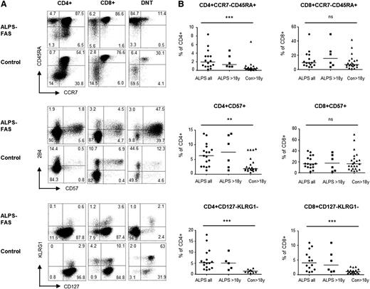
We then asked whether we could identify cells with this distinct phenotype among CD4+ and CD8+ SPT also. ALPS patients had increased percentages of TEMRA SPT, either defined by the CCR7−CD45RA+ phenotype or by CD57 expression (Figure 3A-B). This reached statistical significance for CD4+, but not for CD8+TEMRA cells. Moreover, only the percentage of CD4+, not that of CD8+TEMRA cells, significantly correlated with the total percentage of DNT (data not shown). It presumably reflects the observation that CD4+TEMRA are rarely detectable in healthy donors and are thus more specifically associated with Fas deficiency, whereas CD8+TEMRA are readily detectable albeit variable in both patients and controls. Similar to DNT, 2B4 expression was dim on CD57+TEMRA cells from ALPS patients, whereas it was bright on cells from healthy donors (Figure 3A). Most significantly, ALPS patients had the unusual CD127−KLRG1− population observed in DNT also among their SPT, whereas this population was virtually absent in SPT and DNT from healthy donors (Figure 3A-B).
T cells resembling DNT can be detected among CD4+ and CD8+ T cells of ALPS patients. (A) Representative plots showing the proportions of CCR7-CD45RA+TEMRA, CD57+ and CD127−KLRG1− T cells among ALPS versus control CD4+ T cells and CD8+ T cells. (B) Percentages of these populations in all studied ALPS patients (ALPS all), adult ALPS patients (ALPS >18 y) and controls (Con >18 y) with statistical comparison between all ALPS patients and adult controls. ns, not significant. **P < .01; ***P < .001.
T cells resembling DNT can be detected among CD4+ and CD8+ T cells of ALPS patients. (A) Representative plots showing the proportions of CCR7-CD45RA+TEMRA, CD57+ and CD127−KLRG1− T cells among ALPS versus control CD4+ T cells and CD8+ T cells. (B) Percentages of these populations in all studied ALPS patients (ALPS all), adult ALPS patients (ALPS >18 y) and controls (Con >18 y) with statistical comparison between all ALPS patients and adult controls. ns, not significant. **P < .01; ***P < .001.
To further examine the relationship of these “DNT-like” TEMRA populations to bona fide DNT in ALPS patients, we analyzed their coreceptor expression. Although TEMRA SPT from healthy donors were predominantly CD27− and CD28− and had normal CD4 or CD8 expression, TEMRA cells of ALPS patients consisted of 2 populations, 1 being CD27− CD28− with normal CD4 or CD8 expression (“conventional”), and 1 with high CD27 and CD28 expression and reduced CD4 or CD8 levels (“DNT-like”) (Figure 4A-B). Thus, T cells with a DNT-like phenotype can be detected among both CD4+ and CD8+TEMRA cells of ALPS patients and they show evidence of partial CD4/CD8 downregulation. Most ALPS patients had either increased CD4low or CD8low T cells or both (supplemental figure 2A-B). Differentiation marker expression on these CD4low and CD8low T cells resembled that of DNT in ALPS patients (supplemental figure 2C).
DNT-like cells have reduced CD4 or CD8 coreceptor expression. (A) CCR7−CD45RA+ CD4+TEMRA or (B) CCR7−CD45RA+ CD8+TEMRA cells were gated and analyzed for their CD27 and CD28 expression. Subsequently, CD4 or CD8 expression was analyzed on CD27−CD28−TEMRA cells (gray tinted) vs CD27+CD28+TEMRA cells (black line). For CD8+TEMRA the CD8 expression level (CD8 MFI, mean fluorescence intensity) is compared between CD27−CD28− and CD27+CD28+TEMRA in 8 ALPS-FAS patients vs adult controls. Because very few healthy donors had detectable CD4+TEMRA, only representative plots are shown.
DNT-like cells have reduced CD4 or CD8 coreceptor expression. (A) CCR7−CD45RA+ CD4+TEMRA or (B) CCR7−CD45RA+ CD8+TEMRA cells were gated and analyzed for their CD27 and CD28 expression. Subsequently, CD4 or CD8 expression was analyzed on CD27−CD28−TEMRA cells (gray tinted) vs CD27+CD28+TEMRA cells (black line). For CD8+TEMRA the CD8 expression level (CD8 MFI, mean fluorescence intensity) is compared between CD27−CD28− and CD27+CD28+TEMRA in 8 ALPS-FAS patients vs adult controls. Because very few healthy donors had detectable CD4+TEMRA, only representative plots are shown.
ALPS DNT and “DNT-like” SPT lack T-bet expression
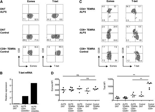
The unusual differentiation pattern of DNT suggested a unique underlying transcriptional profile. Tbox transcription factor expressed in T cells (T-bet) and its paralog eomesodermin (Eomes) represent a pair of counterregulatory transcription factors crucially involved in the determination of CD8+ T-cell fate, phenotype, and function.30 Although T-bet induces terminal differentiation associated with KLRG1 and CD57 upregulation, Eomes promotes long-term memory formation and homeostatic renewal.31-33 Thus, we determined the ratio of T-bet and Eomes expression in DNT. As expected from previous studies,15 ALPS DNT showed high Eomes expression, which was also observed in CD4+ and CD8+TEMRA cells (Figure 5A-C) at similar fluorescence intensity (Figure 5D). In contrast, ALPS DNT almost completely lacked expression of T-bet (Figure 5A). To substantiate this finding, quantitative RT-PCR was performed with sorted T-cell populations in 4 patients and confirmed absent T-bet mRNA in ALPS DNT, although it was highly expressed in memory CD8 T cells (Figure 5B). Interestingly, also CD28+ “DNT-like” CD4+ and CD8+TEMRA cells from ALPS patients were T-bet–negative, whereas CD28− “normal” CD8+TEMRA cells from ALPS patients and control CD8+TEMRA expressed T-bet (Figure 5C-D). Of note, CD4+TEMRA cells were not detectable and therefore could not be analyzed in the tested control donors. These findings indicate abnormal programming, not only of ALPS DNT, but also of their presumed precursors, which can be found among both CD4+ and CD8+ T cells.
ALPS DNT lack T-bet expression. (A) Representative plots showing expression of Eomes and T-bet in ALPS and control DNT (gate: CD3+TCRαβ+CD4−CD8−), and in control CD8+TEMRA cells (gate: CD3+CD8+CCR7−CD45RO−). (B) T-bet messenger RNA was quantified by RT-PCR using sorted DNT, CD8+CD45RA+, and CD8+CD45RA− T-cell populations. CD8+CD45RA− memory cells served as a positive control because the proportions of TEMRA cells were too low. Representative data from 1 of 4 analyzed ALPS-FAS patients are shown. (C) CD4+TEMRA, CD8++TEMRA, and DNT were gated and analyzed for Eomes and T-bet expression in combination with CD28 to distinguish between “DNT-like” (CD28+) and “normal” (CD28−) TEMRA cells. (D) T-bet and Eomes levels were compared between ALPS DNT, control DNT, CD4+CD28+TEMRA, and CD8+CD28+TEMRA from 5 ALPS patients and CD8+TEMRA from 5 healthy donors. The great majority of control CD8+TEMRA were CD28−. MFI, mean fluorescence intensity; ns, not significant. **P < .01.
ALPS DNT lack T-bet expression. (A) Representative plots showing expression of Eomes and T-bet in ALPS and control DNT (gate: CD3+TCRαβ+CD4−CD8−), and in control CD8+TEMRA cells (gate: CD3+CD8+CCR7−CD45RO−). (B) T-bet messenger RNA was quantified by RT-PCR using sorted DNT, CD8+CD45RA+, and CD8+CD45RA− T-cell populations. CD8+CD45RA− memory cells served as a positive control because the proportions of TEMRA cells were too low. Representative data from 1 of 4 analyzed ALPS-FAS patients are shown. (C) CD4+TEMRA, CD8++TEMRA, and DNT were gated and analyzed for Eomes and T-bet expression in combination with CD28 to distinguish between “DNT-like” (CD28+) and “normal” (CD28−) TEMRA cells. (D) T-bet and Eomes levels were compared between ALPS DNT, control DNT, CD4+CD28+TEMRA, and CD8+CD28+TEMRA from 5 ALPS patients and CD8+TEMRA from 5 healthy donors. The great majority of control CD8+TEMRA were CD28−. MFI, mean fluorescence intensity; ns, not significant. **P < .01.
ALPS DNT share TCRβ CDR3 sequences with CD4+ and CD8+TEMRA cells
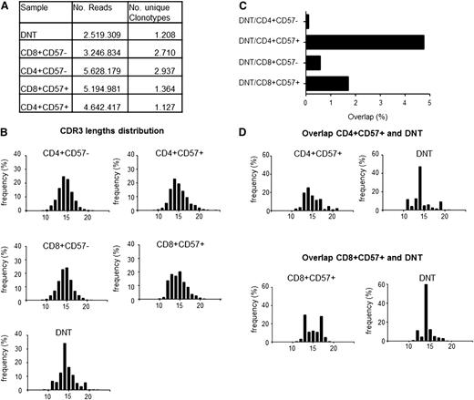
To further corroborate the hypothesis of dual lineage origin of DNT, we performed deep sequencing of TCRβ rearrangements in sorted T-cell populations of 1 untreated ALPS-FAS patient. This allowed the identification of CDR3 sequences shared between individual SPT populations and DNT. Defined amounts of genomic DNA were amplified by a multiplex PCR approach combined with massive parallel sequencing.34 The numbers of unique CDR3 sequences (designated as clonotypes) among the major CD4+CD57− and CD8+CD57− populations were in the expected range of healthy individuals (unpublished data), but were more than 50% lower in CD4+CD57+TEMRA, CD8+CD57+TEMRA and DNT (Figure 6A). CDR3 lengths had a Gaussian distribution among the CD57-negative populations, whereas mild skewing was observed among CD8+CD57+ T cells more than among CD4+CD57+ T cells. Despite a similar number of unique sequences, more pronounced skewing was detected among DNT (Figure 6B).
DNT share CDR3 sequences with CD4+ and CD8+ TEMRA cells. (A) DNT and different SPT-cell populations of 1 ALPS-FAS patient were sorted for TCRβ deep sequencing. Equal amounts of genomic DNA were amplified by multiplex PCR. The number of resulting reads and unique clonotypes are shown for each cell population. (B) CDR3 lengths distribution is shown for all clonotypes within each T-cell population. (C) Percentage of overlapping identical sequences among the different T-cell populations. (D) CDR3 lengths distribution was analyzed among overlapping sequences between CD4+CD57+ and DNT, as well as between CD8+CD57+ and DNT.
DNT share CDR3 sequences with CD4+ and CD8+ TEMRA cells. (A) DNT and different SPT-cell populations of 1 ALPS-FAS patient were sorted for TCRβ deep sequencing. Equal amounts of genomic DNA were amplified by multiplex PCR. The number of resulting reads and unique clonotypes are shown for each cell population. (B) CDR3 lengths distribution is shown for all clonotypes within each T-cell population. (C) Percentage of overlapping identical sequences among the different T-cell populations. (D) CDR3 lengths distribution was analyzed among overlapping sequences between CD4+CD57+ and DNT, as well as between CD8+CD57+ and DNT.
Then, we determined the frequency of unique clonotypes shared between the different T-cell populations. The greatest overlap was found between DNT and CD4+TEMRA (4.75% identical clonotypes). In contrast, only 0.57% identical CDR3 sequences could be detected between CD4+CD57− and CD4+CD57+ T-cell populations. The overlap between DNT and CD8+TEMRA cells was less pronounced (1.71%) (Figure 6C). Consistent with these genetic data, in this patient, almost all CD4+TEMRA cells, but only a part of CD8+TEMRA cells, had a DNT-like phenotype (not shown). These data provide a strong argument for the hypothesis that both CD4+ and CD8+ single positive TEMRA cells can give rise to ALPS DNT. Notably, although the putative parental SPT populations showed moderate oligoclonality, this was more pronounced among DNT (Figure 6D). One CDR3 length dominated the overlapping DNT clonotypes, but this peak still represented 19 unique sequences.
Genetic evidence for the origin of ALPS DNT from CD4+ and CD8+TEMRA cells
In an independent approach, we made use of an unusual genetic constellation in some ALPS patients. In patients with ALPS-FAS-sLOH, LOH presumably occurs in a hematopoetic precursor.5 The percentage of CD4+ or CD8+ T cells carrying this second genetic hit is very small, but it becomes prominent among DNT, in which usually more than 90% of cells have a biallelic FAS mutation. If the mutation is amorphic, this leads to absent Fas expression on most DNT (supplemental figure 3). Therefore, in such ALPS-FAS-sLOH patients, cells with biallelic mutations in a population that is normally Fas-positive can be identified by absent Fas expression. Accumulation of such cells in a given cell population indicates Fas-dependent aberrant expansion and/or defective elimination.
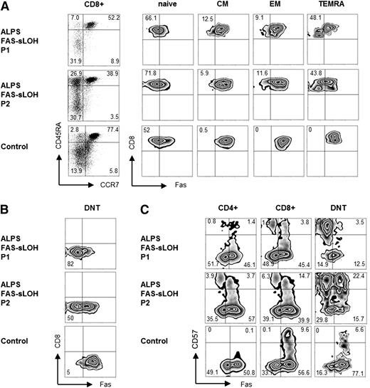
The majority of naïve SPT cells are Fas-negative. Once activated, T cells upregulate Fas and remain positive throughout their differentiation to TEMRA cells (Figure 7A). In ALPS-FAS-sLOH patients, a small proportion of CD8+ and CD4+ CM and EM cells, and more than 50% of CD4+ and CD8+TEMRA cells did not express Fas (Figure 7A and supplemental figure 4). Interestingly, CD4 or CD8 coreceptor expression was slightly reduced on Fas-negative TEMRA cells. As expected, the majority of ALPS DNT were Fas-negative (Figure 7B). Similarly, a significant fraction of CD57+ CD4+ or CD8+ T cells from ALPS-FAS-sLOH patients did not express Fas, whereas all CD57+ T cells in controls were Fas-positive (Figure 7C). To confirm that Fas-negative T cells indeed represented cells with a biallelic mutation, we quantified the percentage of mutated alleles in sorted total DNT vs sorted Fas-CD57+ DNT in 1 ALPS-FAS-sLOH patient (P2). In this patient, 50% of total DNT were Fas-negative (Figure 7C), predicting about 75% mutant alleles (50% monoallelic due to the germ line mutation and 50% biallelic due to the second hit) in this population. Indeed, genetic analysis revealed a frequency of the mutant allele of 70% in total DNT, whereas this was 98% in sorted Fas-CD57+ DNT. These results show that biallelic mutated T cells already accumulate among CD4+ and CD8+TEMRA cells, providing a further argument that subsets of both populations represent DNT precursors.
Double mutated T cells accumulate among CD4 and CD8 TEMRA cells in patients with ALPS-FAS-sLOH. (A) Fas expression is shown on naïve (naïve, CCR7+CD45RA+), central memory (CM, CCR7+CD45RA−), effector memory (EM, CCR7−CD45RA−), and terminally differentiated CD8+ T cells (TEMRA, CCR7−CD45RA+), and (B) on CD3+TCRαβ+CD4−CD8− DNT of 2 patients with ALPS-FAS-sLOH (P1: 14 y, P2: 32 y) compared with an adult control. (C) CD57 and Fas expression on CD4+, CD8+, and DNT of the same patients compared with an adult control.
Double mutated T cells accumulate among CD4 and CD8 TEMRA cells in patients with ALPS-FAS-sLOH. (A) Fas expression is shown on naïve (naïve, CCR7+CD45RA+), central memory (CM, CCR7+CD45RA−), effector memory (EM, CCR7−CD45RA−), and terminally differentiated CD8+ T cells (TEMRA, CCR7−CD45RA+), and (B) on CD3+TCRαβ+CD4−CD8− DNT of 2 patients with ALPS-FAS-sLOH (P1: 14 y, P2: 32 y) compared with an adult control. (C) CD57 and Fas expression on CD4+, CD8+, and DNT of the same patients compared with an adult control.
Discussion
This study combines phenotypic analysis with 2 independent genetic approaches to demonstrate that in the absence of intact Fas signaling, cells with the phenotypic and transcriptional characteristics of ALPS DNT already accumulate among single positive terminally differentiated T cells, and that this occurs among both CD4+ and CD8+ T-cells. These findings provide novel evidence for a role of Fas in human T-cell differentiation beyond the homeostatic regulation of T cells through induction of apoptosis. Furthermore, they provide the first significant evidence that human DNT can also differentiate from CD4+ T cells.
Accumulation of DNT is a hallmark of patients with genetic defects in FAS. Current evidence indicates that FAS mutant DNT arise from chronically activated CD8+ T cells, which cannot be eliminated. In support of this, previous studies have shown that ALPS DNT highly express CD57 and CD45RA.14 Here, we analyzed DNT differentiation in more detail and demonstrated that they have a CCR7−CD45RA+ phenotype similar to TEMRA cells.35 Low CD127 and high expression of the senescence and exhaustion markers CD57 and PD-1 were further signs of terminal differentiation. However, in contrast to conventional TEMRA cells, ALPS DNT highly expressed CD27 and CD2814 and lacked expression of KLRG1, an inhibitory receptor that is normally coexpressed with other inhibitory molecules and CD57 on TEMRA cells.36 This distinct and unusual differentiation phenotype indicated that ALPS DNT might have a particular transcriptional program. Because a previous study had shown increased Eomes and (to a lesser extent) T-bet expression in human ALPS and murine lpr DNT compared with bulk CD4+ or CD8+ T-cell populations,15 we analyzed expression of these 2 transcription factors in ALPS DNT and CD8+TEMRA cells from healthy donors. Surprisingly, although Eomes was present at similar levels in both populations, ALPS DNT completely lacked T-bet, which was highly expressed in TEMRA cells from healthy donors. Because T-bet drives terminal effector differentiation and correlates with CD57 and KLRG1 expression,31-33,37 the unusual lack of KLRG1 on ALPS DNT could thus be linked to their absent T-bet expression. A previous study has clearly defined an essential and nonredundant role of Eomes for DNT development or maintenance in mice.15 Our observations raise the question, whether this is (at least in humans) really an effect of Eomes expression per se or an effect of Eomes in the context of absent T-bet expression. KLRG1 binds cadherins expressed by epithelial cells and a wide range of antigen-presenting cells, and inhibits proliferation of highly differentiated CD8+ T cells.38 Thus, lack of KLRG1 could support the high-proliferative capacity of DNT, despite their TEMRA-like differentiation.
We detected the same abnormal differentiation pattern already in SPT populations, supporting the concept that the characteristic ALPS DNT phenotype is already determined before the step of coreceptor downregulation. Thus, we found T cells phenotypically resembling DNT among CD4+ and CD8+ CCR7−CD45RA+TEMRA cells of ALPS patients. Significantly, “DNT-like” SPT populations also lacked T-bet expression. This indicates that already before and during downregulation of the CD4 or the CD8 coreceptor, a proportion of cells had acquired the “ALPS DNT-like” phenotype. Further support for this concept was provided by our genetic approach illustrating that biallelic mutant T cells in ALPS-FAS-sLOH patients accumulate not only among DNT, but also among CCR7−CD45RA+ or CD57+ CD4+ or CD8+ T cells. More than 50% of TEMRA cells were Fas-negative in the 2 analyzed patients. Fas-negative cells were also present among cells with CM and EM differentiation, but at a frequency <5%. We could not estimate the fraction of biallelic mutant cells by Fas staining in naïve SPT cells, but the percentage of mutated allele in sorted naïve cells was approximately 50%, indicating that the fraction of double-mutated cells among naïve T cells must be small. In this context, it should be stated that accumulation of DNT-like cells among TEMRA cells does not imply a relation to conventional TEMRA cells. This just reflects the fact that expression of terminal differentiation markers on aberrant T cells, presumably after extensive replication, allows their identification among T-cell populations undergoing their normal differentiation sequence.
The lineage origin of DNT has been widely debated and most evidence in mice and humans suggests that DNT arise from CD8+ T cells.7-11,39 Our data challenge this view. DNT-like cells lacking T-bet expression were detected in both CD4+ and CD8+TEMRA populations. Moreover, cells with biallelic mutations accumulated in CD4+ and CD8+TEMRA compartments of ALPS-FAS-sLOH patients, although the relative accumulation in the 2 compartments differed between individuals. Of note, in most patients, the percentage of DNT correlated better with the percentage of CD4+TEMRA and the percentage of CD4low T cells than with the percentage of their CD8+ counterparts. Finally, the detection of a significant fraction of identical TCRβ CDR3 sequences shared by CD4+TEMRA and DNT, as well as by CD8+TEMRA and DNT, strongly argues that also CD4+ T cells can give rise to DNT. Previous observations on similarities in Vβ usage and detection of shared CDR3 sequences of Vβ-Jβ transcripts between DNT and CD8+ T cells have indicated a CD8 origin of ALPS DNT.11 However in that study, CD4+ T cells were not included in molecular analysis, because they did not show the TCR-Vβ skewing characteristic of DNT. Our phenotypic and genetic data are more consistent with a dual lineage origin of DNT in humans. Of note, the stronger correlation of CD4+TEMRA with the percentage of DNT reflects the fact that CD4+TEMRA, although a small population, are more specifically increased in ALPS patients. This does not imply a greater contribution of CD4+ T cells to DNT. The relative contribution of CD4+ vs CD8+ T cells to DNT remains unknown and presumably varies among individuals and in response to intrinsic and/or external stimuli.
How can these data be integrated into a model explaining the role of Fas in T-cell differentiation? We hypothesize that at 1 or at several earlier stages of T-cell differentiation (which may be as early as during thymic selection), impairment of a first Fas-dependent checkpoint determines that a subset of CD4+ and CD8+ T cells is programmed differently, and takes a differentiation pathway that eventually leads to the DNT stage. This Fas-dependent checkpoint most likely involves Fas-mediated apoptosis. However, Fas has additional nonapoptotic signaling properties including activation of nuclear factor kB and mitogen-activated protein kinase pathways, which could contribute to abnormal differentiation and different transcriptional programming.40,41 As suggested by observations in mice, such Fas-regulated cells could be subsets of T cells, which have escaped thymic selection39,42 or peripheral T cells homeostatically proliferating in response to limited self-peptide/major histocompatibility complex contacts.43 Abnormal transcriptional programming of such cells could lead to their uncontrolled proliferation with accumulation of senescent DNT. It is possible, but not required in this model, that impairment of a second Fas-dependent checkpoint prevents apoptosis of DNT, and thus contributes to their accumulation at the single positive TEMRA, and even more at the DNTstage. The questions of which properties define those T cells destined to become DNT in Fas deficiency, which signaling pathways/networks induce abnormal differentiation and function, and if and which external signals/antigens drive their expansion, remain to be answered.
The understanding that Fas regulates T-cell homeostasis by eliminating or reprogramming cells at earlier stages of the natural CD4+ and CD8+ T-cell life cycle, which then undergo aberrant differentiation, rather than by eliminating normally differentiated senescent T cells, has implications for the treatment of ALPS patients. Indeed, disrupting aberrant signaling by mammalian target of rapamycin inhibition with sirolimus has proven to be very successful in treating their lymphoproliferative manifestations. Further dissection of aberrant signaling in Fas deficiency may pave the way to future identification of more specific therapeutic targets.
The publication costs of this article were defrayed in part by page charge payment. Therefore, and solely to indicate this fact, this article is hereby marked “advertisement” in accordance with 18 USC section 1734.
There is an Inside Blood Commentary on this article in this issue.
Acknowledgments
We acknowledge our patients, their families, and referring physicians who made this study possible. We also thank Ursula Warthorst and the technicians of the Center for Chronic Immunodeficiency Advanced Diagnostic Unit for excellent technical assistance, Jan Bodinek for cell sorting, and Dr Steffen Hennig (HS Diagnomics, Berlin) for provision of bioinformatics support.
This work was supported by the German Federal Ministry of Education and Research grant (Bundesministerium für Bildung und Forschung1 EO 0803) to the Center of Chronic Immunodeficiency, the Bundesministerium für Bildung und Forschung grant (01GM1111B) to the Primary Immunodeficiency Network initiative, and the Interdisciplinary Center for Clinical Research Erlangen grant (Interdisziplinäres Zentrum für Klinische Forschung project A58).
Authorship
Contribution: A.R.E., S.V., S.E., A.M., and C.S. designed the research; C.S., A.J., M.A., A.C., S.H., K.K., C.B.C., S.H., I.K., A.P., and M.G.S. repeatedly referred patients; I.F. carried out diagnostic tests; M.R.L. and K.S. performed genetic analysis, A.R.E., S.V., and S.A. collected data; J.R. and M.H. performed TCRβ deep sequencing; A.R.E., S.V., M.R.L., K.S., J.R., A.A., M.H., A.J., C.S., B.B., R.T., H.P., A.M., and S.E. analyzed and interpreted data; A.R.E., S.V., and S.E. wrote the manuscript; and all of the authors edited the manuscript.
Conflict-of-interest disclosure: The authors declare no competing financial interests.
Correspondence: Stephan Ehl, Center for Chronic Immunodeficiency, Breisacher Strasse 117, Freiburg, 79106 Germany; e-mail: stephan.ehl@uniklinik-freiburg.de.
References
Author notes
A.R.E. and S.V. contributed equally to this study.


This feature is available to Subscribers Only
Sign In or Create an Account Close Modal