Abstract
Introduction
Multiple myeloma (MM) is one of the hematologic malignancies characterized as accumulation of monoclonal tumor of plasma cells. Although novel therapeutic agents have significantly improved the survival of MM patients, MM is still a mostly incurable disease. The most common reason of relapsed and refractory MM is that myeloma cells intimately communicate with bone marrow stroma and acquire drug resistance.
Stroma is known to secrete soluble factors and express some adhesion molecules advantageous for progression of MM. Recently, there are accumulating evidences that bone marrow-derived mesenchymal stem cells (MSC) serve as a component of stroma and support growth and drug resistance of myeloma cells. Although the exact etiology has not been clearly defined, it has been suggested that the incidence of MM increases with aging.
In this study, we tried to explore the relationship between MM progression and cellular senescence in MSC.
Methods
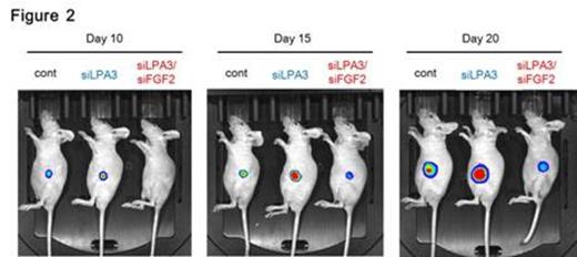
Human myeloma cell lines used in this study were IM-9, OPM-2, and RPMI-8226. Human MSC used in this study were obtained from Texas A&M Health Science Center for the Preparation and Distribution of Adult Stem Cells. To establish a mouse xenograft model of human MM, 1.0 x 10⁶ of IM-9 cells were co-injected with 4.0 x 10⁵ of human MSC subcutaneously into the flank of BALB/c nude mice. Visible and palpable tumors were formed in all animals after 20 days. Tumor specimens were resected surgically and diagnosed pathologically as MM based on marker expression. For in vivo bioimaging, luciferase-expressing IM-9 (IM-9-luc+) cells were co-injected with human MSC subcutaneously into BALB/c-nude mice, and photon emission was detected with a sensitive CCD camera 10, 15 and 20 days later.
Results
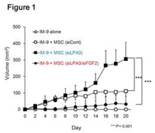
In our earlier research, we have demonstrated that the signaling of lysophosphatidic acid (LPA), a bioactive lipid mediator, modulates cellular senescence in MSC (Kanehira et al. PLoS One. 2012). MSC produced autotaxin (ATX), a key enzyme in LPA synthesis, in response to myeloma cells via Toll-like receptor 4 (TLR4)/NF-kappaB-dependent pathway. To determine the LPA receptor responsible for cellular senescence in MSC, six LPA receptors (LPAR1-6) were individually knocked down using siRNA for each receptor. In BrdU incorpotaion assay, LPAR3 gene-silenced MSC (siLPAR3-MSC) were less proliferative than control MSC, and in cell cycle analysis with 7-AAD and Pyronin Y, siLPAR3-MSC was arrested in G1 phase. Interestingly, siLPAR3-MSC exhibited some characteristics of cellular senescence, such as up-regulation of senescence-associated beta-galactosidase activity, increased cell size, and flattened morphology. In a mouse xenograft model of MM, siLPAR3-MSC promoted progression of MM and also tumor-associated angiogenesis. In in vitro study, we confirmed that siLPAR3-MSC easily transdifferentiated into alpha-SMA+ tumor-associated fibroblast and secreted FGF2 in response to myeloma cells. The MM promoting effect and elevated tumor-associated angiogenesis observed in siLPAR3-MSC co-injection were both completely cancelled by FGF2 gene-silencing in siLPAR3-MSC (Figure 1 and 2).
Conclusion and discussion
In this study, we verified the impairment of LPAR3 signaling may accelerate cellular senescence in MSC. And senesced MSC can provide an advantageous microenvironment for MM progression by FGF2-dependent formation of tumor-stroma milieu. Here, we provide the possibility that LPAR3 signaling could be promising as a therapeutic target in MM.
Fujiwara:Chugai Pharmaceutical CO., LTD: Research Funding. Fukuhara:Gilead Sciences: Research Funding. Harigae:Chugai Pharmaceutical Co., Ltd.: Research Funding.
Author notes
Asterisk with author names denotes non-ASH members.


This feature is available to Subscribers Only
Sign In or Create an Account Close Modal