Abstract
Background: Programmed cell death 1 (PD-1) protein downregulates T cell activation and is related to immune tolerance. PDL1 up regulation, T cell infiltration, and T cell exhaustion are features, which suggest susceptibility to PD-1 blockade antibodies. Blockade of PD-1 or its ligand PD-L1 has shown promising responses in several malignancies. Although little clinical activity has been seen in patients with relapsed multiple myeloma (MM), the role of the PD-1 pathway and T cell exhaustion in newly diagnosed MM has not been explored.
Objective: To determine whether T-cell infiltrate or expression of PD-1 correlates with clinical features and prognosis among patients with newly diagnosed multiple myeloma.
Methods: We screened a clinically annotated database of 341 patients seen at MSKCC between 1998 and 2012 that had Multiple Myeloma, received a bone marrow transplant and were consented to a biospecimen research protocol for availability of pre-treatment bone marrow specimens. A total of 64 bone marrow biopsy specimens were identified. Immunohistochemistry (IHC) was performed in formalin-fixed paraffin-embedded specimens using an anti human CD3 monoclonal antibody (mAb) (Dako, Clone F7.2.38) and an anti human PD-1 mAb (Cell Marque, catalog #315M-95). CD3 and PD1 IHC staining were graded as negative (<5% for CD3, < 1% for PD1), or positive (≥5% for CD3, ≥1% for PD1). Correlative analyses were performed between CD3/PD1 expression and clinical outcome using the following parameters: International Staging System (ISS), cytogenetic risk, progression free survival (PFS), overall survival (OS), and response to treatment. Groups were compared by Fisher's exact test. OS and PFS were assessed by Cox regression and estimated by Kaplan-Meier methods.
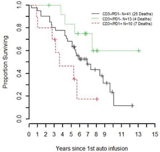
Results: 23 specimens (36%) were CD3 positive and 10 specimens (16%) were PD-1 positive. All PD-1 positive specimens were CD3 positive. 41 specimens (64%) were CD3 negative (<5%) and PD1 negative (<1%). Based on these results, specimens were divided into three groups: Exhausted T-cell infiltrate (CD3+/PD1+), non exhausted T-cell infiltrate (CD3+/PD1-) and no T-Cell infiltrate (CD3-/PD1-). In the exhausted T-cell infiltrate group 30% of patients had ISS stage 3 and 40% had high risk cytogenetics. In the non-exhausted T-cell group 15% of patients had ISS stage 3 and 15% high cytogenetic risk. In the no T-cell infiltrate group 10% had ISS stage 3 disease and 22% high cytogenetic risk. These proportions were not significantly different across the 3 groups. Median OS from 1st auto infusion was 7 years while median PFS was 2.3 years. On univariate analysis, there was no significant difference in PFS between the 3 groups. The presence of CD3 and PD1 T-cells were significantly associated with OS (p-value = 0.04). Median OS from 1st auto infusion was 43 months for the exhausted T-Cell infiltrate group followed by 83 months for the no T-Cell infiltrate group. The non exhausted T-cell group had the highest OS, median not reached; OS by 7-years was 75%. Cytogenetic risk at diagnosis was significantly associated with OS (p-value = 0.03). In a multivariable model, CD3/PD1 staining continued to trend toward an association with OS (p-value = 0.08) and cytogenetic risk remained significant (p-value = 0.05).
Conclusions: The presence of T-cells with PD-1 expression was not associated with higher risk disease at MM diagnosis based on cytogenetics and ISS stage. The presence of PD-1 expressing CD3+ T cells trends toward an association with poorer overall survival in newly diagnosed MM, especially compared to non exhausted T-cell infiltrate, suggesting the possibility that T cell exhaustion represents a novel high risk disease characteristic. Further investigation is necessary to assess if the presence of CD3+PD-1+ T cells is an independent prognostic feature in newly diagnosed MM.
Overall survival by CD3/PD1 Staining in 64 newly diagnosed myeloma patients.
Giralt:JAZZ: Consultancy, Honoraria, Research Funding, Speakers Bureau; TAKEDA: Consultancy, Honoraria, Research Funding; CELGENE: Consultancy, Honoraria, Research Funding; AMGEN: Consultancy, Research Funding; SANOFI: Consultancy, Honoraria, Research Funding. Landgren:International Myeloma Foundation: Research Funding; Onyx: Consultancy; Bristol-Myers Squibb: Consultancy; Celgene: Consultancy; Medscape: Honoraria; Bristol-Myers Squibb: Honoraria; Medscape: Consultancy; BMJ Publishing: Consultancy; Celgene: Honoraria; Onyx: Honoraria; BMJ Publishing: Honoraria; Onyx: Research Funding. Hassoun:Novartis: Consultancy; Takeda: Research Funding; Celgene: Research Funding; Celgene: Membership on an entity's Board of Directors or advisory committees. Lesokhin:Genentech: Research Funding; Aduro: Consultancy; Janssen: Consultancy, Research Funding; Bristol Myers Squibb: Consultancy, Research Funding; Efranat: Consultancy.
Author notes
Asterisk with author names denotes non-ASH members.


This feature is available to Subscribers Only
Sign In or Create an Account Close Modal