Abstract
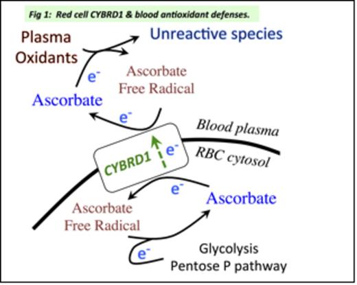
Introduction: The pathology of sickle cell disease (SCD) includes ischemia / reperfusion events in the vasculature, compromised blood antioxidant defenses and elevated risk of cardiovascular complications. Vitamin C (ascorbate) is a major blood antioxidant; ascorbate inside red blood cells (RBC) furnishes an electron (e-) to recycle plasma ascorbate to its active, reduced state (Fig. 1). A key step, transmembrane e- transfer, involves cytochrome b reductase 1 (CYBRD1). Thus, a deficiency or defect in red cell CYBRD1 could plausibly increase oxidative stress and interconnected inflammatory processes, contributing to cardiovascular complications in SCD patients. Testing for CYBRD1 deficiency requires a quantitative assay for the protein in RBC, so we developed a suitable assay and used it to screen a small number of SCD patients and healthy individuals.
Materials and methods: Blood samples were collected from steady state adult SCD patients, healthy African American adults, and steady state pediatric SCD patients before and 2-5 months on hydroxyurea (HU) therapy. Protocols were approved by institutional review.
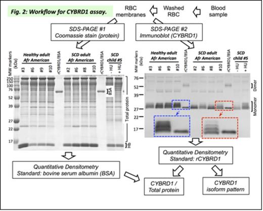
The CYBRD1 assay workflow is shown in Fig. 2. RBC membrane proteins were separated on two SDS-PAGE gels, along with a mixed standard (1.0 µg bovine serum albumin (BSA) + 1.9 ng of His6-tagged recombinant human CYBRD1 (rCYBRD1; PMID 21501687)). One gel was Coomassie-stained for quantitation of total membrane protein minus membrane-bound hemoglobin (Hb) (standard: BSA). Proteins in the other gel were transferred to a nitrocellulose filter, probed with CYBRD1 antibody (GeneTex #104793) and visualized with secondary antibody and chromogenic substrate for quantitation of CYBRD1 (standard: rCYBRD1), which was normalized to membrane protein. Gels, blots and calibrated density step tablets were scanned (transmittance mode) with an Epson V550; digitized images were analyzed using ImageJ. Two independent samples and paired t tests were used to compare between- and within-group mean differences in RBC CYBRD1 levels, respectively.
Results: The Coomassie-stained gels exhibited the usual profile of RBC membrane proteins (Fig. 2). In the immunoblots, the rCYBRD1 standard showed the expected narrow monomer band at ~30 kDa and faint dimer band at ~60 kDa (Fig. 2). However, RBC membrane samples revealed several CYBRD1 monomer bands, indicating the presence of previously unsuspected isoforms of CYBRD1. Further, the CYBRD1 isoform pattern in RBC from healthy controls was quite different from those seen in SCD patients. The functional impact of these differences remains to be investigated.
Assay of progressively diluted control RBC membranes produced a linear response for CYBRD1 from 0.7-7.2 ng (r2 = 0.975) and for total protein from 1 to 10 µg (r2 = 0.993). The sub-nanogram detection of CYBRD1 protein makes the assay usable with as little as 0.25 ml of blood. The coefficient of variation was ~15%, quite reasonable precision for quantitative immunoblotting.
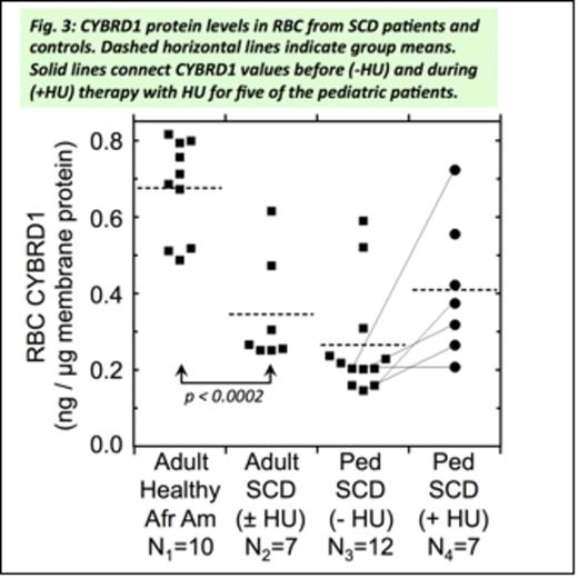
CYBRD1 levels in RBC from healthy African American adults averaged 0.68 ± 0.13 ng/µg membrane protein (N1 = 10; Fig. 3). CYBRD1 levels in RBC of adult SCD patients averaged 0.35 ± 0.14 ng/µg membrane protein (N2=7). This difference in means was highly statistically significant by two-tailed t-test (p < 0.0002), supporting our hypothesis that a deficiency in RBC CYBRD1 contributes to elevated oxidative stress in SCD patients.
RBC CYBRD1 levels in pediatric SCD patients not on HU therapy averaged 0.27 ± 0.14 ng/µg protein (N3=12; Fig. 3). Pediatric patients on HU therapy averaged 0.41 ± 0.18 ng CYBRD1/µg membrane protein (N4=7). For the five pediatric patients examined both before and during HU therapy there was a marginally significant increased mean CYBRD1 level on HU (p < 0.099 by paired, two-tailed t test).
Conclusions: Our newly developed assay allows quantitation of the RBC CYBRD1 protein level and analysis of the CYBRD1 isoform pattern using a small blood sample. The pilot screening results suggest that adult SCD patients have lower levels of CYBRD1 in their RBC, and a different pattern of CYBRD1 isoforms. Analysis of the CYBRD1 protein may be useful in assessing the state of individual SCD patients' blood antioxidant defenses and the antioxidant effects of HU. Thus, it will be worthwhile to examine the RBC CYBRD1 protein and activity in larger numbers of patients and controls, and to explore the origins of any differences between the groups.
No relevant conflicts of interest to declare.
Author notes
Asterisk with author names denotes non-ASH members.


This feature is available to Subscribers Only
Sign In or Create an Account Close Modal