Abstract
During arterial haemostasis, platelets tether to and translocate on disrupted vascular surface via binding of GPIbα to the VWF A1 domain, which triggers activation signals that induce intracellular Ca2+ release and up-regulate integrin αIIbβ3 binding capacity1,2. Inhibition of this signaling pathway or blockade of αIIbβ3 binding has a devastating impact on platelet firm adhesion and aggregate formation1,3,4.
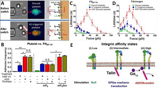
We used a newly developed switch biomembrane force probe (BFP) assay to characterize the cooperativity between GPIbα and αIIbβ3 on single-molecular level. VWF-A1 and fibronectin fragment (FNIII7-10) were separately coated on two different probes, enabling controlled presentation of two ligands in separate space and time (Fig. A). Pulling GPIbα by A1 on the first probe with a durable force activated the discoid platelet (Fig. A, upper panel), as revealed by an intracellular calcium flux. Upon switching to the FNIII7-10-bearing bead, the A1-triggered platelet was interrogated for the activity of its surface αIIbβ3 (Fig. A, lower panel).
A 25-pN force of >2s duration on a single GPIbα-A1 bond was found to activate αIIbβ3 into an intermediate affinity state, as reflected by a ~30% adhesion frequency with a FNIII7-10 bead, significantly higher than binding to low affinity αIIbβ3 on platelets without A1-triggering but much lower than binding to high affinity αIIbβ3 on platelets pre-incubated with ADP or thrombin (Fig. B), two strong platelet-activating agonists. This submaximal activation agreed with the previous immuno-staining findings2.
αIIbβ3 activation and outside-in signaling require the sequential engagement of talin and Gα13 to β3 cytoplasmic tail, respectively5,6. Blocking Gα13-β3 engagement by a synthetic peptide mP6 (gift from Xiaoping Du, UIC)6 had no effect on FNIII7-10 binding of resting and A1-triggered platelets, but significantly suppressed the FNIII7-10 binding of platelets pre-incubated with ADP or thrombin by lowering their adhesion frequencies to a level comparable with A1-triggered platelets (Fig. B). The control peptide mP6Scr showed no effect on the ADP or thrombin stimulated platelets (Fig. B).
In addition to binding affinity, bond lifetimes were measured by force-clamp experiment. Platelets with no treatment, A1 triggering and ADP stimulation formed αIIbβ3-FNIII7-10 (Fig. C) and αIIbβ3-fibrinogen (Fig. D) bonds with short, intermediate and long lifetimes, respectively. Interactions with fibrinogen exhibited catch-slip bonds regardless of the αIIbβ3 affinity state. By comparison, only high affinity αIIbβ3 exhibited a catch-slip bond with FNIII7-10 as slip-only bonds were observed for FNIII7-10 interactions with intermediate and low affinity αIIbβ3. These data suggest the existence of three distinct integrin states that correspond to the three affinities and force-dependent bond lifetime patterns, in which the intermediate state was induced by the GPIbα mechanotransduction (Fig. E). Gα13 engagement was not required for the intermediate state, but necessary for the high affinity state (Fig. E).
References:
1. Nesbitt, W. S. et al. The Journal of biological chemistry277, 2965-2972, doi:10.1074/jbc.M110070200 (2002).
2. Kasirer-Friede, A. et al. Blood103, 3403-3411, doi:10.1182/blood-2003-10-3664 (2004).
3. Savage, B., Saldivar, E. & Ruggeri, Z. M. Cell84, 289-297 (1996).
4. Warwick, S. N. et al. Nature Medicine15, doi:10.1038/nm.1955 (2009).
5. Gong, H. et al. Science327, 340-343, doi:10.1126/science.1174779 (2010).
6. Shen, B. et al. Nature503, 131-135, doi:10.1038/nature12613 (2013).
No relevant conflicts of interest to declare.
Author notes
Asterisk with author names denotes non-ASH members.


This feature is available to Subscribers Only
Sign In or Create an Account Close Modal