Abstract
It is known that SCF/c-kit signaling pathway is one of the most important pathways in regulating of cell proliferation, differentiation and apoptosis, which can be continuously activated by C-KIT mutation and then lead to cell proliferation increase or apoptosis decrease. Furthermore, we have proven in our previous study that amyloid precursor protein (APP) gene, which is reported to increase cell proliferation in some solid cancer, is correlated with C-KIT mutation and adversely affects the disease outcome in AML1-ETO-positive acute myeloid leukemia (AML1-ETO-positive AML). In this study we focus on the regulation of cell proliferation and apoptosis in AML1-ETO-positive leukemia by APP gene and its mechanism.
APP and C-KIT expression in bone marrow cells before the first chemotherapy from the 65 patients with AML1-ETO-positive AML (Blood. 2014,124:942) was simultaneously assessed by quantitative reverse transcriptase (QRT)-PCR method, meanwhile, the correlation of APP expression with peripheral WBC count, and the rate of bone marrow cellularity and blasts were observed. Furthermore, kasumi-1 cell line was chosen as cell model, and APP gene was knocked down using siRNA technology. The correlation of cell proliferation, differentiation and apoptosis with APP expression, as well as the regulation of SCF/c-kit signaling pathway by APP gene was analyzed on the cell line.
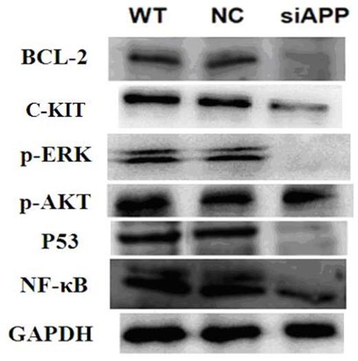
WBC count and bone marrow cellularity, but not bone marrow blasts, was correlated with APP expression, in that a higher WBC count (29.2±3.9·109/L) was observed in APP-H patients when compared with 17.9±2.9·109/L in APP-L patients (P=0.008), and a higher percentage of bone marrow cellularity in APP-H patients (86.7%±1.7%) versus 80.3%±2.3% in APP-L cases (P=0.031). Moreover, the level of C-KIT mRNA expression was positively correlated with APP expression (r=0.349, P=0.011). In kasumi-1 cell line, compared with wild type and negative control cells, cell apoptosis, both early and late, increased significantly when APP gene was knocked down (Figure 1)., however, not apparently change was observed in cell proliferation and differentiation. What's more, C-KIT expression at both transcription (as evidenced by RTQ¨CPCR analysis) and translation (as confirmed by CD117 assay) levels, as well as BCL-2, C-KIT, p-ERK, p-AKT, P53 and NF-κB (as evidenced by western blot analysis, Figure 2), decreased significantly when APP was knocked down.
In conclusion, APP gene involves in the regulation of cell apoptosis but not proliferation in AML1-ETO-positive leukemia via SCF/c-kit signaling pathway.
Flow cytometry analysis of cell apoptosis in kasumi-1 cells with different APP expression. Both the rate of early (Q4-2) and late apoptosis (Q2-2) in si-APP kasumi-1 cell increased significantly when compared with the rate in wild type and NC kasumi-1 cell (Early apoptosis rate: si-APP: 29.00%±0.98%, wild type: 21.43%±0.86%, NC: 21.67%±0.78%, F=71.927, P<0.001; Late apoptosis rate: si-APP: 19.80%±1.51%, wild type: 12.33%±0.75%, NC: 12.90%±1.25%, F=35.239, P<0.001).
Flow cytometry analysis of cell apoptosis in kasumi-1 cells with different APP expression. Both the rate of early (Q4-2) and late apoptosis (Q2-2) in si-APP kasumi-1 cell increased significantly when compared with the rate in wild type and NC kasumi-1 cell (Early apoptosis rate: si-APP: 29.00%±0.98%, wild type: 21.43%±0.86%, NC: 21.67%±0.78%, F=71.927, P<0.001; Late apoptosis rate: si-APP: 19.80%±1.51%, wild type: 12.33%±0.75%, NC: 12.90%±1.25%, F=35.239, P<0.001).
Western blot analysis of the expression of BCL-2, C-KIT, p-ERK, p-AKT, P53 and NF-κB with different APP expression.
Western blot analysis of the expression of BCL-2, C-KIT, p-ERK, p-AKT, P53 and NF-κB with different APP expression.
No relevant conflicts of interest to declare.
Author notes
Asterisk with author names denotes non-ASH members.


This feature is available to Subscribers Only
Sign In or Create an Account Close Modal