Abstract
The JAK2 V617F gain-of-function mutation is found in hematopoietic cells of nearly all patients with myeloproliferative diseases (MPDs). Ruxolitinib (Ruxo), a potent oral JAK1/JAK2 inhibitor, attenuates cytokine signaling by inhibition of JAK signaling, leading to anti-proliferative and pro-apoptotic effects. In patients with myelofibrosis (MF), effects of Ruxolitinib on splenomegaly, symptom improvement and possibly survival were successfully demonstrated in the two COMFORT studies. Furthermore, resolution of bone marrow fibrosis after long-term treatment with Ruxo was recently reported (Wilkins et al., Haematologica 2013). However, bone marrow stromal cells (BMSC), which contribute to fibrosis, do not harbor the JAK2 mutation or other chromosomal abnormalities that can be found in hematopoietic stem/progenitor cells. Therefore, the current study aimed to investigate possible effects of Ruxo on these non-hematopoietic cells.
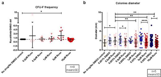
Bone marrow mononuclear cells were harvested from consenting donors and Ruxo effects on stroma progenitor cells were investigated using the standard CFU-F (colony-forming unit, fibroblast) assay. Our data showed that CFU-F numbers were not decreased by Ruxo at doses ranging from 0.2 to 10 μM (n = 9, p = 0.41, one-way ANOVA), but were even found to be increased at the 5 μΜ dose as indicated by a Ruxo/DMSO ratio of over one (p = 0.04, multiple comparisons test, control-vs-5 μΜ) (Figure 1). In contrast, statistically significant differences were observed in CFU-F colony size with a tendency to decreased sizes with higher doses of Ruxo (p < 0.0001, one-way ANOVA) (Figure 1). Next, we tested if Ruxo affected standard culture-derived bone marrow stromal cell (BMSC) growth, both, in short-term (6 h, 12 h, 24 h and 48 h, n = 3) and in long-term exposure experiments (up to a total of 21 days, n = 3). Neither short-term nor long-term exposure with Ruxo at 0.2 μΜ, 0.5 μΜ, 1 μΜ, 5 μΜ and 10 μΜ caused significant changes of BMSC numbers when compared to their corresponding DMSO controls (p > 0.05, two-way ANOVA), however, a trend to lower BMSC counts with higher Ruxo doses was observed. BMSC were reduced by maximum 25.3±5.7 % (mean ± SD) after 24-hour exposure with 10 μΜ Ruxo. After 21 days drug exposure, ratios of Ruxo-treated BMSC relative to their corresponding DMSO controls were 1.1±0.2, 1.1±0.3, 0.9±0.2, 0.7±0.4%, and 0.6±0.2 for 0.2 μΜ, 0.5 μΜ, 1 μΜ, 5 μΜ and 10 μΜ Ruxo, respectively.
These data indicated that Ruxo might have a cytotoxic effect on BMSC, however, only at very high concentrations. We therefore went on to study Ruxo effects on JAK signaling in BMSC after stimulation with IL-6, in order to mimic the inflammatory environment in MPDs. Western blot assays revealed that IL-6 treatment (100 ng/ml) induced pJAK2 in BMSC, which was not detectable in unstimulated BMSC. Furthermore, exposure of IL-6 stimulated BMSC to Ruxo (1 μΜ) diminished pSTAT3 and reduced downstream STAT targets such as pAKT and pMAPK. Furthermore, preliminary results on the cytokine expression profile of supernatants from IL-6-activated BMSC treated with Ruxo showed significant differences compared to controls. Specifically, IL-23 and CCL4 were elevated in Ruxo-treated/IL-6 activated BMSC whereas MCP-1 was strongly reduced compared to the non-Ruxo treated controls.
Taken together, these data show that clinically-relevant doses of Ruxo did not affect the clonogenic potential and proliferation of primary marrow stroma cells, indicating that Ruxo most likely has no or little direct effect on the fibrosis-causing cells in MPD. However, Ruxo considerably affects JAK-STAT signaling in activated BMSCs, leading to an altered cytokine expression profile which potentially contributes to the amelioration of fibrosis, and, accordingly, ongoing experiments address this question.
Effects of increasing doses of Ruxo on a) clonogenic bone marrow stroma cells expressed as ratio of the number of Ruxo-treated CFU-F and corresponding DMSO controls, and b) colony size (*p < 0.05, **p < 0.01, ***p < 0.001)
Effects of increasing doses of Ruxo on a) clonogenic bone marrow stroma cells expressed as ratio of the number of Ruxo-treated CFU-F and corresponding DMSO controls, and b) colony size (*p < 0.05, **p < 0.01, ***p < 0.001)
No relevant conflicts of interest to declare.
Author notes
Asterisk with author names denotes non-ASH members.


This feature is available to Subscribers Only
Sign In or Create an Account Close Modal