Abstract
Introduction. Ruxolitinib is the first JAK1/JAK2 inhibitor to be approved by the US Food and Drug Administration for the treatment of myelofibrosis (MF) and polycythemia vera (PV). Ruxolitinib is beneficial in relieving constitution symptoms and reducing splenomegaly. The main mechanism is the inhibition of T cell differentiation and function. Ruxulitinib inhibits Th1, TH17, Treg differentiation , and proliferation both in vivo and in vitro. Several clinical trials are under way to identify other Jak2 inhibitors to further improve efficacy and reduce adverse events. Notably momelotinib, a JAK1 and JAK2 inhibitor and pacritinib, a JAK2 and Fms-like tyrosine kinase 3 (FLT- 3) inhibitor that does not inhibit JAK1, have shown promise. Hence, we compared these three agents in their ability to interfere with T cell differentiation and function.
Materials and Methods. Peripheral mononuclear cells (MNC) from patients with myeloproliferative neoplasms (MPNs) and normal volunteer controls were isolated through Ficoll-paque density-gradient columns and cultured for 5-7 days with CD3/CD28 microbeads (Invitrogen Corp). The cells were treated with ruxolitinib (300 nM; SelleckChem), momelotinib (500 nM; SelleckChem), pacritinib (10 μM; SelleckChem), or an equivalent volume of their vehicle, DMSO, every other days for 5-7 days. Assays for TH1, TH17, Treg, and T cell proliferation by CFSE were performed. Cultured cells were stimulated with PMA (20 ng/ml) for 4h in the presence of ionomycin (1 µg/ml) and monensin (1 µM) before being harvested. TH1 were measured as IFN-γ secreting cells which were immunostained with IFN-γ catch and detection reagents for flow cytometry (IFN-γ secretion Assay kit, Miltenyi Biotec). Treg cells were detected using Treg Detection Kit (Miltenyi Biotec, Auburn, CA) for CD4+ CD25+ FoxP3+ cell quantification. Quantitation of Th17 cells was utilizing Human Th17 Flow Kit (BioLegend) for CD3+ CD4+ IL-17+ cell quantification by flow cytometry.For CFSE cell proliferation assay, viable 106 MNC cells were labeled with carboxyfluoresceinsuccinimidyl ester (CFSE, Invitrogen) cultured, and treated with either ruxolitinib, momelotinib, pacritinib, or DMSO in the presence of anti-CD3CD28-coated microbeads for 5-7 seven days at 37°C in 5% CO2 incubator. Cells were then washed twice and CD4+ cell division was analyzed by flow cytometry. Cells undergoing division were identified by the percentage of CFSEdim cells.
Statitiscal Anaysis. Statistical analyses were performed with paired or unpaired Student's t-test and one-way ANOVA for comparison of groups. P<0.05 were considered to be significant.
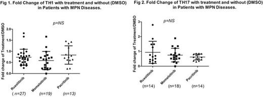
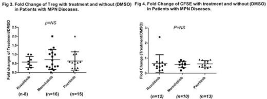
Results: The results were expressed as fold changes in the percentage of cells treated over those untreated. All three compounds showed inhibitory effects on the T cell differentiation to TH1, TH17, Treg, and CFSE. No statistical difference was found among all three compounds as shown in the following Figure 1,2,3,4.
Conclusion. No statitistical differenceswerefound among ruxolitinib, momelotinib, and pacritinib on the TH1, TH17, Treg differentiation, and T cell proliferation by CFSE assay.
Wang:Celgene: Research Funding.
Author notes
Asterisk with author names denotes non-ASH members.


This feature is available to Subscribers Only
Sign In or Create an Account Close Modal