Abstract
Therapy-related myelodysplastic syndromes and acute myeloid leukaemia (t-MDS/AML) occur as a late complication of cytotoxic chemotherapy and/or radiation for neoplastic and non-neoplastic disorders and are a major cause of non-relapse mortality. The prognosis of t-MDS/AML is poor because of an increased incidence of adverse cytogenetic features and relative resistance/intolerance to conventional therapies. TP53 is a tumour suppressor and cell cycle regulatory protein. Mutations in the TP53 gene are known to be associated with adverse outcomes in a variety of cancers including haematological malignancies.
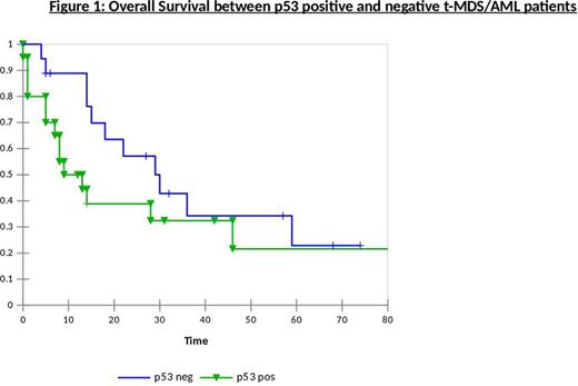
We examined p53 expression using immunohistochemical staining, as a surrogate marker for TP53 mutation, in marrow biopsies of 39 patients with t-MDS/AML from a single institution and correlated p53 expression with survival. Formalin fixed, paraffin embedded marrow trephine samples at diagnosis of t-MDS/AML were stained with DO-7 mouse p53 monoclonal antibody. Positive expression was defined as per Modified Quick Score. Of 39 patients, 35 had t-MDS and 4 had t-AML. 51% of patients had marrows at diagnosis that showed p53 positivity and 49% were negative. In a control group of 47 MDS patients in our centre, 22% were p53 positive and 78% were p53 negative. Of the t-MDS/AML group, age at diagnosis and gender distribution was similar in p53 positive and negative patients. Median latency (time from diagnosis of primary malignancy to diagnosis of t-MDS/AML) was similar between p53 positive and negative patients (66 vs. 52 months, p=0.51). A similar distribution was noted for the type of primary tumours (haematological vs. solid tumour) among the two groups, but all patients with more than one prior malignancy were noted to be p53 positive (N=4) whereas all patients who developed t-MDS after cytotoxic therapy for non-neoplastic disorders were p53 negative(N=3). There was no difference in p53 expression based on the type of primary therapy received (chemotherapy and/or radiotherapy) but a greater proportion (71%) of patients who received combined chemotherapy and radiation was p53 positive. A higher proportion of p53 negative patients had lower risk cytogenetics and normal karyotype whereas in p53 positive group there was lower incidence of good risk and a higher proportion to intermediate risk cytogenetics. However, equal numbers of patients with higher risk cytogenetics were found between p53 positive and negative groups. These findings were again reflected by IPSS-R risk groups (Table 1). Median overall survival was 10.5 months in p53 positive group compared to 22.5 months in p53 negative patients, p= 0.208. (Figure 1)
In conclusion, our study demonstrated that a higher proportion of patients with t-MDS/AML were positive for p53 than in de novo MDS which may be related to poor outcomes in this group. p53 positive t-MDS/AML patients tend to have lower incidence of favourable karyotypes and showed poor survival compared to their counterparts who were p53 negative. Analysis of p53 expression by immunohistochemistry is a readily accessible, cost-effective method of assessment without the need for expensive gene sequencing and is a clinically useful prognostic tool. It may be particularly useful in patients with t-MDS/AML.
p53 expression in Therapy-related Myeloid Neoplasms
| . | p53 Positive (N=20, 51%) . | p53 Negative (N=19, 49%) . |
|---|---|---|
| Primary Tumour Haematological (22) Solid Tumour (10) Autoimmune disease (3) More than one disorder (4) | 45% 60% 0 100% | 55% 40% 100% 0 |
| Primary Therapy Chemotherapy alone (27) Radiotherapy alone (5) Combined therapy (7) | 44% 60% 71% | 56% 40% 29% |
| Cytogenetic Risks Low (very good, good) (14) Intermediate (8) High (poor, very poor) (12) | 29% 75% 50% | 71% 25% 50% |
| IPSS-R Lower (very low, low)(12) Intermediate (4) Higher (high, very high) (16) | 33% 75% 50% | 67% 25% 50% |
No relevant conflicts of interest to declare.
Author notes
Asterisk with author names denotes non-ASH members.


This feature is available to Subscribers Only
Sign In or Create an Account Close Modal