Abstract
Background
Patients with HGL achieving complete remission (CR) after first-line combination chemotherapy undergo regular surveillance to detect early signs of relapse. As follow-up imaging is associated with increased radiation-related risk and minimal benefit in asymptomatic (aSx) patients, imaging as surveillance is no longer routine. (Eichenauer Ann Oncol 2014, Tilly Ann Oncol 2015) Regular laboratory testing (Labs) still feature in internationally-recognised surveillance guidelines (eg NCCN), despite limited evidence in the modern era for their use in relapse detection. Studies conducted prior to modern imaging (PET/CT) and use of rituximab suggested LDH and ESR may be useful, and more recently the absolute lymphocyte count (ALC) and lymphocyte-monocyte ratio (LMR) have shown promise in small series (Weeks JCO 1991, Henry-Amar Ann Int Med 1991,Wei Leuk Lymph 2015, Porrata Leukemia 2010). However large scale data are lacking hence there is no consensus on the value and optimum frequency of specific laboratory parameters for use in routine surveillance of HGL.
Methods
We conducted a retrospective analysis of patients with histologically-proven cHL, DLBCL, T-cell lymphoma or Burkitt lymphoma receiving curative-intent treatment over a 15-year period. Eligible patients were aged≥16years, in CR for ≥3 months & had >12months follow-up. Primary CNS lymphoma, HIV-associated lymphomas and transformation from indolent subtypes were excluded. Data were collated from patient records. Laboratory results were considered abnormal if a) FBE, ALC, neutrophil count, absolute monocyte count (AMC), ESR or LDH fell outside local laboratory normal limits b) the derangement was not present previously and c) could not be explained by a concurrent medical condition. Abnormal Lab results were investigated at clinician discretion. Labs were evaluated based on their independent ability to detect relapse within 3 months of confirmation. Kaplan-Meier method was used for survival analysis and compared by the log-rank test between groups.
Results
Between 2000-2015, 346 eligible patients underwent 3048 outpatient visits. Median follow-up was 30 months (range 3-184). Labs were performed at 90% of appointments: FBE in 99.7%, LDH in 78% (of NHL), ESR in 38% (of HL) with abnormalities in 26%, 12% and 16% respectively. Fifty-five patients relapsed (16%). Routine Labs detected aSx relapse in 3/55 (5%); routine imaging detected 9/55 relapses (16%) and clinical symptoms/signs lead to diagnosis of relapse in 43/55 (78%; 19/43 were unscheduled visits). Unscheduled appointments due to patient-reported symptoms (3% of all visits) showed a significantly stronger association with relapse than scheduled visits (OR 50.4, p<0.001).
Table 1 presents sensitivity, specificity, PPV and NPV of Labs, demonstrating in particular a low PPV and high NPV. ALC, AMC and LMR were very poor markers for relapse, with area under the curve of 0.517, 0.529 and 0.576 respectively. In addition, an abnormal Lab result prompted a change in management for only 16% of aSx patients, (37 imaging, 38 repeat Labs, 22 earlier scheduling of next review), compared to 59% with symptoms/signs. In 43 episodes, abnormal Labs lead to further investigations which were subsequently negative (25 imaging, 6 biopsies, 11 repeat Labs).
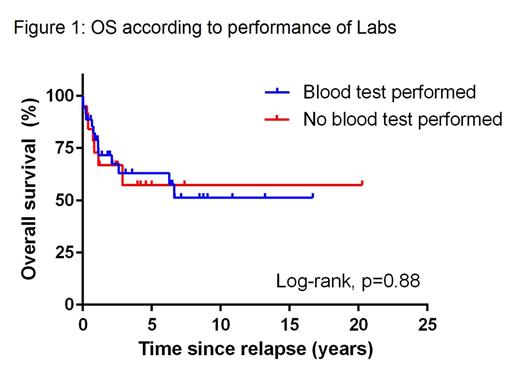
There was no significant difference in OS between patients who had Labs performed ≥3 months prior to documented relapse versus patients who did not (p=0.88, figure 1).
Conclusion
Although normal Lab results have a high NPV for relapse, most HGL relapses are detected by patient-reported symptoms, often at unscheduled visits. Abnormal Labs in aSx patients infrequently trigger a change in management. Routine Labs rarely detect relapse in aSx patients with HGL in CR after primary curative-intent chemotherapy, demonstrate unacceptably poor performance characteristics, have no impact on survival and have limited value in routine surveillance.
Hawkes:BMS: Other: travel expenses, Research Funding; Takeda: Other: travel expenses; Merck Serono: Research Funding.
Author notes
Asterisk with author names denotes non-ASH members.


This feature is available to Subscribers Only
Sign In or Create an Account Close Modal