Abstract
Top of Form
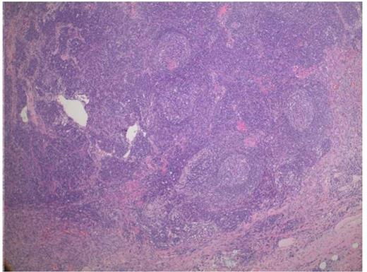
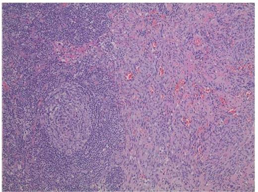
Kaposi's Sarcoma (KS) and Multicentric Castleman's Disease (MCD) can infrequently occur simultaneously in immunocompromised patients. Here we present a rare case of HIV negative, HHV8 positive, EBV positive MCD with simultaneous pure nodal KS in an immunocompetent patient. A 69 year old female with no past medical history presented in June 2015 with neck pains, night sweats, and 20 pound weight loss. Physical examination revealed bilateral cervical, inguinal lymphadenopathy. She also had bulky bilateral axillary lymphadenopathy. No skin lesions were noted. Excisional lymph node axillary biopsy performed 7/16/15 revealed follicular hyperplasia with intense follicular expansion of plasma cells (figure 1, 2). Some follicles showed transformed germinal centers with expanded mantle zones and occasionally more than one germinal cell within a single mantle (figure 1, 2). By Immunohistochemical stains, CD20 highlighted the follicular B-cells and expanded mantle zone B-cells. CD3 highlighted small interfollicular T-cells. CD138 highlighted interfollicular expansion of plasma cells that were positive for kappa and lambda light chains in the interfollicular areas by in-situ hybridization. The plasmablastic cells in the mantle zone showed lambda light chain restriction and HHV-8 positivity. HHV-8 additionally highlighted the nuclei of the spindle cell proliferation (figure 3). PET CT 7/14/15 showed bilateral axillary adenopathy with SUV 8.0, mild hilar adenopathy with SUV 6.0 (Figure 4) and prominent adenopathy of pelvic sidewall and inguinal regions with SUV ranging 3.0-8.0. Baseline labs 7/14/15: HIV (-), HHV8 DNA (-), EBV IgG 7.7 AI (+), EBV DNA (+), IL-6 elevated 7.91 pg/mL and CRP 3.64 mg/dL (high). CBC and CMP were normal. Serum immunoglobulins: IgA 653m/dL (high), IgG 3210 mg/dL (high), IgM 33 mg/dL (low). SPEP with immunofixation showed hypergammaglobulinemia with slight peak asymmetry of the gamma globulins without evidence of monoclonality. LDH was normal at 127 U/L. The patient was given Rituximab 375m/m2 IV day 1 and Liposomal Doxorubicin 20 mg/m2 IV day 1 (R-Dox) every 3 weeks for 4 cycles between 8/7/15 - 10/5/15 with complete resolution of lymphadenopathy clinically and by PET CT 10/15/15. Patient has been doing well without evidence of recurrence as of last clinic visit 3/14/16.
Low power (5X magnification) showing targetoid pattern of follicles and prominent interfollicular stroma with prominent capillaries consistent with Castleman's and a spindle cell proliferation towards the bottom consistent with Kaposi Sarcoma
Low power (5X magnification) showing targetoid pattern of follicles and prominent interfollicular stroma with prominent capillaries consistent with Castleman's and a spindle cell proliferation towards the bottom consistent with Kaposi Sarcoma
Higher magnification (10X) shows a prominent follicle with concentric layering of peripheral lymphocytes that resembles onion-skin. To the right, the KS shows spindle cells forming slits with extravasated red blood cells.
Higher magnification (10X) shows a prominent follicle with concentric layering of peripheral lymphocytes that resembles onion-skin. To the right, the KS shows spindle cells forming slits with extravasated red blood cells.
HHV-8 nuclear immunostain highlights KS cells with nuclear staining.
HHV-8 nuclear immunostain highlights KS cells with nuclear staining.
No relevant conflicts of interest to declare.
Author notes
Asterisk with author names denotes non-ASH members.


This feature is available to Subscribers Only
Sign In or Create an Account Close Modal