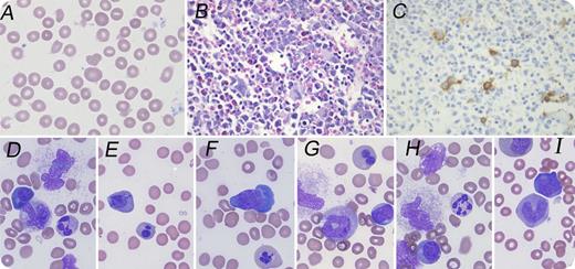
An 11-month-old girl presented with fatigue and vomiting. Complete blood count revealed severe macrocytic anemia (hemoglobin, 2.8 g/dL; mean corpuscular volume, 117 fL), thrombocytopenia (platelets, 117 × 109/L), and normal white blood cell count (13.6 × 10⁹/L). Peripheral blood smear revealed anisocytic erythrocytes with frequent macroovalocytes (panel A; original magnification ×1000, Wright's stain). Serum cobalamin and erythrocyte folate were within normal limits. Bone marrow (BM) biopsy showed hypercellular marrow with frequent megaloblasts and few micromegakaryocytes (panel B; original magnification ×400, hematoxylin and eosin stain), which were easily seen on CD61 immunostain (panel C; original magnification ×400). BM smears showed >50% of the erythroblasts with dysplastic features, including nuclear-cytoplasmic asynchrony, nuclear lobation or irregular shapes, and frequent giant bands or neutrophils with abnormal nuclear lobation (panels D-I; original magnification ×1000, Wright-Giemsa stain). Cytogenetic study revealed normal female karyotype. Exome next-generation sequencing revealed 2 variants of the TCN2 gene (22q12.2), a 2-base deletion, and a nonsense mutation, consistent with transcobalamin II (TCII) deficiency.
TCII deficiency is a rare autosomal-recessive disease characterized by defective intestinal absorption of cobalamin and decreased cobalamin availability. The early recognition of this condition is critical because early parenteral cobalamin therapy can result in complete resolution of symptoms and normal growth; neurologic damage may become permanent if initiation of treatment is delayed. TCII deficiency should be considered in infants with megaloblastic anemia and/or hematopoietic dyspoiesis with normal cobalamin/folate levels.
For additional images, visit the ASH IMAGE BANK, a reference and teaching tool that is continually updated with new atlas and case study images. For more information visit http://imagebank.hematology.org.


This feature is available to Subscribers Only
Sign In or Create an Account Close Modal