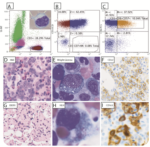
An 80-year-old man with a 20-year history of T-cell large granular lymphocytic leukemia (T-LGL), receiving low-dose neupogen for neutropenia, presented with new-onset fever, pancytopenia, and tachycardia. Flow cytometry of the peripheral blood showed aberrant clonal T cells (6% to 8% of total lymphocytes; panels A-C), CD3+, CD8+, CD7−, CD4−, CD57+ (panel A inset LGL, 100× objective), supporting T-LGL. Bone marrow (BM) biopsy (panel D, 40× objective) and smear (panel E, 100× objective) showed large pleomorphic cells with prominent nucleoli and cytoplasmic vacuoles CD22+ (panel F), PAX5+, CD79A+, MUM1+, CD30+, and CD10− (not shown). T-LGL cells were persistent in the BM, CD3+, CD8+, and CD57+. EBER1 in situ hybridization–positive cells (panel G) supported Epstein-Barr virus (EBV) diffuse large B-cell lymphoma (EBV+ DLBCL), non–germinal center type. Interphase fluorescence in situ hybridization was negative for BCL2, BCL6, and MYC rearrangements. Hemophagocytosis (HLH) was seen on BM smear (panel H, 100× objective), highlighted by CD163 (panel I) on biopsy. Ferritin was 27 519 ng/mL (reference, 30-400 ng/mL); aspartate aminotransferase, 59 U/L (reference, 1-35 U/L); lactate dehydrogenase, 1041 U/L (reference, 100-200 U/L); and fibrinogen, 99 mg/dL (reference, 175-450 mg/dL), supporting HLH.
T-LGL is a rare chronic clonal mature T-cell neoplasm, commonly coexisting with clonal B-cell processes. In this patient, EBV+ DLBCL most likely caused secondary HLH, an ultimate cytokine storm, and immune dysregulation. The patient died soon after.
For additional images, visit the ASH Image Bank, a reference and teaching tool that is continually updated with new atlas and case study images. For more information, visit http://imagebank.hematology.org.


This feature is available to Subscribers Only
Sign In or Create an Account Close Modal