Background: Anti-CD38 monoclonal antibodies (MoAbs), including daratumumab and isatuximab, are introduced in the treatment of multiple myeloma (MM). However, CD38 is also expressed on red blood cells (RBCs), so that free anti-CD38 MoAbs in patient sera sometimes binds to test RBCs in pretransfusion blood-compatibility tests. Consequently, panreactive agglutination arises in the standard indirect antiglobulin test (IAT) used for antibody screening and cross-matching. Several methods, including pretreatment of RBCs with dithiothreitol (DTT), have been introduced to overcome the interference. However, the optimum concentration and treatment time for these reagents have not been well elucidated (Chapuy et al, Hosokawa et al). One reason is that evaluation for RBC agglutination during the IAT relies on visual judgement. In this study, we quantified CD38 on RBCs before and after DTT treatment using a newly-devised flow cytometric antibody binding assay (FABA; Takeshita et al) by employing the D-value in the Kolmogorov-Smirnov (K-S) test, and compared with the results from IAT and a conventional flow cytometric method using fluorescent labelled anti-CD38 antibody and isotype-matched control antibody. We then assessed the optimum DTT concentration and treatment time for CD38 inactivation on RBCs to enhance MoAb therapy for MM.
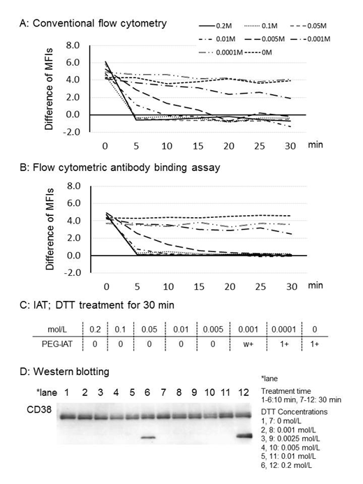
Methods: RBCs from healthy donors were treated with 0.2-0.0001 mol/L of DTT for 0-30 min at 37°C. The effect of DTT treatment was evaluated using flow cytometetry (FCM, Navious, Beckman Coulter, Tokyo) and IAT. For conventional FCM analysis, untreated or DTT-treated RBCs were incubated with fluorescein isothiocyanate (FITC)-labeled anti-CD38 antibody or anti-IgG1 antibody for 30 min, and then analyzed. For FABA, untreated or DTT treated RBCs were incubated with FITC-labelled anti-CD38 antibody, in the presence or absence of a 100-fold or more excess of unlabeled anti-CD38 antibody and then analyzed by FCM. Dissociation of CD38 positive and control histograms were determined from the mean fluorescence intensity (MFI) and the D-value in the K-S test, which is the maximum vertical displacement between two cumulative frequency distributions for two histograms. Statistical analyses were performed by paired t-test (SPSS, Tokyo), with significance set at p<0.05. The amount of CD38 following DTT treatment was also evaluated by Western blotting.
Results: In conventional FCM using FITC-labelled isotype-matched antibody as control, the difference of measured MFIs between CD38 positive and control were considerably volatile in repeated analysis (Fig. A). By contrast, in FABA using an excess concentration of unlabeled anti-CD38 antibody as control, the values were more consistent than those from conventional FCM (Fig. B). The D-value in the K-S test is reliable by numerical modeling in the analysis of the difference between CD38 positive and control histograms. Analysis showed that 0.005 mol/L DTT for 30 min is sufficient to inactivate CD38 on RBCs. These results correlated with those of IAT (Fig. C). Western blotting identified CD38 at DTT concentrations of 0.01-0.001 mol/L, but denaturation of CD38 was observed at 0.2 mol/L of DTT (Fig. D).
Conclusions: Our newly-devised flow cytometric antibody binding assay, FABA, facilitates the detection of small numbers of cell surface antigens. The method is also useful as an objective way of evaluating the efficacy of DTT treatment for CD38 on RBCs. Our results show 0.005 mol/L DTT for 30 min is sufficient to inactivate CD38 on RBCs in plasma sampled after treatment with the anti-CD38 MoAbs.
Takeshita:Chugai Pharmaceutical Co.Ltd.: Research Funding; Pfizer Japan Inc: Research Funding; Astellas Pharma Inc.: Research Funding; Takeda Pharmaceutical Co.Ltd.: Research Funding; Bristol-Myers Squib Co.: Research Funding.
Author notes
Asterisk with author names denotes non-ASH members.


This feature is available to Subscribers Only
Sign In or Create an Account Close Modal