Background and Aims
A part of lower-risk myelodysplastic (LR-MDS) patients progress to higher-risk MDS or acute myeloid leukemia (AML). The progression may be predicted in some of these patients by determining mutations associated with myeloid malignancies at the time of diagnosis. We focused to find out dysregulated pathways caused by a well-defined mutation associated with disease progression.
Methods
We examined 158 samples from LR-MDS patients at diagnosis using TruSight Myeloid Sequencing Panel containing 54 genes (Illumina) to identify the mutational profile. We applied RNA-seq (NEB; Illumina) on bone marrow CD34+ cells of 5 LR-MDS patients with RUNX1 mutation and 6 LR-MDS patients without mutations. We performed differential gene expression analysis, gene set enrichment analysis (GSEA), Reactome biological pathway and Gene Ontology (GO) annotations.
Results
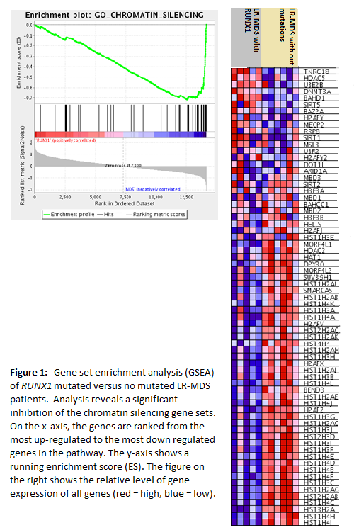
Forty out of 158 patients (25%) progressed during the follow-up period (median: 45.1 months). Mutation in RUNX1 gene was detected in 10 patients. Using RNA-seq, significantly dysregulated expression was observed in 986 genes (FDR<0.05; FC >2). Patients with mutation in RUNX1 gene had significant down regulation of expression genes involved in cellular senescence induced by DNA Damage/Telomere Stress (FDR=1.4e-11) and Oxidative Stress (FDR=3.6e-9), HDACs deacetylate histones (FDR =1.1e-13) and Senescence Associated Secretory Phenotype (FDR=1.4e-11) according to Reactome pathway database. There were 21 genes involved in the regulation of these pathways, of which 10 were part of all described pathways and belonged to the Nucleosome Assembly according to GO biological process with 33 down regulated genes (FDR=6.3e-28). According to GSEA, GO chromatin silencing gene set (Figure 1) was down regulated in the patients with RUNX1 mutation (p<0.001; NES=-1.8) same as Reactome apoptosis (p<0.001; NES-1.7). In these patients, expression of 750 genes was up-regulated (FDR<0.05; FC >2); however, they did not form a specific pathway. The up-regulated genes were classified as so-called leukemia-associated antigens (LAAs) such as PRAME (adjusted p=6.9e-09; logFC=6.8), WT1AS (adjusted p=4.4e-07; logFC=5.7), BAALC (adjusted p=3.5e-06; logFC=2.1), WT1 (adjusted p=6.8e-07; logFC=3.2), FLT3 (adjusted p=0.004; logFC=1.3) and LEF1 (adjusted p=5.3e-08; logFC=-4.2).
Conclusions
To study the transcriptome of the hematopoietic stem cells of LR-MDS patients, we used gene expression profiling and identified biological processes in relation to mutations in RUNX1 gene that were detected in patients with disease progression. According to our results, we suppose that cells of LR-MDS patients with no mutations are maintained in senescent state or apoptosis in opposite to RUNX1 mutated cells. Finding out of dysregulated senescence was reinforced by other dysregulated pathways such as HDACs deacetylate histones, chromatin structural changes and senescence-associated secretory phenotype. About cellular senescence is known that may act as a strong tumor suppression mechanism that restrains proliferation of cells at risk for malignant transformation. We suppose that in precancerous cells of LR-MDS patients apoptosis or cellular senescence limits the replicative capacity of cells, thus preventing the proliferation. The disturbed state of cellular senescence in RUNX1 gene mutated hematopoietic stem cell can escape proliferation control and then can contribute to the disease progression in these patients.
Supported by grants NV18-03-00227 and 00023736 from the Ministry of Health of the Czech Republic.
No relevant conflicts of interest to declare.
Author notes
Asterisk with author names denotes non-ASH members.


This feature is available to Subscribers Only
Sign In or Create an Account Close Modal