Introduction
Multicolor flow cytometry (MFC) has been frequently adopted as a method for minimal residual disease (MRD) detection, and is also a promising technique to detect post-transplant lymphoproliferative disorder. Some abnormal donor origin cells might be found when detecting MRD following an allogeneic hematopoietic stem cell transplantation (allo-HSCT). To minimize the effects from donor cells, using MFC prior to allo-HSCT to screen donor peripheral blood (PB) or bone marrow (BM) might be feasible.
Methods
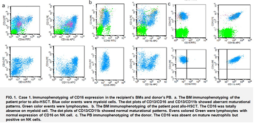
We performed 3395 allo-HSCTs between January 2013 and December 2019 at Lu Daopei Hospital in Langfang, China. MRD was detected in recipients' BMs according to a conventional two-tube 8 or 9-color MFC panel. Abnormal cells were observed in BMs from three patients in complete remission (CR) one to four months post allo-HSCT. Abnormal neutrophils lacking CD16 expression were found in a patient with secondary acute myeloid leukemia (AML) that developed from a myelodysplastic/ myeloproliferative neoplasm (MDS/MPN). After ruling out MDS and paroxysmal nocturnal hemoglobinuria (PNH), we hypothesized that an Fcγ receptor IIIB (FcγRIIIB) gene deletion was the most likely reason. Abnormal natural killer (NK) cells were detected in the BM from an allo-HSCT recipient with T-cell acute lymphoblastic leukemia (ALL), and monoclonal B lymphocytosis (MBL) in allo-HSCT recipient with B-cell ALL. These three patinets' PBs were detected using MFC after the new finding to decide the cell origin. Besides, 4.54%(in WBC) CD4+ and CD8+ double positive T- cells which were monoclonal cells of the TCRVβ repertoire were detected in a PB sample from a donor prior to allo-HSCT. To evaluate the incidence rate The immunophenotypings were studied in the BMs from 79 NK lymphoma patients.
Results
Identical phenotypes were recognized in PBs obtained from the three respective donors. The fourth donor did not donate her cells for allo-HSCT, yet. The incidence rate of abnormal cells in donor samples was 0.1% (4/3395 cases), but this rate might be underestimated because MFC screening was not a routine procedure for donors. Additionally, only abnormal immunophenotyping related to patient diagnosis might have been found using an MRD panel as this panel only included markers related to diagnosis. Among general population, the incidence rate of suspicious FcγRIIIB deletion was 0.2% (11/5256 cases), the incidence rate of NK cells without CD2+ and homogeneously expressed CD159c was 0.05% (1/2000 cases) and none among the 79 NK lymphoma samples. The rate of MBL was 0.75% (15/2000 cases) and 1.36% in older than 40 years old people and the rate of monoclonal CD4/CD8 DP T-cells was 0.05% (1/2000 cases). All of these abnormal cells or polymorphism could be analyzed using a two tube MFC panel-- ckappa/clambda/(CD34)/CD19/ CD5/CD20/CD38 /CD45/CD56 and CD16/(CD117)/CD3/ CD4/CD5/ CD8/CD56/CD45/CD2.
Conclusion
Donor original abnormal cells or phenotypic polymorphisms could have an effect on MFC-based MRD or PTLD detection of recipients following allo-HSCT. These patients might be mis-diagnosed as being MRD positive or having PTLD if the technician lacks experience. To avoid mis-diagnosis and minimize the risk of allo-HSCT, it might be promising to utilize a suitable MFC panel to screen donor PB or BM samples prior to transplantation.
No relevant conflicts of interest to declare.
Author notes
Asterisk with author names denotes non-ASH members.


This feature is available to Subscribers Only
Sign In or Create an Account Close Modal