Del(17p) in chronic lymphocytic leukemia (CLL) and plasma cell myeloma has a unique genomic profile leading to refractoriness to conventional therapies and poor overall survival. Detection is generally by fluorescence in situ hybridization (FISH) on a slide with analysis of up to 200 nuclei, not necessarily all of being neoplastic cells. The small cell sample analyzed, and high threshold for a positive result (>5% positive cells) makes FISH a low precision test. This means false negative results are inevitable, especially if del(17p) is only in a minor clone, potentially leading to suboptimal treatment. To address this, we developed an automated FISH method with analysis on an imaging flow cytometer, an instrument with the functionality of a standard flow cytometer which generates high-resolution digital images of each cell. By combining immunophenotyping with FISH, on whole cells in a single test, we can detect chromosomal defects in cells with a specific phenotype.
Aims: Our aim was to determine the capability of this automated integrated immunophenotyping-FISH imaging flow cytometry method to detect del(17p) in CLL and myeloma, the two commonest hematological malignancies, and in which this genetic defect encodes poor prognosis. We hypothesized that it would be more sensitive and specific than standard FISH.
Methods: Bone marrow and/or blood samples in EDTA anticoagulant from 19 cases of CLL or myeloma, at diagnosis or on therapy, were studied. After red cell lysis, cells were incubated with fluorochrome-conjugated antibodies to CD3, CD5, CD19, CD38, and CD138 antigens (fluorochromes: BV480, BV605, AF647). Following fixation, cell membranes were permeabilized and double-stranded DNA denatured (78oC for 5 mins). FISH probes to the centromere of chromosome 17 (CEP17, Spectrum Green) and 17p12 locus (Orange-Red) were added and hybridized for 24 hours at 37oC. Nuclei were stained with SYTOX AADvanced. Data for up to 200,000 cells was collected on the Amnis® ImageStream®XMk II imaging flow cytometer. Digital images (x60) and quantitative data derived from computational algorithms (IDEAS software) were used to assess FISH signals overlying the nuclei of CD5/CD19-positive CLL cells or CD38/CD138-positive plasma cells for each probe. Digital images and quantitative data were assessed for FISH signals within immunophenotyped cells.
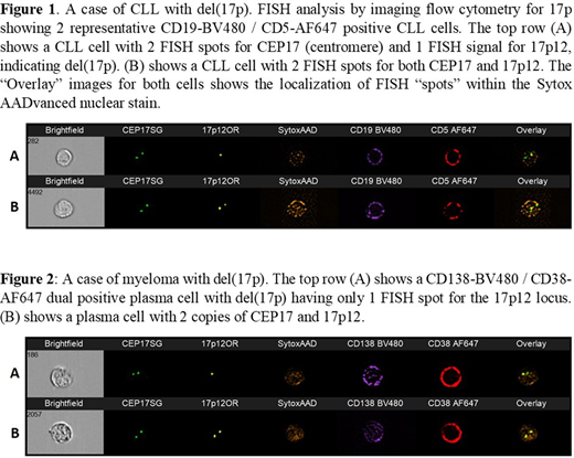
Results: Between 10,000 and 200,000 (mean 60,000) cells were analyzed per sample. The FISH signals were seen on the digital images and confirmed by quantitative mean channel fluorescence intensity of the probes. There were 12 CLL cases with one 17p FISH signal in the CD5/CD19-positive population, with the number of del(17p) CLL cells ranging from 2 - 35% (or 0.4 - 23% of all cells analyzed) (Fig 1). This amounted to between 270 and 35,441 cells in the analysed sample with del(17p). The lowest del(17p) burden was in a patient on cytoreductive therapy. All CLL cells had normal diploid spots for the control CEP17 probe, and the CD3/CD5-positive T cells had dual signals for both CEP17 and 17p12 probes.
There were 5 myeloma cases with 1 FISH signal for 17p overlying the nucleus of the CD38/CD138-positive plasma cells (Fig 2). In these cases, 52-90% of cells (15,600-18,000 cells) had a plasma cell phenotype and 13-19% of these (or 2,340-2,964 CD38/CD138-positive cells) showed del(17p). This represented 1-5% of all cells in the sample. All gated plasma cells had 2 FISH spots for CEP17.
Conclusion: This imaging flow cytometry method that integrates FISH with immunophenotyping could detect del(17p) in CLL and myeloma with a lowest limit of detection of 0.4% and 1% respectively. The high sensitivity was achieved as many thousands of cells were analyzed, and 17p was only assessed in gated cells with the phenotype of interest (i.e. CD5/CD19 or CD38/CD138). This method, that includes positive cell identification by phenotype, precludes the need for prior cell sorting or purification. Imaging flow cytometry for del(17p) by "immuno-flowFISH" brings a new dimension to FISH analysis at diagnosis and for disease monitoring. Its high precision and specificity will enable detection of del(17p), even when only present in minor sub-clones, for therapeutic decision making and prognostic stratification. This technique has a real place in clinical assessment of del(17p) in CLL and myeloma and could be applied for other significant chromosomal defects of therapeutic and prognostic significance.
Augustson:Roche: Other: Support of parent study and funding of editorial support. Cheah:Celgene, F. Hoffmann-La Roche, Abbvie, MSD: Research Funding; Celgene, F. Hoffmann-La Roche, MSD, Janssen, Gilead, Ascentage Pharma, Acerta, Loxo Oncology, TG therapeutics: Honoraria.
Author notes
Asterisk with author names denotes non-ASH members.


This feature is available to Subscribers Only
Sign In or Create an Account Close Modal