Abstract
Since the approval of ruxolitinib (JAK1/JAK2) in 2011, two additional inhibitors, fedratinib (JAK2/FLT3) and pacritinib (JAK2/FLT3/IRAK1/CSF1R), have received FDA approval for myelofibrosis, and a new drug application for a fourth inhibitor, momelotinib (JAK1/JAK2/ACVR1/TBK1) has been submitted in June 2022. Although all four inhibitors potently suppress JAK2, the targeting of additional auxiliary targets may contribute to distinct responses observed in the clinic. Here, we take a comprehensive approach to assess the unique proliferative, transcriptional, and signaling pathways altered by these four JAK2 inhibitors in myeloproliferative neoplasms (MPN).
In three human JAK2 mutant leukemic cell lines (HEL, UKE-1, SET2) and murine Ba/F3 EpoR-JAK2VF cells, ruxolitinib consistently showed the lowest IC50. Following, CD34+ hematopoietic stem/progenitor cells (HSPCs) from 8 MPN patients and 3 healthy donors were seeded for colony formation assays. Erythroid colonies were highly sensitive to ruxolitinib, fedratinib, and pacritinib inhibition. In contrast, momelotinib demonstrated greater relative sparing of erythroid colonies, consistent with clinical anemia amelioration, albeit potentially via a ACVR1/ALK2 and hepcidin-independent manner. Overall, greatest potency was demonstrated by pacritinib ex vivo, followed by ruxolitinib, fedratinib, and momelotinib. These findings were somewhat distinct from in vitro assays, likely reflecting additional underlying biology of human HSPCs.
We then performed high dimensional mass cytometry to assess signaling changes at single cell resolution across different cell populations in 9 MPN patient samples and 3 healthy donors treated ex vivo with JAK inhibitors. We observed potent suppression of pSTAT1, pSTAT3, and pSTAT5 in CD34+ cells, with the 4 JAK2 inhibitors exerting similar degrees of inhibition. In B cells, pacritinib suppressed IκBα, pERK, and pCREB levels, while momelotinib inhibited pTBK1. Similar observations were noted in T cells, which in addition exhibited suppression of pSTAT1 and pSTAT3. These findings indicate distinct suppression responses across specific cell types and further highlight nuances beyond sole suppression of JAK-STAT signaling by these inhibitors.
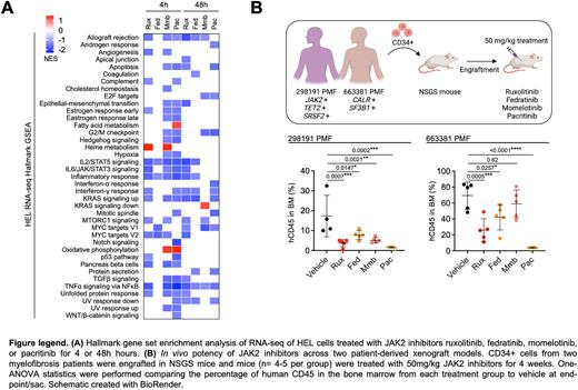
RNA-sequencing of HEL cells treated with the 4 JAK2 inhibitors also identified unique alteration of expression profiles and pathways (Fig. A). Differential gene expression (DEG) analysis revealed pacritinib inducing the greatest transcriptional alterations after 4 hours (3,121 genes) and ruxolitinib (1,969 genes) following 48 hours treatment duration. Only 82 and 103 DEGs relative to control were shared across the 4 JAK2 inhibitor treatment at 4 and 48 hours, respectively. Additional pathway analysis revealed at least 3 out of 4 inhibitors demonstrating capacity to suppress 1) JAK-STAT signaling, 2) inflammatory response, and 3) TNF signaling via NFκB pathways. Upregulation of multiple metabolic pathways was observed such as oxidative phosphorylation by momelotinib and pacritinib, potentially reflecting adaptive transcriptional feedback, as Seahorse Mito Stress assays revealed strongest suppression of oxygen consumption rate of PBMCs mediated by pacritinib and fedratinib.
Lastly, we compared the in vivo potency of JAK2 inhibitors using patient-derived xenograft (PDX) models (Fig. B). Engrafted immunodeficient NSGS mice were treated with JAK2 inhibitors for 4 weeks across 2 sets of PDX experiments with CD34+ cells from unique JAK2 and CALR mutant donors. Pacritinib demonstrated greatest potency in reduction of peripheral blood and bone marrow human CD45+ leukemic cells, followed by ruxolitinib, and fedratinib/momelotinib. Vehicle treated mice were moribund by week 3 of treatment and survival was significantly prolonged with all JAK2 inhibitor treatments, with all animals surviving in the pacritinib treatment group after 4 weeks of treatment at end point.
Despite all four JAK2 inhibitors demonstrating affinity in suppressing JAK-STAT signaling, our assays reveal varying degrees of alteration of transcriptional, proteomic, and metabolic profiles. In conjunction with comparative assessment of in vivo activity in PDX models, these findings provide insights with potential relevance to distinct responses observed with these inhibitors in the clinic and may help guide the use of specific JAK2 inhibitors in combination studies.
Disclosures
Oh:CTI BioPharma: Consultancy, Membership on an entity's Board of Directors or advisory committees; Disc Medicine: Consultancy, Membership on an entity's Board of Directors or advisory committees; Geron: Consultancy, Membership on an entity's Board of Directors or advisory committees; Incyte: Consultancy, Membership on an entity's Board of Directors or advisory committees; Kartos: Consultancy, Membership on an entity's Board of Directors or advisory committees; Sierra Oncology: Consultancy, Membership on an entity's Board of Directors or advisory committees; PharmaEssentia: Consultancy, Membership on an entity's Board of Directors or advisory committees; Celgne/BMS: Consultancy, Membership on an entity's Board of Directors or advisory committees; Novartis: Consultancy, Membership on an entity's Board of Directors or advisory committees; Constellation: Consultancy, Membership on an entity's Board of Directors or advisory committees; Blueprint Medicines: Consultancy, Membership on an entity's Board of Directors or advisory committees; AbbVie: Consultancy, Membership on an entity's Board of Directors or advisory committees.
Author notes
Asterisk with author names denotes non-ASH members.


This feature is available to Subscribers Only
Sign In or Create an Account Close Modal