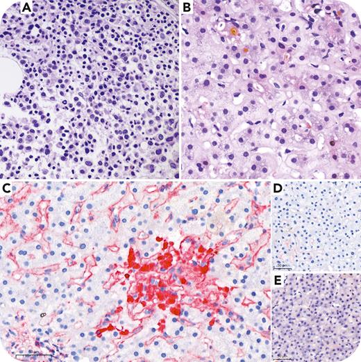
A 76-year-old woman presented with rapidly worsening renal function and cholestatic liver injury. Infection, medication toxicity, and autoimmune disease were excluded. Free κ monoclonal gammopathy was found in the urine by immunofixation, but not in the serum. The patient had no anemia, hypercalcemia, or bone lesions on computerized tomography scan. A bone marrow biopsy confirmed the diagnosis of multiple myeloma (panel A, hematoxylin and eosin stain [H&E], ×100 objective). A liver biopsy showed cholestasis, feathery degeneration, and foci of foamy cell aggregation (panel B, H&E, ×100 objective). The κ stain shows diffuse deposition in the sinusoidal and vascular walls (panel C, ×100 objective). The Congo red and λ stain (panels D and E, respectively, ×100 objective) were negative. The patient had progressive hyperbilirubinemia despite treatment with bortezomib, thalidomide, prednisolone, and daratumumab. She died of liver failure.
Light chain deposition disease is characterized by deposition of immunoglobulin light chains in organs, resulting in progressive damage. These light chains are mostly of the κ type and have high affinity for basement membrane because of the increased hydrophobicity caused by somatic mutations. The majority of patients have an underlying lymphoproliferative disorder, mainly multiple myeloma. Organs that can be affected include the kidneys (most frequent), lungs, heart, and nerves. Severe hepatic involvement is rare. In the liver, the disease typically has a perisinusoidal distribution, as opposed to the parenchymal distribution in amyloidosis.
For additional images, visit the ASH Image Bank, a reference and teaching tool that is continually updated with new atlas and case study images. For more information, visit http://imagebank.hematology.org.


This feature is available to Subscribers Only
Sign In or Create an Account Close Modal