Introduction
In JAK2-V617F (JAK2-VF) mutated cells, over-activation of β1/β2 integrin inside-out signaling has been described (Edelmann-Stephan et al., 2017, JCI). Integrins play a fundamental role in cell interaction and cell invasion during inflammatory processes. In patients suffering from Myeloproliferative Neoplasms (MPNs), highly elevated pro-inflammatory cytokine levels including IL-1β play an important role in pathophysiology. Importantly, Rai et al. (2023, BioRχ) have shown that IL-1β promotes initiation of MPN. In this study, we investigated a potential link between integrin over-activation and elevated pro-inflammatory cytokines including IL-1α and IL-1β in JAK2-VF induced disease.
Methods
We employed (1) murine myeloid progenitor 32D cells co-transfected with EPO-R and JAK2-WT or JAK2-VF, (2) granulocytes and hematopoietic progenitor cell populations from total bone marrow of Vav1-Cre x JAK2 +/+ and Vav1-Cre x JAK2 VF/+ mice (described by Mullally et al. 2010), and (3) human granulocytes isolated from PB of JAK2-VF positive patients and healthy volunteers. Cells were allowed to adhere on immobilized VCAM-1/ICAM-1 or on BSA (control) and subsequently subjected to further analysis including (1) qPCR analysis and ELISA of a panel of inflammatory cytokines (IL-1β, IL-1α, TNF-α, IL-6, CXCL10), (2) Western-blotting monitoring integrin outside-in signaling pathways, (3) FLICA assays assessing adhesion-induced active caspase-1, (4) RNAseq of mouse and human granulocytes (3 JAK2-VF patients, 2 controls).
Results
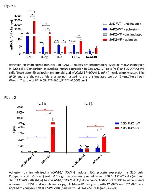
In the 32D cell model, adhesion on mVCAM-1/mICAM-1 strongly induced mRNA expression of IL-1α, IL1-β, IL-6, TNF-α and CXCL10. Interestingly, cytokine RNA induction was significantly higher in 32D JAK2-VF cells as compared to the 32D JAK2-WT cells (Figure 1). By applying a panel of integrin signaling inhibitors we could show an essential involvement of pivotal signaling nodes such as PI3K, Syk, FAK and NFκB, respectively. Next, cytokine proteins were investigated by ELISA. Adhesion on mVCAM-1/mICAM-1 increased intracellular IL-1α and IL-1β cytokine levels. Again, this effect was strongly amplified by the JAK2-VF mutation (Figure 2). Importantly, both cytokines were found in their mature form, indicating intact protein processing.
RNAseq of bone marrow (BM) derived mouse granulocytes (including immature pro-and pre-neutrophils) also showed significant upregulation of a number of pro-inflammatory genes including Nlrp3, Il1a, Il1b, Tnf, Cxcl2 and Cxcl10 upon adhesion on mVCAM-1/mICAM-1. However, no significant differences between WT and VF cells were observed. In contrast, RNAseq of VCAM-1/ICAM-1 stimulated human JAK2-VF positive PB granulocytes (which represent terminally differentiated cells) revealed no cytokine up-regulation. Based on this finding, we hypothesize that the adhesion-induced cytokine phenotype is restricted to more immature cell populations as observed in the 32D cell model. Therefore, we are currently investigating (by qPCR and intracellular IL-1 immunophenotyping) hematopoietic progenitor cell populations from Vav1-Cre x JAK2 +/+ and Vav1-Cre x JAK2 VF/+ mice.
Since active IL-1β requires processing by the inflammasome, FLICA assays for active caspase-1 were performed using mouse hematopoietic progenitor cells. Importantly, CD41+ cells and megakaryocyte progenitors (MKP) showed an increased FLICA signal upon adhesion on mVCAM-1/mICAM-1. This indicates that caspase 1 is activated upon adhesion in distinct progenitor cell populations. Finally, to support our hypothesis that JAK2-VF augments induction of pro-inflammatory cytokines upon adhesion to VCAM-1/ICAM-1, Vav1-Cre x JAK2 VF/+ mice (n=8) were treated in vivo with anti-β1/β2-integrin antibodies for 1 week. As expected we found a strong reduction of IL-1α concentration in serum of antibody treated mice (528.7 ± 82.2 pg/ml) as compared to PBS-treated control mice (1302.3 ± 370.4 pg/ml, p = 0.0541)
Conclusion
Here, we show a functional link between elevated cytokine levels and integrin activation in JAK2-VF induced disease. Adhesion to VCAM-1/ICAM-1 promoted the expression of a number of pro-inflammatory cytokines which are known to play important roles in initiation, clinical symptoms and prognosis of MPNs. Further studies aimed at precisely defining the primary cell populations involved in the mouse MPN disease models as well as in humans are currently ongoing.
Disclosures
Mougiakakos:Miltenyi: Honoraria.


This feature is available to Subscribers Only
Sign In or Create an Account Close Modal