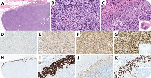
A 58-year-old man with a history of chronic lymphocytic leukemia (CLL) presented with 2 months of abdominal pain and night sweats. Imaging showed hypermetabolic abdominal, cervical, and axillary lymphadenopathy. Excisional biopsy of a 2.7-cm right axillary lymph node showed effacement of the lymph node architecture by a diffuse infiltrate of small lymphocytes with scattered proliferation centers (panel A, hemotoxylin and eosin [H&E] stain, 4× lens objective; panel B, H&E stain 40× lens objective). The sinusoids contained large atypical cells, including “hallmark” cells (panel C, H&E stain, 40× lens objective; inset, 100× lens objective). By immunohistochemistry, the small cells were positive for CD20 (weak; panel D, 40× lens objective), PAX5, CD5 (panel E, 40× lens objective), CD23 (panel F, 40× lens objective), and LEF1 (panel G, 40× lens objective) and negative for cyclin D1 (panel G, 40× lens objective; inset). The large cells were positive for CD30 (panel H, 4× lens objective; panel I, 40× lens objective) and weakly positive for CD2 and CD4 (panel J, 40× lens objective). Ki67 showed a high proliferation rate (panel K, 40× lens objective). They were negative for CD20, CD79a, PAX5, CD138, CD3, CD5, CD8, TIA1, granzyme B, and ALK (panel K, 40× lens objective; inset). Epstein-Barr virus encoded RNA was negative. Fluorescence in situ hybridization for DUSP22 and TP63 rearrangements was negative. These features indicated a composite lymphoma comprising CLL and ALK-negative anaplastic large cell lymphoma (ALCL).
Clinical progression in patients with CLL raises concern for transformation to aggressive B-cell lymphoma (Richter syndrome). De novo ALCL complicating CLL is uncommon but may occur. Some ALCLs present with a predominantly sinusoidal pattern, and pathological features may be subtle. Careful morphologic and immunophenotypic evaluation is essential for accurate diagnosis, which is critical because ALCL requires distinct therapy.
For additional images, visit the ASH Image Bank, a reference and teaching tool that is continually updated with new atlas and case study images. For more information, visit https://imagebank.hematology.org.


This feature is available to Subscribers Only
Sign In or Create an Account Close Modal