Abstract
Administration in the drinking water of the orally-active iron chelator 1,2-diethyl-3-hydroxypyridin-4-one (CP94) to C57BL/10ScSn mice caused the development of hepatic protoporphyria. This was detected after 1 week and continued as long as the chelator was given (15 weeks). The more hydrophilic 1,2-dimethyl- and 1-hydroxyethyl,2-ethyl-analogues (CP20 and CP102) were also tested, but they were both inactive in inducing accumulation of protoporphyrin in the liver. Restriction of in vivo iron supply for ferrochelatase seemed a likely mode of action, but an approximately 30% decrease in activity of this enzyme was also observed when measured in vitro. Extracts of livers from mice given CP20, CP94, and CP102 showed no potential to inhibit mouse ferrochelatase, in contrast to the findings with an extract from mice treated with the known porphyrogenic chemical 4-ethyl - 3 , 5 - diethoxycarbonyl - 2 , 6 - dimethyl - 1 , 4 - dihydropyridine, -indicating that ferrochelatase inhibition did not occur by the formation of an N-ethyl-protoporphyrin derived from metabolism by cytochrome P450. CP20, CP94, CP102, and CP117 (the pivoyl ester of CP102) all caused significant depression of the levels of ferritin-iron and total nonheme iron, but only CP94 caused the significant accumulation of protoporphyrin. Protoporphyria did not occur with iron overloaded C57BL/10ScSn mice or in SWR mice that had elevated basal iron status. Although the protoporphyrin had only a small effect on the total levels of the hemoprotein cytochrome P450 in C57BL/10ScSn mice, the activity of the CYP2B isoforms of cytochrome P450 was actually induced in both strains. The results show that CP94 could cause protoporphyria in individuals of low iron status, perhaps through specifically targeting particular iron pools available to ferrochelatase and by concomitantly stimulating heme synthesis.
IRON CHELATORS are used to treat a variety of hematological disorders in which iron overload develops and in which phlebotomy is undesirable or impossible.1,2 The most commonly treated disorder is iron overload resulting from the transfusion treatment of β-thalassemia, but chelation is also used in the management of other congenital or acquired anemias. Thalassemias are among the most common genetic diseases with many people affected worldwide, especially throughout the Mediterranean region, North Africa, the Middle East, India, and Southeast Asia.3 Chelation of iron has been proposed not just as a treatment for iron overload, but also in individuals with normal iron status when restriction of certain iron pools might protect against propagation of free radical reactions or cell proliferation.2 Such circumstances include reperfusion injury, rheumatoid arthritis, cerebral malaria, cancer, and toxicity of chemicals like paraquat. The chelator that has found the widest use is desferrioxamine, which has a high-affinity for iron and a proven therapeutic record in treating patients.4 However, a number of disadvantages are associated with the use of desferrioxamine. For instance, it lacks activity by the oral route and thus has to be administered by injection or via an intravenous pump. Since frequent administrations are necessary, noncompliance is a significant problem with some patients. Another disadvantage of desferrioxamine is that it is relatively expensive.1 Since many potential patients to be treated with iron chelators are in countries of low economic status, cost, as well as ease of use, becomes an important factor over a lifetime of use.
To overcome these problems many laboratories have been searching for alternatives to desferrioxamine, which are active orally and relatively nontoxic. Particularly promising are the 3-hydroxypyridin-4-one, bidentate iron chelators.5 Many of these have been synthesized and candidates selected for their efficiency and specificity of chelation, lipophilicity, and low toxicity. A number of studies in humans have been performed with 1,2-dimethyl-3-hydroxypyridin-4-one (CP20)6,7 and 1,2-diethyl-3-hydroxypyridin-4-one (CP94).5,8-10 Unfortunately both the compounds suffer rapid conjugation with glucuronic acid in man and are lost as chelators.8,11 Hydroxyalkyl derivatives, for example CP102, lack this limitation and consequently have clear advantages over the rapidly metabolized hydroxypyridinones.12 An important criterion for chelators is that they should remove excess iron from hemosiderin and “low molecular weight” iron pools without affecting normal cell function.13 In overloaded cells, this may be relatively easy to achieve. The risk of untoward iron removal may become greater, however, if chelators are used in other pathological situations, such as cerebral malaria or as an anticancer agent where the cellular iron content is not increased.2 In support of this proposal, the present study demonstrates that the chronic oral administration of CP94 to C57BL/10ScSn mice can cause disruption of a major pathway of iron utilization, the biosynthesis of heme. This complication is not observed in another strain of mice that had an elevated liver iron content.
MATERIALS AND METHODS
Chemicals.The N-derivatized-3-hydroxypyridin-4-ones CP20, CP94, CP102, and CP117 were synthesized and characterized by high performance liquid chromatography (HPLC) and nuclear magnetic resonance (NMR) as described previously.14 Their structures are shown in Fig 1. 4-Ethyl-3,5-diethoxycarbonyl-2,4-dimethyl-1,4 -dihydropyridine (ET-DDC), ie, the 4-ethyl analogue of 3,5-diethoxycarbonyl-1,4-dihydrocollidine was prepared as described15 and characterized by mass spectrometry and NMR. Iron-dextran solution (100 mg Fe/mL) and protoporphyrin IX were purchased from Sigma Chemical Co, Ltd (Poole, UK).
Mice and treatments.Male C57BL/10ScSn and SWR mice were purchased from Harlan-Olac Ltd (Bicester, UK) or bred from parents obtained from this breeder. Mice were housed, maintained, and treated with chemicals in accordance with the U.K. Animals (Scientific Procedures) Act, 1986. Mice received chelators dissolved in the drinking water at concentrations varying between 2 and 5 mg/mL. Iron overload of C57BL/10ScSn mice was produced by a single subcutaneous injection of iron-dextran solution at a dose of 600 mg of Fe/kg 1 week before treatment of mice with chelator. Mice were given the 4-ethyl analogue of 3,5-diethoxycarbonyl-1,4-dihydrocollidine (50 mg/kg) as an oil solution (10 mL/kg) by intraperitoneal injection and killed after 48 hours. Animals were killed by cervical dislocation except when blood was collected, which occurred under terminal anesthesia induced by isofluorane.
Porphyrin analyses.Livers were homogenized in water (1:4 wt/vol) and an aliquot (0.5 mL) was mixed with 10 mL of 1 mol/L perchloric acid/ethanol (1:1 vol:vol). Porphyrins were estimated by spectrofluorometry16 and either expressed in terms of protoporphyrin or calculated by the method of Grandchamp et al,17 which distinguishes between uroporphyrin, coproporphyrin, and protoporphyrin types. Protoporphyrin contents of erythrocytes were quantitated similarly using samples spiked with protoporphyrin. HPLC of extracts was performed using LDC/Milton Roy ConstatMetric 3000 pumps (Thermal Separation Products, Stone, Staffs, UK) and a Spherisorb 5 μ ODS-1 column (Phase Separations, Deeside, Clwyd, UK).18 Protoporphyrin was eluted with a gradient constructed from 1 mol/L ammonium acetate, acetonitrile, and methanol.19
Measurement of liver protoheme ferro-lyase.Liver protoheme ferro-lyase activity (ferrochelatase) was measured anaerobically by the method of Cole et al20 using mesoporphyrin and Fe2SO4 (37.5 mmol/L) as substrates and a 2% (wt/vol) tissue homogenate equivalent to 16 mg wet weight of liver. Ferrochelatase protein was examined by Western blotting using a rabbit antimouse ferrochelatase antibody kindly supplied by Prof H.A. Dailey (University of Georgia, Athens). Mitochondrial preparations of mouse liver were prepared as 12,000g fractions and subjected to sodium dodecyl sulfate-polyacrylamide gel electrophoresis (SDS-PAGE). Affinity for the ferrochelatase antibody was revealed by chemiluminescence using Amersham Life Sciences ECL Western blotting detection reagents (Buckinghamshire, UK).
Inhibitor studies.A total of 500 to 800 mg of liver were homogenized and extracted by the method reported by De Matteis et al.21 The residue was exhaustively dried under nitrogen and dissolved in 200 μL dimethyl sulfoxide (DMSO) for the determination of its inhibitory activity toward protoheme ferro-lyase. Enzyme activity was measured aerobically using CoCl2 and mesoporphyrin as substrates as described previously in Tephly et al22 with the following modifications. Change in absorbance (498 to 511 nm) was measured against time in a Cary 3 spectrophotometer (Varian Associates Ltd, Surrey, UK), using a 6-position thermostatted cell holder and a fast slew speed between wavelength pairs. In this way a pseudo-double–monochromator assay could be performed on six cuvettes simultaneously. To maintain a low turbidity, a submitochondrial particle preparation23 was used as the source of enzyme, diluted so as to give an activity of 4.2 nmol mesoporphyrin used/minute in the control samples, preincubated with DMSO alone. The preparation was stable for at least 1 year when stored in aliquots at −80°C, and under these conditions, activity was linear with protein content to 14 nmol mesoporphyrin used/min. A unit of inhibitor is defined as that amount which will inhibit enzyme activity by 50% under these standard conditions.21
Iron and ferritin levels.Nonheme iron levels were estimated by an adaptation of the methods of Bothwell et al24 and Carthew et al25 using wet tissue or ferritin extracts. Ferritin was extracted from liver by modification of the methods described by Massover26 and Dean et al.27 Portions of livers were homogenized in water (9:1 wt/vol) and centrifuged at 3,000g for 30 minutes. Ferritin was quantitated by immunoblotting using a polyclonal antibody raised in guinea pigs against mouse ferritin that had been purified from C57BL/10ScSn mice with iron overload.
Cytochrome P450 activity.Benzyloxyresorufin dealkylation by hepatic C57BL/10ScSn and SWR mouse microsomes and total cytochrome P450 contents were measured as described previously.28
Statistics.Data were analyzed by the analysis of variance and results are expressed ± standard deviation (SD). Results are deemed significant if P < .05.
RESULTS
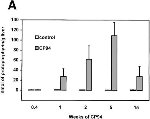
Induction of protoporphyria.Administration of the 3-hydroxypyridin-4-one iron chelator CP94 in the drinking water to C57BL/10ScSn mice caused the accumulation of porphyrin in their livers. This was detected after 1 week and was still present after 15 weeks of treatment (Fig 2A). Preliminary analysis by the spectrofluorometric method of Grandchamp et al17 suggested that the porphyrin accumulating was protoporphyrin IX, the last precursor in the biosynthesis of heme. Confirmation of this finding was obtained by HPLC (Fig 2B). Higher levels of CP94 in the drinking water did not cause a more marked effect (data not shown). The daily intake of CP94 given at the usual dose of 2 mg/mL was estimated to be equivalent to a dose greater than 200 mg/kg/d. Analysis of blood showed no significant elevation of protoporphyrin levels in erythrocytes after a 3-week exposure to CP94 (<1% of control). The accumulation of hepatic protoporphyrin suggested that the rate of its conversion into heme, a reaction that involves the insertion of iron by the enzyme ferrochelatase, might be inhibited in vivo because of the shortage of iron substrate. However, when ferrochelatase activity of livers from the mice administered CP94 for 5 weeks was determined in vitro in the presence of saturating amounts of iron substrate, a decrease of approximately 30% was still observed (control 45.7 ± 2.9, CP94 31.4 ± 1.1 nmol/h/mg protein; n = 6, significantly different). Protoporphyria cannot, therefore, be explained entirely on the basis of insufficent iron substrate in the mitochondria in vivo. On the other hand, in a separate study, CP94 was administered for 3 weeks and then mitochondrial fractions of liver were isolated. After Western blotting using an antibody against mouse ferrochelatase, no decrease in protein levels was detected (results not shown).
Development of protoporphyria in C57BL/10ScSn mice after CP94. (A) Accumulation of hepatic porphyrins expressed as protoporphyrin. Mice were administered CP94 in the drinking water (2 mg/mL) for up to 15 weeks. Results are mean ± SD from 3 to 8 mice per group. (B) HPLC of liver porphyrins after 2 weeks of exposure to CP94. (a) control, (b) CP94, and (c) standard protoporphyrin.
Development of protoporphyria in C57BL/10ScSn mice after CP94. (A) Accumulation of hepatic porphyrins expressed as protoporphyrin. Mice were administered CP94 in the drinking water (2 mg/mL) for up to 15 weeks. Results are mean ± SD from 3 to 8 mice per group. (B) HPLC of liver porphyrins after 2 weeks of exposure to CP94. (a) control, (b) CP94, and (c) standard protoporphyrin.
In a further study, CP94 was compared with two less lipophilic hydroxypyridinones, the dimethyl homologue CP20 and the N-hydroxyethyl analogue CP102. Table 1 shows that in this experiment only CP94 caused the accumulation of protoporphyrin in the liver and that no other type of porphyrin was observed.
Influence of 3-hydroxypyridin-4-ones on the Accumulation of Porphyrin Types in Livers of C57BL/10ScSn Mice
| Treatment . | Porphyrin Type and Concentration (nmol/g liver) . | ||
|---|---|---|---|
| . | Uroporphyrin . | Coproporphyrin . | Protoporphyrin . |
| Control | 0.05 ± 0.01 | 0.01 ± 0.01 | 0.27 ± 0.05 |
| CP20 | 0.04 ± 0.01 | 0.01 ± 0.00 | 0.25 ± 0.01 |
| CP94 | 0.22 ± 0.09 | 0 | 40.9 ± 13.2* |
| CP102 | 0.04 ± 0.01 | 0 | 0.32 ± 0.08 |
C57BL/10ScSn mice were administered CP20, CP94, or CP102 at 2 mg/mL in the drinking water for 2 weeks. Livers were analyzed by spectrofluorometry to distinguish between the three main porphyrin types as described by Grandchamp et al.17 Values are means ± SD from 3 to 6 mice per group.
Significantly different from other groups, P < .05.
Inhibition of ferrochelatase.The metabolism of 3,5-diethoxycarbonyl-1,4-dihydrocollidine (DDC) and its 4-ethyl homologue (Et-DDC) by hepatic cytochrome P450 causes the formation of N-alkyl-protoporphyrins through a suicidal reaction with the heme moiety of the cytochrome.21 These adducts are potent inhibitors of ferrochelatase and treatment of mice with the parent chemicals causes protoporphyria. To determine whether an inhibitor of ferrochelatase, such as an N-alkyl porphyrin formed during the metabolism of hydroxypyridin-4-ones, was responsible for the loss of enzyme activity, extracts were prepared from livers of mice treated with CP94 and CP20. The ability of these extracts to inhibit a semipurified preparation of mouse ferrochelatase was tested and compared with findings obtained with an extract of livers from mice administered Et-DDC. A significant inhibition of ferrochelatase was observed with extracts from the liver of Et-DDC–treated mice, as expected, but no inhibitory activity greater than controls was detected with liver extracts from mice given either of the chelators (Table 2).
Analysis of Liver Extracts for the Presence of a Ferrochelatase Inhibitor
| Treatment . | Inhibitory Units . |
|---|---|
| Control (water) | 4.6 ± 0.3 |
| CP94 | 3.9 ± 0.5 |
| CP102 | 4.1 ± 0.1 |
| Control (oil) | 4.1 ± 1.2 |
| Et-DDC | 21.4 ± 1.2* |
| Treatment . | Inhibitory Units . |
|---|---|
| Control (water) | 4.6 ± 0.3 |
| CP94 | 3.9 ± 0.5 |
| CP102 | 4.1 ± 0.1 |
| Control (oil) | 4.1 ± 1.2 |
| Et-DDC | 21.4 ± 1.2* |
C57BL/10ScSn mice were administered CP94 or CP102 in the drinking water (at 2 mg/mL) for 2 weeks. Et-DDC was administered by intraperitoneal injection in oil at 50 mg/kg and mice were left for 48 hours. Liver extracts were prepared and inhibitory activity estimated as described and defined in Materials and Methods. Inhibitory activity in control livers has been reported previously.22 Values are the means of 3 mice per group ± SD.
Significantly different from control group, P < .05.
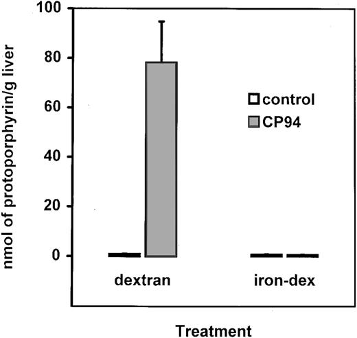
Comparison of iron status.In a further experiment, CP20, CP94, CP102, and CP117 were compared for their ability to decrease hepatic nonheme iron levels, ferritin, and ferritin iron of C57BL/10ScSn mice (Table 3). All variables were significantly reduced by each of the chelators. Examination of ferrochelatase activities showed that only CP94 caused a statistically significant depression, although CP20 may also induce a small effect. Again only CP94 caused significant accumulation of protoporphyrin. CP117, the more hydrophobic pivoyl ester of CP102, also failed to induce porphyria, despite reducing iron levels to some extent. The previously described experiments show that the protoporphyria caused by CP94 probably occurs through restriction of mitochondrial iron supply in vivo, but that somehow this also results in decreased ferrochelatase activity as measured in vitro. When iron-loaded C57BL/10ScSn mice received CP94, protoporphyria did not occur (Fig 3) illustrating that under these circumstances, there was no apparent interference in cellular iron pools. Young adult male SWR mice have liver and spleen iron stores threefold to fivefold greater than C57BL/10ScSn mice (B. Clothier and A.G. Smith, unpublished data, November 1995), eg, for the liver SWR 153 ± 7 mg/g of wet tissue and C57BL/105c5n 54 ± 2 mg/g. This phenomenon is not explained, but it is under investigation and appears to have a genetic basis. Administration of CP94 to SWR mice for 2 weeks had very little effect on protoporphyrin production (Fig 4) showing that the protoporphyric response could also be reduced by a genetically determined elevation in hepatic stores, as already described for experimentally induced iron overload.
Comparison of the Removal of Iron and Porphyrin Accumulation in the Liver Induced by Hydroxypyridinone Chelators
| Treatment . | Nonheme Iron (μg/g liver) . | Ferritin . | Ferritin Iron (μg/g liver) . | Ferrochelatase Activity (nmol/h/mg) . | Protoporphyrin (nmol/g liver) . |
|---|---|---|---|---|---|
| . | . | (% of control) . | . | . | . |
| Control | 55 ± 5 | 100 ± 27 | 6.8 ± 1.4 | 12.9 ± 3.9 | 0.4 ± 0.1 |
| CP20 | 33 ± 13-150 | 72 ± 153-150 | 1.8 ± 0.33-150 | 8.2 ± 3.9 | 3.2 ± 3.9 |
| CP94 | 36 ± 23-150 | 68 ± 103-150 | 2.1 ± 0.23-150 | 8.1 ± 1.73-150 | 17.1 ± 15.93-150 |
| CP102 | 44 ± 73-150 | 78 ± 223-150 | 2.9 ± 0.63-150 | 14.9 ± 5.1 | 0.3 ± 0.1 |
| CP117 | 36 ± 23-150 | 60 ± 73-150 | 3.1 ± 0.83-150 | ND | 0.6 ± 0.1 |
C57BL/10 ScSn mice received CP20, CP94, and CP102 in the drinking water at 2 mg/mL and CP117 at 3 mg/mL for 2 weeks. Values are the means of 5 mice per group.
Abbreviation: ND, not determined.
Significantly different from control, P < .05.
Influence of iron overload on the development of protoporphyria. C57BL/10ScSn mice received CP94 for 2 weeks. Mice were injected with dextran or iron-dextran 1 week before starting CP94 treatment. Results are mean ± SD from 3 mice per group. Results in the CP94 group given dextran were significantly different from its controls.
Influence of iron overload on the development of protoporphyria. C57BL/10ScSn mice received CP94 for 2 weeks. Mice were injected with dextran or iron-dextran 1 week before starting CP94 treatment. Results are mean ± SD from 3 mice per group. Results in the CP94 group given dextran were significantly different from its controls.
Comparison of response to CP94 in C57BL/10ScSn and SWR mice. Mice received CP94 in the water (2 mg/mL) for 2 weeks. Results are significantly different in the C57BL/10ScSn group, but not in the SWR mice (n = 3 or 4 per group).
Comparison of response to CP94 in C57BL/10ScSn and SWR mice. Mice received CP94 in the water (2 mg/mL) for 2 weeks. Results are significantly different in the C57BL/10ScSn group, but not in the SWR mice (n = 3 or 4 per group).
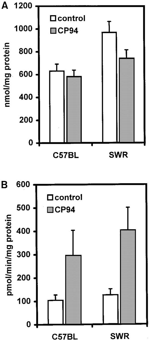
Influence on cytochrome P450.To determine whether restriction of the heme biosynthetic pathway had any effects on a major pool of hepatic hemoproteins, total microsomal cytochrome P450 was measured in C57BL/10ScSn and SWR mice after treatment with CP94. Small decreases were observed especially with the SWR strain (Fig 5A), although this was not susceptible to the protoporphyric action of CP94. In contrast, with benzyloxyresorufin as a model substrate for some isoforms of cytochrome P450 (CYP2B isoforms, in particular),29 a twofold to threefold induction of dealkylase activity (BROD) was observed with both strains (Fig 5B). This was confirmed by immunoblotting with anti-CYP2B1 antibody (data not shown) illustrating that in mice, at least, CP94 can act as a phenobarbital-type inducer of cytochrome P450.
Influence of CP94 on microsomal total cytochrome P450 content (A) and BROD activity (B) in C57BL/10ScSn and SWR mice after 2 weeks' exposure. Results are mean ± SD of 5 mice per group. BROD activity is the oxidation of benzyloxyresorufin to resorufin catalyzed in particular by CYP2B isoforms of cytochrome P450.
Influence of CP94 on microsomal total cytochrome P450 content (A) and BROD activity (B) in C57BL/10ScSn and SWR mice after 2 weeks' exposure. Results are mean ± SD of 5 mice per group. BROD activity is the oxidation of benzyloxyresorufin to resorufin catalyzed in particular by CYP2B isoforms of cytochrome P450.
DISCUSSION
The major application for acceptable orally active iron chelators would be as a more compliant treatment regime than present ones in iron overload associated with thalassemias and related conditions.1,2 However, their use to scavenge iron in conditions without overt iron overload has also been suggested. These uses include ameliorating damage caused by bleomycin and paraquat damage, reducing postischemic reperfusion injury, and in the treatment of malaria, porphyria cutanea tarda, rheumatoid arthritis, and cancer.10 The greatest clinical experience has been with CP20, but its use is controversial and undesirable side effects have been observed.30,31 Extensive studies have shown the diethyl compound CP94 to be very promising in terms of both relatively low toxicity and good iron chelating ability.5,8 The demonstration in this report that the chronic use of CP94 causes protoporphyria (a block in heme biosynthesis) in C57BL/10ScSn mice illustrates the possible complications that may arise from chelation of iron pools concerned with normal cellular function. Our results show that, as might be expected, this response is particularly noticeable in mice with low basal iron levels while SWR mice, with relatively high basal iron levels, were much less affected. If there are individuals in the human population who have low basal iron for genetic or environmental reasons, they could be at risk from untoward iron depletion leading to critically reduced levels. In man, CP94 and CP20 undergo extensive hepatic metabolism to form glucuronides, which cannot function as chelators.11 This problem has been addressed by the development of the N-hydroxyethyl analogue of CP94, namely CP102, which is poorly glucuronidated in the liver32 and thus a greater proportion of the hydroxypyridinone dose is available for iron chelation. Unfortunately, the introduction of a hydroxyl function on this class of molecule leads to an appreciable reduction of the Kpart value (water/n-octanol), which in turn, reduces the efficiency of extraction of iron from the liver. This is an undesirable change in properties as the liver is a major iron storage organ. To overcome this decrease in lipophilicity, hydrophobic esters have been synthesized (for instance, CP117). Such modification permits efficient delivery to the hepatocyte, where the ester is hydrolyzed to CP102 intracellularly.33 In our experiments, CP20, CP94, CP102, and CP117 all significantly lowered total hepatic nonheme iron levels (60% to 80% of controls) and iron present in ferritin (25% to 45% of controls), but only CP94 caused a significant elevation of protoporphyrin levels.
What is the mechanism of the hepatic protoporphyria caused by CP94? An obvious explanation is that mitochondrial iron pools or the delivery of iron to mitochondria are depressed thus inhibiting in vivo the action of the enzyme ferrochelatase, which inserts iron into protoporphyrin to form heme. A complication of this proposal is that measurement of ferrochelatase activity in vitro under conditions of large excesses of substrates showed a small (30%), but significant, decrease with CP94 and a nonsignificant decrease with CP20. As far as we could ascertain, the decrease was real and not an artifact of assay in the presence of some residual protoporphyrin. Certain chemicals such as DDC and Et-DDC are well known to cause inhibition of ferrochelatase and protoporphyria in mice. In these cases, the chemicals are metabolized by the cytochrome P450 system during which suicidal inactivation of the hemoprotein occurs with the formation of N-alkyl protoporphyrin from the heme moiety.21 N-methyl and N-ethyl protoporphyrins are high-affinity inhibitors of ferrochelatase, and inhibition can still be detected in vitro. However, our experiments comparing the effects of extracts of livers from CP94 and Et-DDC–treated mice on mouse ferrochelatase have shown that formation of an N-alkylprotoporphyrin is not apparently essential for the development of protoporphyia by the iron chelating drug. We cannot eliminate the possibility that protoporphryrin generating free radicals could be causing ferrochelatase loss, although we did not observe any protein depletion.34
Another possible explanation for the effects of CP94 on ferrochelatase has recently emerged. Both human and mouse ferrochelatases have an iron-sulphur cluster located at the C-terminal region of the proteins.35,36 This cluster is not apparently required for catalytic activity (in the sense that ferrochelatase from lower organisms are capable of functioning in its absence), but may be important in regulation of activity. The protoporphyrin accumulation in the liver and ferrochelatase inhibition that we observed in vitro may be due not only to depletion of iron as a substrate for the enzyme, but also to removal of iron from the iron-sulphur cluster [2Fe-2S]. This would be consistent with the apparent lack in changes of protein levels. Recently, inactivation of ferrochelatase by NO-disruption of the iron-sulphur cluster has been reported.37 Disruption of other iron-sulphur clusters are probably to be discovered. A [4Fe-4S] iron-sulphur cluster is found in the cytosolic iron responsive element-binding protein.13 The reason why CP94, but not the other chelators, may have this effect on mitochondrial iron and ferrochelatase could depend on the greater lipophility of CP94, thereby permitting a greater transmitochondrial passage. This problem would be avoided by the lipophilic CP117, as it is rapidly converted to the more hydrophilic CP102 in the cytoplasm of the hepatocyte.33 Future studies could address the depletion of particular iron pools targeted by different chelators, especially mitochondrial iron. The data in Fig 2 suggest that the response to CP94 fell after 15 weeks, perhaps reflecting a lowered response with age.
It seems likely that some factor, in addition to ferrochelatase activity, influences the potential for protoporphyrin accumulation. Liem et al38 observed that in rats desferrioxamine caused a 50% inhibition of ferrochelatase activity, but with no elevation of hepatic porphyrins. The main controlling enzyme of heme biosynthesis, 5-aminolevulinate synthase, and the heme degrading enzyme, heme oxygenase, were also unaffected. However, under in vitro conditions with isolated hepatocytes from C57BL/10ScSn mice, desferrioxamine depressed cytochrome P450 contents suggesting that restriction of iron can affect heme utilization in some circumstances.39 In the present in vivo study in C57BL/10ScSn mice, CP94 caused accumulation of hepatic protoporphyrin, but only a slight depletion of total cytochrome P450 and even induction of CYP2B isoforms as occurs with phenobarbital. Previous studies have shown that phenobarbital and many other drugs acting as similar inducers of cytochrome P450 have the property of stimulating the formation of porphyrins in the presence of a partial block in heme synthesis by the synergistic induction of 5-aminolevulinate synthase.40 This was observed in studies with Et-DDC when there was apparent inhibition of hepatic ferrochelatase at the same time as induction of certain CYP isoforms.41 The induction of cytochrome P450 activities by chronic administration of CP94 appears to be the first demonstration that these chelators can act in this way and this may have to be taken into account if their clinical use is expanded. Thus, the ability of CP94 to cause porphyia in the liver, but not erythrocytes, may be a combination of both depletion of certain iron pools to depress ferrochelatase activity and its ability to stimulate concomitantly the hepatic heme biosynthetic pathway exacerbating the restriction.
ACKNOWLEDGMENT
We thank Prof H.S. Dailey for the gift of antibody and D. Judah for help.
F.DeM. thanks the Italian government for MURST funds (40 and 60%).
Address reprint requests to Andrew G. Smith, PhD, MRC Toxicology Unit, Hodgkin Building, University of Leicester, Lancaster Rd, PO Box 138, Leicester LE1 9HN, UK.


This feature is available to Subscribers Only
Sign In or Create an Account Close Modal