Abstract
A controversy persists in autologous transplantation as to which source of progenitor cells, bone marrow (BM) or peripheral blood (PB), contains the lowest number of contaminating lymphoma cells, and how mobilization procedures affect these numbers. To accurately measure the number of non-Hodgkin's lymphoma (NHL) cells harboring the bcl-2/immunoglobulin H (IgH) rearrangement in progenitor cell grafts, we developed a nested quantitative competitive polymerase chain reaction assay (QC-PCR). DNA from lymph nodes of four patients with NHL were cloned into the pSK(+) vectors to generate four internal controls (ICs) (two with major breakpoint region [MBR] and two with minor cluster region [mcr] rearrangements). The kinetics of amplification of ICs paralleled those of bcl-2/IgH rearranged genomic DNA. When used in a QC-PCR assay, these ICs were accurate at a 0.2-log level and provided reproducible results, as shown by low intrarun and interrun variability. An excellent correlation between predicted and observed lymphoma cell content (r = .99) was observed over a range of at least 5 logs of rearranged cells. This approach was used to measure involvement by NHL cells at the time of progenitor cell harvest in 37 autologous transplant patients. The number of bcl-2/IgH rearranged cells in BM, PB, and mobilized PB (mPB) was found to vary from 1 to 1.1 × 10⁵ per million cells. The number of lymphoma cells present in BM was significantly higher than in PB (P = .0001), with a median difference in lymphoma cell content between BM and PB of 0.48 log of cells (range, −0.7 to 5 logs). In contrast, we found no difference in the concentration of bcl-2/IgH rearranged cells present in BM versus PB progenitor cells mobilized with cyclophosphamide and granulocyte colony-stimulating factor (G-CSF) (mPB) (P = .57). In conclusion, the QC-PCR assay described in this study could measure accurately and reproducibly the number of bcl-2/IgH rearranged cells among normal cells. Differences in levels of contamination by lymphoma cells between BM and PB were of less than one log (10-fold), and no differences in lymphoma cell concentrations were observed between BM and mobilized PB. As more cells are usually infused with mPB than with BM grafts, mPB progenitor cell grafts may actually be associated with higher levels of contamination by lymphoma cells. Furthermore, this QC-PCR assay should provide an important tool to assess the prognostic impact of lymphoma cell burden both in progenitor cell grafts and in vivo.
THE USE OF peripheral blood (PB) as a source of progenitor cells has been a major contributor to the rapid expansion of high-dose therapy with autologous progenitor cell transplantation (PCT) because it facilitates progenitor cell collection, decreases the time to neutrophil and platelet engraftment, and is associated with decreased procedure-related toxicity.1,2 However, a major concern with autologous PB or bone marrow (BM) transplantation is the risk associated with the reinfusion of occult malignant cells present in the progenitor cell graft.3-5 Indeed, previous studies have shown that non-Hodgkin's lymphoma (NHL) cells are detectable in the BM of patients with high-risk disease, even after intensive therapy.6-8 In addition, in patients with leukemia, gene-marking studies showed that the progenitor cell graft could contain cells involved in disease relapse.9,10 Moreover, purging of NHL cells below levels detectable by the polymerase chain reaction (PCR) resulted in improved disease-free survival, particularly in patients with low-grade lymphoma.11,12 Interestingly, a few studies suggested that PB is less frequently infiltrated by lymphoma cells than BM, and may thus be a better source of progenitor cells than BM.13,14 In contrast, other groups showed that bcl-2/immunoglobulin H (IgH) rearranged cells could be found with similar frequencies in PB and BM.15-17 In patients with various solid tumors, it has been shown that mobilized PB (mPB) can contain neoplastic cells,3,4,18,19 and that mobilization regimens could enhance the release of tumor cells into the PB.19
In most previous studies, PCR has been the primary tool used to detect lymphoma cells by targeting the 14;18 translocation, which is present in the majority of follicular lymphomas and approximately a third of NHL with diffuse histologies.20,21 PCR approaches are extremely sensitive and enable the detection of as few as one lymphoma cell in a million normal cells.11,15 22 However, these assays are usually qualitative, and the number of rearranged cells in a PCR positive sample could vary by more than five logs of lymphoma cells. Such large differences in the number of PCR-positive cells limit the interpretation of results and their prognostic and therapeutic implications.
In this study, we developed a QC-PCR methodology to accurately measure small differences in the number of bcl-2/IgH rearranged cells in clinical samples from patients with NHL. Four internal controls (ICs) generating PCR amplification products of different lengths for both major breakpoint region (MBR) and minor cluster region (mcr) translocations were isolated and cloned. The kinetics of amplification, as well as reproducibility and sensitivity of the test were evaluated to optimize the assay, and to determine the reliability, and particularly the precision of this approach. This QC-PCR methodology was then used to measure the level of contamination by NHL cells in BM, PB, and mobilized PB collections from patients undergoing autologous transplantation to determine the optimal source of progenitor cells in terms of tumor cell contamination.
MATERIALS AND METHODS
Patient population.
Clinical protocols were approved by the Human Subjects Protection Committee of the Maisonneuve-Rosemont Hospital and samples were obtained with the informed consent of the patients. Eighty-nine patients with B-cell NHL undergoing autologous BM (n = 50) or PB (n = 39) progenitor cell transplantation (PCT) were evaluated for the presence of a t(14;18). Overall, 38 patients were retained for the analysis because they satisfied the following two conditions: (1) the presence of a bcl-2/IgH rearrangement in BM, PB, or mobilized PB (mPB) at the time of progenitor cell harvest, and (2) both BM and corresponding PB samples (28 patients), or BM and mPB samples (17 patients), or samples from all three sources (7 patients) were available for analysis. All patients had relapsed or failed to achieve a first complete remission after eight cycles of “curative intent” chemotherapy and reached either a minimal residual disease (MRD) status or a partial response (PR). All patients had 5% or less of the intratrabecular space infiltrated by lymphoma cells on pathologic examination. Steady-state PB and BM samples were obtained at the time of marrow harvest or immediately before PB progenitor cell mobilization. Cyclophosphamide (1.5 g/m2) was administered on the first day of mobilization, followed by granulocyte colony-stimulating factor (G-CSF) (10 μg/kg/d) from days 1 to 12, and leukapheresis was performed on 2 consecutive days when the white blood cell count was above 8 × 10⁹/L (days 10 to 12).23
Clinical samples.
Lymph node biopsies were obtained from four patients with NHL presenting a t(14;18). The tissue was stored at −80°C before DNA extraction. Samples of PB and BM from patients undergoing PCT and healthy volunteer donors, used as controls, were collected with preservative-free heparin, and mononuclear cells (MNC) isolated by ficoll-hypaque density gradient centrifugation (Ficoll-Paque; Pharmacia, Piscataway, NJ). All cell samples were cryopreserved in 10% dimethyl sulfoxide (DMSO) using standard techniques and stored in the vapor phase of liquid nitrogen.24
Lymphoma cell lines.
The cell lines RL and H2 were derived from patients with NHL and harbored a bcl-2 translocation involving MBR, while DHL-16 cells had an mcr translocation. RL cells were a kind gift from Dr W. Urba (National Cancer Institute, Frederick, MD) and H2 and DHL-16 cells were kind gifts from Dr J.G. Gribben (Dana-Farber Cancer Institute, Boston, MA).
DNA extraction.
DNA was extracted by conventional cell lysis and proteinase K (Sigma, St Louis, MO) digestion in 100 mmol/L NaCl, 10 mmol/L Tris-HCl pH 7, 25 mmol/L EDTA, and 0.5% sodium dodecyl sulfate (SDS) at 50°C overnight.22 25 DNA was purified by a phenol-chloroform extraction and precipitated with 3 mol/L sodium acetate and ethanol. The concentration and quality of DNA was measured by spectrophotometry at 260 and 280 nm wavelength.
Construction of internal controls.
Internal controls (ICs) were generated by amplification of DNA from lymph nodes of patients with NHL. Patient DNA was PCR amplified with corresponding MBR or mcr outer primers,11,22 and amplification products were cloned into pSK(+) vectors [pCR-Script SK(+) Cloning Kits; Stratagene, La Jolla, CA]. Four different plasmids were obtained: two with a MBR (MACM and GU1M) and two with a mcr (R11 and MD6) rearrangement. Large-scale preparation of the cloned ICs was performed by lysing bacteria with the alkali method and purifying by equilibrium centrifugation in cesium chloride-ethidium bromide gradients (Boehringer-Mannheim, Mannheim, Germany).25Inserts were sequenced to confirm that inner and outer primer binding regions were conserved (Sequenase 1.0; United States Biochemical Inc, Cleveland, OH). The plasmids were then linearized with EcoRI (LifeTechnologies, Inc, Gaithersburg, MD). Plasmid DNA concentration was determined spectrophotometrically and the copy number calculated. A precise series of dilutions of the ICs, ranging from 0.1 ng to 0.05 fg, were prepared for competition assays.
Nested competitive PCR amplification.
Conditions for the nested competitive PCR reaction at the MBR translocation site were optimized using the ICs GUIM and MACM so that one copy of IC in 2.5 × 10⁵ normal MNCs was reproducibly detectable. Similar results were subsequently obtained with the other ICs.
For the first amplification, different amounts of IC were added to 1.5 μg of purified sample DNA and were amplified in a 100-μL reaction volume containing 25 pmol of each oligonucleotide primer, 200 mmol/L each of deoxyribonucleoside triphosphates (dNTPs), 2.5 U of Taq DNA polymerase (Boehringer, Mannheim, Germany), 10 mmol/L Tris-HCl (pH 8.3), 2.5 mmol/L MgCl2, 50 mmol/L KCl 0.1% gelatin.11,22 Oligonucleotides for the first amplification included MBR: 5′-CAGCCTTGAAACATTGATGG, or mcr 5′-CGTGCTGGTACCACTCCTG, and JH: 5′-ACCTGAGGAGACGGTGACC.11,22 DNA was subjected to 27 cycles of amplification in a Perkin Elmer Cetus 9600 thermal cycler (Cetus, Emeryville, CA). Cycle conditions included a 94°C denaturation step (15 seconds), a 55°C annealing step (15 seconds), and a 72°C extension step (15 seconds). The initial denaturation step was prolonged to 2 minutes and the final extension step to 5 minutes. On completion of the first amplification round, 10 μL of the amplified mixture was then reamplified for another 27 cycles using oligonucleotide primers internal (i) to the original primers (nested PCR) MBR-i: 5′-TATGGTGGTTTGACCTTTAG, or mcr-i: 5′-GGACCTTCCTTGGTGTGTTG, and JH-i: 5′-ACCAGGGTCCCTTGGCCCCA.11 22 This second PCR amplification was performed with the same parameters as described above, except for the addition of 3.5 U of Taq polymerase and 50 pmol of each inner primer.
Aliquots of 20 μL from the first and the second PCR amplification were analyzed by electrophoresis in 6% acrylamide-7 mol/l urea or 2% agarose gels (Boehringer) containing 0.5 mg/mL of ethidium bromide in Tris-borate buffer and visualized under UV light. The intensity of fluorescence for each PCR product was measured with a gel documentation system (Alpha Innotech Corp, San Leandro, CA). Estimations derived from the QC-PCR assay were multiplied by a correction factor (size of ICs' PCR product [bp]/size of patient's PCR product [bp]).26 27
In all instances, at least four aliquots of each sample were evaluated to determine PCR positivity for bcl-2/IgH rearrangement. With each experiment, one positive control consisting of IC only and a negative control sample consisting of water instead of DNA were always used. To confirm that DNA was amplifiable, negative samples were amplified with commercial primers for the β-globin gene (Gene Amplimer, Cetus). PCR procedures included the following measures to avoid false-positive results due to contamination.22 28 Processing of samples was accomplished with disposable pipettes in a biological hood never exposed to post-PCR material. Reagents and samples were mixed in sterile tubes with dedicated pipettors and filter pipette tips (Molecular Bio-Products Inc, San Diego, CA) and all reagents aliquoted for single use. Post-PCR products were manipulated in a different laboratory.
Kinetics of IC.
To determine the kinetics of amplification of the ICs, JH and JH-i primers were labeled by direct phosphorylation kinase reaction with (γ32P) ATP using T4 polynucleotide kinase (Boehringer). Radiolabeled primers were added to unlabeled primers in a proportion of 1 in 10 to the PCR reaction. A total of 30 μL of each PCR product was migrated in triplicate on a 2.5% agarose gel, each band was excised and radioactivity measured.26
Statistical analysis.
Although the distributions of log-transformed values were fairly normal, all data were analyzed using nonparametric techniques because many comparisons involved small subsamples. Comparisons between various measures of infiltration in BM, PB, and mobilized PB were performed using the Wilcoxon match-pairs rank test, with a Bonferroni-corrected alpha level of .05. Mann-Whitney U tests were used for between-groups comparisons. All analyses were considered significant when reaching the .05 alpha level.
RESULTS
Generation of internal controls.
To precisely determine the number of target lymphoma cells, the amplification of target DNA templates is compared with that of known amounts of ICs in a competitive PCR assay. For optimal competitive PCR amplification, ICs should ideally be amplified by the same set of primers as the target template and be of similar size.26 29To satisfy these objectives, ICs were generated from lymph nodes heavily infiltrated with lymphoma cells harboring the bcl-2/IgH rearrangement. Taking advantage of the inherent diversity in the breakpoint of the t(14,18) between different patients, lymph nodes from four patients were selected to obtain PCR amplification products of various sizes. DNA was extracted and amplified with the outer PCR primers. PCR products were purified, inserted into pSK(+) vectors, and cloned. Two ICs with MBR translocations were generated: GU1M and MACM, which on reamplification with internal primers, provided a second PCR product of 224 bp and 174 bp, respectively (Table1). Similarly, two ICs with mcr rearrangements, MD6 and R11, provided second PCR products of 516 bp and 1045 bp, respectively. Sequence analysis at inner and outer primer homologous binding regions showed that no nucleotide was lost or modified during the cloning of ICs (data not shown).
Characteristics of ICs
| IC . | Breakpoint . | Size of First PCR Product (outer primers) (bp) . | Size of Second PCR Product (inner primers) (bp) . |
|---|---|---|---|
| GU1M | MBR | 825 | 224 |
| MACM | MBR | 801 | 174 |
| MD6 | mcr | 631 | 516 |
| R11 | mcr | 1,068 | 1,045 |
| IC . | Breakpoint . | Size of First PCR Product (outer primers) (bp) . | Size of Second PCR Product (inner primers) (bp) . |
|---|---|---|---|
| GU1M | MBR | 825 | 224 |
| MACM | MBR | 801 | 174 |
| MD6 | mcr | 631 | 516 |
| R11 | mcr | 1,068 | 1,045 |
Kinetics of amplification of the IC.
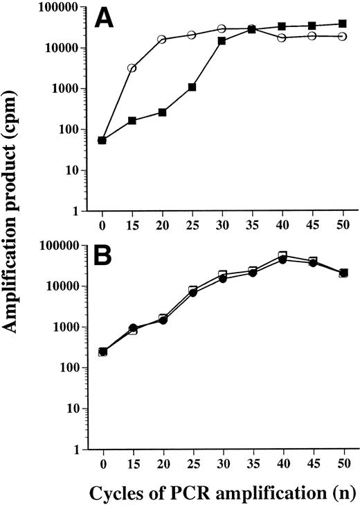
The first round of PCR amplification should remain in the exponential phase to prevent a loss of sensitivity and specificity and to avoid the formation of hybrid PCR products in the subsequent amplification with internal primers.26 To define the kinetics of amplification of the IC during the first round of PCR, 5 × 10⁴copies of IC (GU1M) were amplified for an increasing number of cycles with 32P end-labeled JH primers. This high number of rearranged cells was chosen to ensure that saturation of the amplification curve would not occur. PCR amplification products were electrophoresed, cut out of the gel, and levels of radioactivity were plotted against the number of cycles of amplification (Fig1A). During the first round of PCR amplification, the exponential phase occurred between 20 and 30 cycles of amplification. To determine the kinetics of amplification of the total nested PCR reaction, 32P end-labeled inner JH primers were added to the second PCR reaction. In these experiments, the number of cycles of amplification was maintained identical for both rounds of PCR amplification. At these high concentrations of DNA target template, a plateau was observed after 20 cycles of amplification, but a loss of efficiency occurred only after 35 cycles. Thus, we limited each round of PCR to 27 cycles. With this approach, one bcl-2/IgH rearranged cell among 250,000 normal cells was consistently detected and generated a signal 100-fold above the detection threshold of the gel (data not shown).22 Using this strategy, no bcl-2/IgH rearrangements were identified in PBMNCs from 19 normal controls.
Kinetics of amplification of ICs. (A) Amounts of amplification products (cpm) of the IC MACM (5 × 10⁴copies per PCR reaction) were measured after the first round (▪) and second round of nested PCR (○), and plotted against the number of PCR cycles. Results are means of two experiments in triplicate. (B) Products generated after PCR amplification of 5 × 10⁴copies of plasmid IC (MACM) (•) were compared with those generated after amplification of genomic DNA (5 × 10⁴ RL cells) (□).
Kinetics of amplification of ICs. (A) Amounts of amplification products (cpm) of the IC MACM (5 × 10⁴copies per PCR reaction) were measured after the first round (▪) and second round of nested PCR (○), and plotted against the number of PCR cycles. Results are means of two experiments in triplicate. (B) Products generated after PCR amplification of 5 × 10⁴copies of plasmid IC (MACM) (•) were compared with those generated after amplification of genomic DNA (5 × 10⁴ RL cells) (□).
The kinetics of amplification of our plamid IC was then compared with that of genomic DNA (RL cells) (Fig 1B). The same number of copies (5 × 10⁴) of IC and genomic rearranged DNA were PCR amplified with 32P-labeled outer primers and the amount of radioactivity was measured. The shape of the PCR amplification curve, as well as the level of PCR products measured, were identical for the IC and genomic DNA, confirming that the kinetics of amplification of plasmid IC and genomic rearranged DNA were identical.
Nested competitive PCR assay.
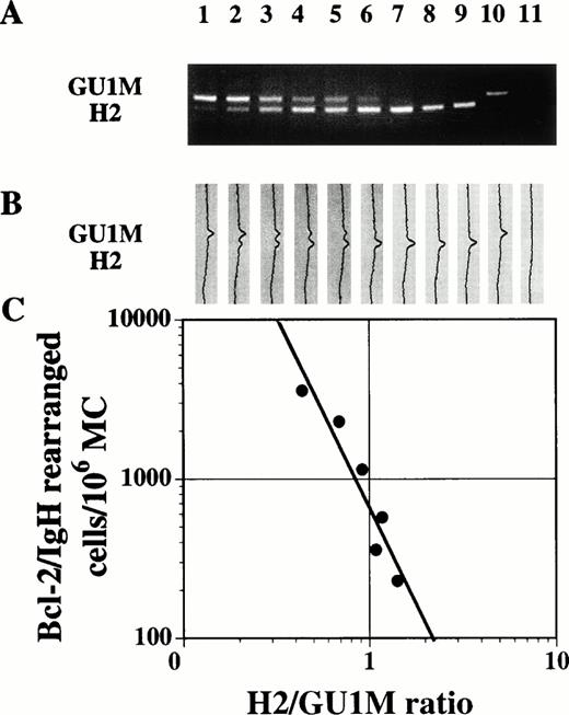
We then evaluated the capacity of IC to measure the number of bcl-2/IgH rearranged cells in a nested competitive PCR assay. Increasing amounts of ICs were added to aliquots of a simulated patient sample, consisting of DNA from 500 H2 cells added to one million normal mononuclear PB cells. Following nested PCR and electrophoresis, the intensity of PCR products corresponding to GU1M IC and H2 cells was measured (Fig2). For each concentration of IC, the ratio of H2/GU1M signals was calculated and plotted as a function of the number of copies of IC. The point where the ratio of patient and IC products was equal to 1 corresponded to the number of bcl-2/IgH rearranged copies in the sample.26 29
ICs were used in a nested competitive PCR assay to quantitate rearranged NHL cells. (A) The following number of copies of IC (GU1M): (1) 5,000; (2) 3,200; (3) 1,600; (4) 800; (5) 500; (6) 320; (7) 160; (8) 50; (9) 0 were added to aliquots of H2 cells mixed with normal PBMC at a ratio of 5 × 10³/106. Each aliquot, a positive (10: IC only) and a negative control (11: water only) were amplified by nested PCR, run on a 2.5% agarose gel, and stained with ethidium bromide. (B) Amounts of PCR products were measured by multidensitometric analysis. (C) Fluorescence ratios (•) were plotted against the concentration of IC (corrected for differences in molecular weight). The estimated number of bcl-2/IgH rearranged cells in the sample corresponded to a ratio of 1 (correlation coefficient = −.945).
ICs were used in a nested competitive PCR assay to quantitate rearranged NHL cells. (A) The following number of copies of IC (GU1M): (1) 5,000; (2) 3,200; (3) 1,600; (4) 800; (5) 500; (6) 320; (7) 160; (8) 50; (9) 0 were added to aliquots of H2 cells mixed with normal PBMC at a ratio of 5 × 10³/106. Each aliquot, a positive (10: IC only) and a negative control (11: water only) were amplified by nested PCR, run on a 2.5% agarose gel, and stained with ethidium bromide. (B) Amounts of PCR products were measured by multidensitometric analysis. (C) Fluorescence ratios (•) were plotted against the concentration of IC (corrected for differences in molecular weight). The estimated number of bcl-2/IgH rearranged cells in the sample corresponded to a ratio of 1 (correlation coefficient = −.945).
Reproducibility.
To assess the reproducibility of this test, we prepared a single sample of 500 H2 cells (enumerated in a counting chamber) in 106normal mononuclear PB cells. Six aliquots of the initial mixture were amplified in the same PCR run using the QC-PCR approach described above. The mean number of rearranged cells evaluated by QC-PCR was 867 ± 90 (± standard deviation [SD]) cells. Different aliquots of the same original cell mixture were then evaluated by QC-PCR in six different nested PCR runs, with different master mixes, over 6 different days and using six different preparations of the IC (GU1M). Even in these extreme conditions, the mean number of cells measured (829) was almost identical to that obtained in intrarun conditions, and the SD (303) was only one third of the mean number of copies. Both agarose and polyacrylamide gels were also used to evaluate PCR products, and similar estimations were obtained with both types of gels (data not shown).
Coamplification of IC and NHL DNA.
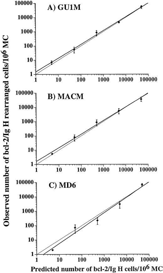
We then determined the linearity of our QC-PCR assay over the whole range of bcl-2/IgH cells that can be detected in patient samples. For this purpose, RL and H2 cells (harboring a MBR rearrangement) and DHL-16 cells (mcr rearrangement) were mixed with normal mononuclear PB cells at ratios varying from 5 to 5 × 10⁴/106, the latter level of infiltration was selected to correspond to infiltration by 5% lymphoma cells. These samples were coamplified with the ICs, MACM, GU1M, and MD6, respectively. When experimental results were plotted against those predicted, the slope of the regression curve was almost identical to that of a theoretical line of identity (Fig3). In addition, for each sample evaluated, only small variations were observed between different experiments and for all ICs. Therefore, these experiments show that our ICs can be used in this QC-PCR assay to measure NHL cell content over a large range of cell concentrations and with high levels of accuracy.
Correlation between the number of bcl-2/IgH rearranged cells predicted and observed. H2 (A), RL (B), and DHL-16 (C) cells in concentrations ranging from 5 to 5 × 10⁴ cells, were added to 106 normal PBMC and then evaluated with the ICs (thick line): GU1M, MACM, and MD6, respectively. Results, mean ± SD of three experiments, are compared with an ideal line of identity (thin line). Correlation coefficients of observed versus predicted results were greater than .99 for all three ICs.
Correlation between the number of bcl-2/IgH rearranged cells predicted and observed. H2 (A), RL (B), and DHL-16 (C) cells in concentrations ranging from 5 to 5 × 10⁴ cells, were added to 106 normal PBMC and then evaluated with the ICs (thick line): GU1M, MACM, and MD6, respectively. Results, mean ± SD of three experiments, are compared with an ideal line of identity (thin line). Correlation coefficients of observed versus predicted results were greater than .99 for all three ICs.
Bcl-2/IgH rearranged cells in BM and PB samples.
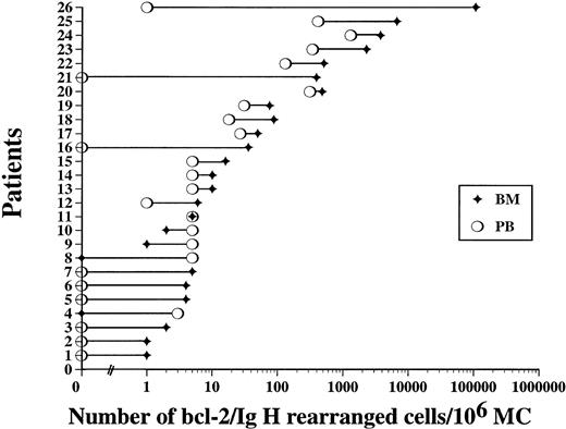
We used this QC-PCR approach to measure the number of lymphoma cells in BM and steady-state PB samples of patients with high-risk NHL before autologous transplantation at the time of progenitor cell harvest. Among the 28 patients where both BM and PB cells were available, all but two patients had at least one site with a bcl-2/IgH rearrangement detectable by PCR, and within this latter group, lymphoma cells were detectable in 69% of PB and 92% of BM specimens. When measured by QC-PCR, 11 patients (42%) had less than 5 bcl-2/IgH rearranged cells per million BM and PB cells evaluated, 5 patients (19%) had between 5 and 49 rearranged cells, 5 patients (19%) had between 50 and 499 rearranged cells, 3 patients (12%) had between 500 and 4,999 rearranged cells, and 2 patients (8%) had more than 5,000 and 50,000 rearranged cells per million cells (Fig 4). We found no differences in the number of rearranged cells in patients with MBR or mcr rearrangements (data not shown).
QC-PCR evaluation of the number of bcl-2/IgH rearranged cells measured in BM (⧫) and PB (○) from 26 different patients at the time of progenitor cell harvest. Lymphoma cell numbers were significantly higher in BM than in PB (P = .0001).
QC-PCR evaluation of the number of bcl-2/IgH rearranged cells measured in BM (⧫) and PB (○) from 26 different patients at the time of progenitor cell harvest. Lymphoma cell numbers were significantly higher in BM than in PB (P = .0001).
Overall, the number of lymphoma cells detected in BM was significantly higher than in PB (P = .0001). The number of lymphoma cells in BM was higher than in PB in 21 patients, lower in 3 patients, and both results were equal in 2 patients. In individual patients, differences in the number of lymphoma cells between BM and PB samples were of 1 log (10-fold) or less in 23 patients, with a median difference of 0.48 log, corresponding to a threefold higher number of lymphoma cells in BM (range, −0.7 to 5 logs). Differences of more than 1 log of NHL cells (1.2, 2.6, and 5 logs) were observed in only three patients, and in each instance, lymphoma cells were more prominent in BM than in PB.
Numbers of NHL cells in the BM and mobilized PB grafts.
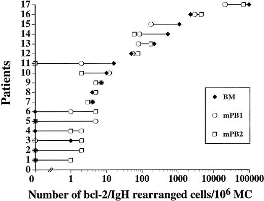
The widespread use of PB progenitor cell mobilization before autologous PCT warranted an evaluation of their lymphoma cell content and a comparison with levels of BM infiltration. At the time of harvest, bcl-2/IgH rearrangements were documented in 71% of BM samples and 82% and 88% of mobilized PB samples collected on the first (mPB1) and second (mPB2) day of leukopheresis, respectively (Fig5). From 0 to 98,608 rearranged cells per million cells were measured in BM, while 0 to 68,362 cells per million were observed in mPB products.
Quantification of lymphoma cells present in the BM (⧫) and the first (○) and second (□) mobilized PB collections from 17 different patients listed according to the number of rearranged cells detectable before autologous PBPCT. No difference in lymphoma cell burden was identified (P = .57).
Quantification of lymphoma cells present in the BM (⧫) and the first (○) and second (□) mobilized PB collections from 17 different patients listed according to the number of rearranged cells detectable before autologous PBPCT. No difference in lymphoma cell burden was identified (P = .57).
The distribution of the contamination level in this patient population (Fig 5) was similar to that observed in the previous group of patients (Fig 4). However, differences in lymphoma cell content between BM and mobilized PB were of only 0.06 log (0 log with mPB1 and 0.11 log with mPB2), without any significant difference (P = .570).
Clinical parameters versus infiltration by NHL cells.
To determine whether patient or disease characteristics were associated with the degree of infiltration by NHL cells, we compared the numbers of NHL cells measured by our QC-PCR assay with various clinical parameters (Table 2). We found no correlation between patient age, sex, stage of disease, remission status and number, and levels of rearranged cells. However, patients with follicular lymphoma had higher levels of infiltration by bcl-2/IgH rearranged cells in BM and mobilized PB than patients with lymphoma of diffuse histology (P = .041 and .034, respectively). This difference was not observed in steady-state PB. In addition, the presence of bone marrow infiltration by pathologic examination, at any time before transplantation, was associated with higher levels of NHL cells by QC-PCR in BM and PB (P = .015 and .012, respectively) than patients without detectable lymphoma cells on pathology. However, when only evaluated at the time of harvest, BM biopsy results did not correlate with the number of lymphoma cells measured by QC-PCR.
Impact of Clinical and Disease Characteristics on Lymphoma Cell Content in BM, PB, and mPB
| Clinical Parameters . | Two-Tailed P Values Versus the No. of Lymphoma Cells (QC-PCR) in . | ||
|---|---|---|---|
| BM (n = 37) . | PB (n = 26) . | Mobilized PB (n = 17) . | |
| Age | |||
| ≤45 v>45 | .703 | .271 | .769 |
| Sex | |||
| Male vfemale | .801 | .783 | .922 |
| Stage | |||
| III vIV | .208 | .094 | .460 |
| Histology | |||
| Follicular vdiffuse | .041 | .929 | .034 |
| Remission status | |||
| CR v PR | .964 | .513 | .207 |
| No. of remissions | |||
| 1* v ≥2 | .397 | .126 | .365 |
| BM infiltration† at any time | |||
| Yes vno | .015 | .012 | .148 |
| BM infiltration† at harvest | |||
| Yes v no | .082 | .136 | .30 |
Differences in the number of lymphoma cells, as measured by our QC-PCR assay, for each clinical parameter were analyzed with Mann-Whitney U Test. Two-tailed P values were corrected for ties. Statistically significant differences (P < .05) are represented in bold.
All patients in first remission failed to achieve a complete remission after completing the initial chemotherapy.
Identified on pathologic examination.
Effect of mobilization on NHL cells.
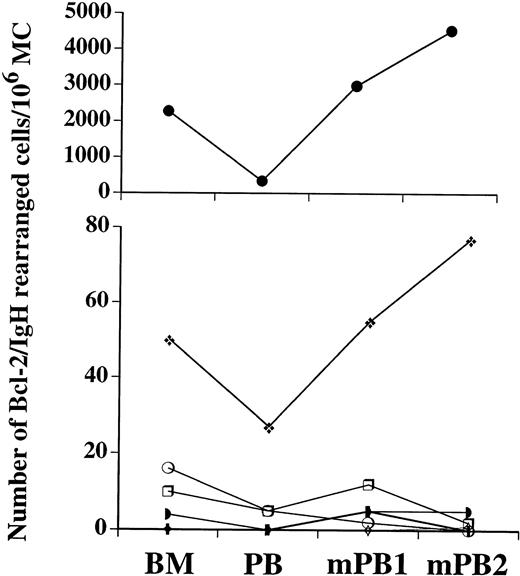
Lymphoma cell content could be evaluated in BM and sequentially in PB obtained before and after chemotherapy and G-CSF progenitor cell mobilization in seven patients. The number of bcl-2/IgH rearranged cells in PB increased following mobilization in six of seven patients (Fig 6). Although increases were limited in most patients, the number of NHL cells in two patients' PB more than tripled after mobilization.
Effect of mobilization on lymphoma cell levels. The number of bcl-2/IgH rearranged cells detectable in BM and PB obtained immediately before mobilization and in mobilized PB collections (1 and 2) are plotted for each of seven individuals.
Effect of mobilization on lymphoma cell levels. The number of bcl-2/IgH rearranged cells detectable in BM and PB obtained immediately before mobilization and in mobilized PB collections (1 and 2) are plotted for each of seven individuals.
DISCUSSION
To accurately measure the number of NHL cells present in minimal residual disease states, we developed a nonradioactive competitive quantitative PCR approach. After generating appropriate internal controls, we defined the kinetics of amplification, as well as sensitivity, reproducibility, and accuracy of this methodology. To identify which hematopoietic sampling site contains the least NHL cells and is the best source of progenitor cells in terms of tumor cell contamination, we used this QC-PCR to measure the number of bcl-2/IgH translocated cells in BM, PB, and mobilized PB. Although extremely variable between patients, the concentration of NHL cells found in BM was higher than in steady-state PB, but identical to that in mobilized PB.
Competitive PCR determines the amount of target DNA by comparing its signal to that of known quantities of an exogenous template that is amplified in the same PCR reaction and with the same primers.26,27,29 This approach, which is probably the most accurate quantitative PCR methodology, necessitates the synthesis of internal controls satisfying strict size and structure requirements. To satisfy these requirements, we cloned several bcl-2/IgH rearranged sequences directly from patient lymphoma cells. In contrast to previous studies that used cell lines as ICs,30 31 we could take advantage of the inherent diversity in the translocation sites of various NHL cells to obtain ICs of various sizes, which can be discriminated from patient PCR products for both MBR and mcr rearrangements, while remaining of similar size for accurate quantitative assessment. Moreover, the generation of such clones of ICs is simple and, importantly, ensures the preservation of intact sequences complementary to both outer and inner primers.
In this study, efforts of optimization and standardization were especially important to develop a quantitative approach capable of detecting extremely small variations in lymphoma cell content and to accurately determine the reliability and precision of such measures. We first ensured, by sequence analysis, that no nucleotides were lost or modified during the cloning of ICs, particularly in the outer primer homologous sequences, as the primers used for cloning were the same as those used for quantification. The first round of amplification also occurred in the exponential phase, while the second round was stopped before reaching the saturation phase, even with high levels of initial target template. In addition, kinetic studies confirmed that amplification of plasmidic IC paralleled that of genomic DNA. In these conditions, lymphoma cells could be measured over a large range, at least 5 logs of cells, and for each IC, this relation was linear, accurate (within 0.2 log of cells) and reproducible. Because the competition phenomenon is log-log linear only when DNA content is near equimolarity,26 we narrowed the number of different concentrations of ICs within one log of the target value, an approach that enabled us to monitor differences of less than one log (10-fold) with unequalled precision and standardization.
In patients with NHL undergoing PCT, it has been postulated that the level of infiltration of the progenitor cell graft by residual malignant cells plays an important role in disease relapse.11,14 A few studies have used sensitive detection methods to determine whether lymphoma cells were present in PB versus BM.13-15,30,32 Gribben et al13 evaluated 45 patients with high-risk NHL and found that bone marrow samples from all of these patients were infiltrated by lymphoma cells, while corresponding PB samples were positive in only half of these patients. Similarly, Sharp et al14, using an in vitro culture system, showed that lymphoma cells grew less frequently from PB than BM samples. In contrast, other investigators found similar numbers of PB and BM samples positive for bcl-2/IgH rearrangements.15-17,30 32 These studies provided important correlations between the presence of lymphoma cells in BM and or PB cells, yet only quantitative comparisons could clarify the issue. To our knowledge, this is the first large scale study to accurately measure the number of lymphoma cells present at these various sites and also in mobilized PB.
Using the QC-PCR approach described in this study, we showed wide variations, ranging from 1 to 107,245 bcl-2/IgH rearranged cells per million MNCs evaluated, in BM, PB, and mPB of patients in complete or partial remission. With such large variations, an evaluation of lymphoma cell content in BM and PB could only be performed by comparing samples obtained from the same patient and at the same time. We found that BM samples had a higher number of contaminating lymphoma cells than PB, but that differences in NHL cell mass between BM and PB were of one log or less in most (88%) patients. Such small (10-fold) differences in lymphoma cell numbers may explain why, in most qualitative PCR studies, similar frequencies of PCR positivity were detected in BM and PB samples.15-17,30,32 The lower frequency of infiltration by NHL cells in PB than in BM, identified by a few groups,13 14 could be attributable to the study of larger numbers of patients with threshold levels of lymphoma cells, where the 10-fold lower level of infiltration in PB identified in this study could result in a negative test in PB and a positive result in BM. This also suggests that PB should be a reliable sampling site to quantitatively evaluate lymphoma cell burden in a majority of patients. In contrast, we found no significant difference in the concentration of bcl-2/IgH rearranged cells present in BM and mobilized PB. Thus, although steady-state PB may offer an advantage in terms of the proportion of lymphoma cells, this difference with BM is very slight and completely abolished after mobilization with growth factor and chemotherapy. Moreover, as the total number of cells infused is usually larger with mPB than BM, mobilized PB grafts could actually contain higher numbers of lymphoma cells than conventional BM grafts. It is noteworthy that in a few individuals, differences in the number of NHL cells found in BM versus PB could reach up to 5 logs (105-fold differences). Such large differences again underline the critical importance of measuring the number of NHL cells, and not only PCR positivity, before stem cell harvesting to identify the importance of lymphoma cell infiltration.
Immunohistochemical studies in patients with breast and lung carcinoma showed that the number of malignant cells can increase in PB following mobilization procedures, and that this phenomenon seems related to BM involvement.19 We analyzed levels of NHL cells before and after mobilization with chemotherapy and G-CSF and found that lymphoma cell content in PB also increased with mobilization in a majority of patients. These preliminary results, along with results obtained by comparing lymphoma cell levels in BM and mPB, suggest that in vivo progenitor cell mobilization may not be appropriate as a purging strategy unless differences in the kinetics of release of lymphoma and normal progenitor cells can be identified. This seems unlikely because in this study, NHL cell levels were increased at the time when CD34+ cells are usually at optimum levels for collection. The fact that various types of tumor cells are mobilized into PB suggest that dissemination probably involves a common pathway related directly or indirectly to the mobilizing agents used. The monitoring of this effect should be important in the study of normal stem cell physiology, as well as of mechanisms implicated in tumor cell release. In addition, different numbers of tumor cells in progenitor cell grafts may have a different impact on patient outcome. For example, Sharp et al5 showed that patients with detectable lymphoma cells in BM grafts had a lower disease-free survival than patients without such colony growth. However, this lymphoma colony assay is less sensitive than nested PCR and possibly identifies patients with higher levels of tumor cell infiltration. Our results clearly show that major variations in lymphoma cell content can be observed in different patients and even in different harvest sites. The definition of clinically significant threshold levels of infiltration is thus clearly needed. It will be important to follow our patient population to prospectively determine whether the amount of lymphoma cells in progenitor cell grafts affects relapse and if a minimum level of bcl-2/IgH rearranged cells could be transplanted without impairing patient prognosis.
We found that patients with NHL of follicular histology had higher levels of infiltration than patient with diffuse NHL, independent of disease stage. This finding could explain why purging strategies seem particularly beneficial in patients with follicular lymphomas.11,12 It also reinforces the importance of developing strategies to purge NHL cells from mobilized PB grafts, and not only BM grafts. Fortunately, various strategies using monoclonal antibodies, either with complement, immunomagnetic beads, or immunotoxins or other purging agents can eliminate several logs of malignant cell line cells, while sparing normal hematopoietic progenitors.11,31 33-35 This QC-PCR assay may be an appealing and easily reproducible approach to devise purging strategies to enhance the elimination of lymphoma cells.
The preponderance of lymphoma cells in BM versus PB is compatible with the existence of a blood-marrow barrier.36,37 In addition, the almost constant one log difference in lymphoma cells between these sites strongly suggests that a gradient rather than a threshold phenomenon is involved. The elimination of such a gradient following stem cell mobilization with chemotherapy and growth factors may be attributable to direct toxic effects from cytotoxic agents. Indeed, Shirota and Tavassoli37 showed that cyclophosphamide administration altered the permeability of murine sinusoidal cells in BM endothelium. On the other hand, cytokines were also shown to modify the proliferation and regulation of cell adhesion molecules in normal hematopoietic progenitor cells following in vivo mobilization procedures.38 Such alterations at the level of either lymphoma cells or marrow stromal cells could result in enhanced circulation between PB and BM compartments.
In this study, 50% of NHL patients had below 10 lymphoma cells detectable per million cells. As described in Materials and Methods, extremely rigorous methods were used to minimize the likelihood of false positive results. In addition, 19 healthy volunteer donors were tested at different times with our assay and, in all instances, these samples were PCR negative. Nevertheless, a few investigators have reported the presence of bcl-2/IgH rearranged cells in normal invidividuals.39 40 However, this rearrangement was only documented at very low levels, mostly in older individuals, and in highly enriched B-cell populations with a frequency of 1 in 105 or less circulating B cells. These findings raise numerous questions regarding the biologic significance of low levels of bcl-2/IgH rearranged cells in normal individuals and NHL patients. The present QC-PCR assay may be useful to determine the evolution of these cells over time, their proliferation and clonogenicity, and also help identify instances where additional oncogenic events are implicated.
In addition to the above applications, accurate measurements of lymphoma cell numbers should be useful for the evaluation and optimization of (1) chemotherapy and immunotherapy protocols, (2) progenitor cell mobilization regimens, and (3) myeloablative regimens. Precise quantitative monitoring of lymphoma cells in vivo may also help to resolve one of the most crucial issues in lymphoma management: the impact of molecular detection of lymphoma cells on disease relapse.
ACKNOWLEDGMENT
The authors thank Dr F. Coutlée for helpful discussions, Dr M. Dumont for statistical analysis, Dr S. Cousineau for his hematopathologic expertise, C. Le Houillier for technical assistance, and M.J. Guertin and O. Marchand for their help with patient samples.
Supported by the Cancer Research Society of Canada. L.B. and D.C.R. are Scholars of the Fonds de la Recherche en Santé du Québec, Canada.
Address reprint requests to Denis-Claude Roy, MD, Division of Hematology-Immunology, Maisonneuve-Rosemont Hospital Research Center, 5415, L'Assomption Blvd, Montreal, Quebec, Canada, H1T 2M4.
The publication costs of this article were defrayed in part by page charge payment. This article must therefore be hereby marked “advertisement” in accordance with 18 U.S.C. section 1734 solely to indicate this fact.

This feature is available to Subscribers Only
Sign In or Create an Account Close Modal