Abstract
Pharmaceutical preparations of normal human immunoglobulin (IgG) are known to contain high-avidity and neutralizing antibodies (Ab) to the cytokines interleukin (IL)-1α, IL-6, and interferon (IFN)α. To test for other cytokine Ab, 23 batches of IgG were tested for saturable binding to eight 125I-labeled recombinant cytokines. All batches bound granulocyte-macrophage colony-stimulating factor (GM-CSF) with high avidity (Kav ≈ 10 pmol/L) and capacities of up to 5 μmol GM-CSF/mol IgG. Only 1 of 15 batches bound IL-5, also with high avidity, whereas 13 of 15 batches bound to IL-10 but with lower capacities and avidities. None of the IgG preparations bound IL-1 receptor antagonist (IL-1ra), IL-2, IL-3, IL-4, or G-CSF. Cross-binding and absorption analyses revealed identical or slightly stronger binding of recombinant GM-CSF, IL-5, and IL-10 than their native counterparts. GM-CSF–IgG complexes did not bind to cellular GM-CSF receptors, but Fc-dependent binding occurred to blood polymorphonuclear cells. Increased binding of GM-CSF to patient sera correlated positively with the binding capacities of infused IgG preparations. Patient and normal sera did not interfere with the binding of Ab to GM-CSF. From these and previous experiments, we conclude that pools of normal human IgG contain variable amounts of specific and high-avidity Ab to some cytokines, and that Ab to GM-CSF constitute a dominant anti-cytokine activity in these preparations. These Ab are available for reactionin vivo following IgG therapy.
NORMAL HUMAN immunoglobulin suitable for intravenous use (IgG) are used for treatment of an increasing number of infectious and immunoinflammatory diseases.1 These preparations usually contain IgG from several thousand individuals and hence a broad range of binding specificities and idiotypes. The mechanisms by which pooled IgG influence immunoinflammatory reactions are incompletely understood but may include antigen neutralization, Fc-receptor blockade, attenuation of complement activation, anti-idiotypic interactions, and binding to other molecules involved in immunoinflammatory processes.2
Cytokines constitute a large group of signal peptides produced during infections and other immunoinflammatory conditions. Several reports have described the presence of antibodies (Ab) against cytokines in patients and in healthy individuals.3,4 Pharmaceutically prepared IgG from normal donors has also been shown to contain specific and high-avidity Ab against the cytokines interleukin (IL)-1α, IL-6, and interferon (IFN)α, and these Ab block receptor binding of the respective cytokines.5-7 Human natural Ab to IL-1α have recently been cloned and expressed, and their high-affinity binding has been confirmed.8
In the present study, we tested for binding of granulocyte-macrophage colony-stimulating factor (GM-CSF), granulocyte colony-stimulating factor (G-CSF), IL-1 receptor antagonist (IL-1ra), IL-2, IL-3, IL-4, IL-5, and IL-10 to several batches of pooled human IgG from three manufacturers. High-affinity Ab against GM-CSF were present in all batches, whereas Ab against IL-5 and IL-10 appeared less frequently and/or with lower activity. The other cytokines bound in a nonsaturable manner. The anti–GM-CSF Ab blocked receptor binding of GM-CSF by simple competition. These observations strengthen the notion that anti-cytokine Ab contained in pharmaceutically prepared human IgG may contribute to the immunomodulatory activities of these preparations.
MATERIALS AND METHODS
Sera, Plasma, and Human IgG Preparations
Sera and citrate plasma samples obtained from healthy adults were from the Blood Bank (Rigshospitalet, Copenhagen, Denmark). Serum samples from two patients with systemic lupus erythematosus and mixed connective tissue disease were obtained immediately before and after 3 days of infusions with pooled IgG. Informed consent was received from the patients.
Preparations of normal human IgG, pharmaceutically prepared for intravenous use, were as follows: 9 batches of Sandoglobulin (Sandoz, Basel, Switzerland), 13 batches of Gammagard (Baxter, Allerød, Denmark), and 5 batches of Nordimmun (Novo Nordisk, Bagsværd, Denmark). Sandoglobulin and Gammagard were from plasma pools of over 8,000 Swiss and North American donors, respectively. Both were produced by alcohol precipitation, and the IgG of Sandoglobulin was further treated at pH 4 with traces of pepsin. Nordimmun was prepared by polyethylene glycol precipitation of a plasma pool of at least 2,000 Danish donors and contains no chemically or enzymatically modified immunoglobulins. The IgG contents of the stock solutions of pharmaceutic IgG and of patients sera were determined by turbidimetry using rabbit Ab to human IgG, code Q 331 (Dako, Copenhagen, Denmark) and HOO-03 reference serum from Janssen Biochemical (Beerse, Belgium).
Cytokines, Soluble Receptors and Ab
The nonglycosylated human cytokines were all expressed in Escherichia coli.
The following were used: IL-1α (Dainippon, Osaka, Japan), IL-1ra (Upjohn, Kalamazoo, MI), IL-2 (Kendall A. Smith, Cornell Medical College, New York, NY), IL-3 (Genzyme/Bie & Berntsen, Rødovre, Denmark), IL-4 (DNAX, Palo Alto, CA), IL-5 (Genzyme), IL-6 (Sandoz), IL-10 (Schering-Plough, Kennilworth, NJ), GM-CSF (PeproTech/TriChem, Virum, Denmark, and Sandoz/Schering-Plough, Copenhagen, Denmark [Leucomax]), G-CSF (Hoffmann La-Roche, Basel, Switzerland [Neupogen]), and IFNα2A (Hoffmann La-Roche [Roceron-A]).
Glycosylated human recombinant cytokines.
IL-5 expressed in T ni cells (Pharmingen, San Diego, CA), or in Sf-21 insect cells (R&D Systems/TriChem), or in Sw 25 murine myeloma cells (NIBSC, Potters Bar, Hertfordshire, England), and GM-CSF expressed in CHO cells (Sandoz/Schering-Plough), or in yeast (Genzyme).
Native human cytokines.
IL-5 and GM-CSF, generated in vitro by Ficoll-Hypaque (Nycomed, Oslo, Norway)-isolated blood mononuclear cells (MNC) stimulated over 4 days with 5 μg/mL of phytohemagglutinin (PHA)-P (Difco, Detroit, MI). IL-10 was generated by MNC challenged over 2 days with PHA-P plus 1 μg/mL of E coli endotoxin Westphal method 055:B5 (Difco).
Human soluble cytokine receptors.
The IL-1 receptor type I (IL-1RI) and the IL-4 receptor (IL-4R) were both expressed in COS cells (Immunex Corp, Seattle, WA).
Ab to human cytokines and GM-CSF receptor.
Neutralizing and monospecific Ab against E coli-derived recombinant IL-2, IL-3, and IL-4 were generated by immunizing rabbits with the purified cytokines, similarly as described.9Monoclonal Ab (MoAb) against IL-10 (MoAb 9D7 and MoAb 12G8) were from Schering-Plough. A blocking MoAb to the α-chain of the human GM-CSF receptor (GM-CSF-R) was from Genzyme (code 80-2977-01).
Radiolabeled Cytokines
All recombinant cytokines labeled with 125I were expressed in E coli, except IL-5, which was expressed in Sf-21 cells. Radiolabeled IL-2 (code IM 227), IL-3 (code IM 220), IL-4 (code IM 242), IL-5 (code IM 265), GM-CSF (code IM 224), and G-CSF (code IM 262) were kindly donated by Amersham (Birkerød, Denmark). IL-1ra was radioiodinated using Bolton Hunter reagent, and IL-10 and GM-CSF were radioiodinated using chloramine-T, as previously described.5 10
All radioiodinated cytokines were separated by molecular size chromatography on Sephadex G-75 superfine columns (Pharmacia, Uppsala, Sweden) and refractionated if necessary to obtain higher purity of the tracer, see below. RPMI 1640 or 0.02 mol/L phosphate buffer, 0.125 mol/L NaCl, pH 7.4 (PBS), both containing 0.5% to 1% (vol/vol) bovine serum albumin (BSA)(Sigma, St Louis, MO), were optimal for purification of 125I-labeled IL-2, IL-10, and G-CSF. When purifying the other radiolabeled cytokines, BSA was substituted by 0.025% to 0.05% (vol/vol) gelatin type A: from porcine skin (Sigma) plus 0.1% (vol/vol) Triton X-100 (Sigma) without influence on the chromatographic behavior or binding activities of the cytokines.
Validation of radiolabeled cytokines.
The 125I-labeled cytokines were validated by their ability to bind to specific Ab and/or to soluble and cellular receptors. Briefly, monomeric 125I-GM-CSF obtained from Amersham and E coli-derived GM-CSF labeled in our laboratory both expressed binding capacities >90% and specific activities of 1 to 2 × 10⁵ cpm/ng when tested with specific Ab contained in pharmaceutic IgG. Specific binding of monomeric125I-G-CSF to human polymorphonuclear granulocytes (PMN) showed a binding capacity >50% with specific activity 0.7 to 2 × 10⁵ cpm/ng. Monomeric 125I-IL-1ra expressed the highest binding capacity >90%, to soluble IL-1RI with specific activity 1 to 2 × 10⁵ cpm/ng. Maximal binding capacity, 80% to 90%, was obtained for monomeric 125I-IL-2 to a rabbit Ab with specific activity 0.3 to 1 × 10⁵ cpm/ng. Monomeric 125I-IL-3 expressed binding capacity >90% to rabbit Ab and specific activity 0.4 to 0.8 × 10⁵ cpm/ng. Highest binding capacity, 80% to 90%, was obtained with monomeric125I-IL-4 to IL-4R and rabbit Ab with specific activities 1 to 3 × 10⁵ cpm/ng. Dimeric 125I-IL-5 had a binding capacity >90% to Ab identified in IgG and specific activity 3 to 5 × 10⁴ cpm/ng. Dimeric 125I-IL-10 reacted stronger than monomeric 125I-IL-10 with MoAb 9D7 and MoAb 12G8, binding capacity >90% (specific activity 0.5 to 1 × 10⁵ cpm/ng).
Chromatographic Experiments
All separations were carried out at 4°C. Molecular size chromatography was performed in 0.9 × 32 cm columns containing Sephadex G-75 superfine or Sephacryl 300 HR (Pharmacia). Samples, 300 μL, were separated at a flow rate of 2.6 mL/h with collection of fractions every 9 minutes. The columns were calibrated with a mixture of molecular weight markers (Bio-Rad, Richmond, CA) in PBS. Elution buffers were RPMI 1640 with 0.15% BSA (natural cytokines) or PBS with 0.025% gelatin and 0.1% Triton X-100 (recombinant cytokines). Columns of 0.9 × 10 cm containing Sephadex G-75 superfine were used for purification of the 125I-labeled cytokines and for binding assays. As running buffers RPMI with 0.5% BSA or PBS with 0.05% gelatin and 0.1% Triton X-100 were used, depending on the tracer (see above).
Affinity chromatography for determination of 125I-cytokine bound to IgG was carried out in columns containing 0.5 mL protein G or protein A Sepharose (Pharmacia). A maximum of 300 μL was applied followed by 100 μL washing buffer and 15 minutes of incubation. The columns were then washed with 3 mL PBS with 0.5% BSA, and the bound material was eluted with 3 mL 0.1 mol/L glycine, pH 2.4.
Binding of Cytokines to IgG and Serum
If not otherwise stated, the samples were incubated at 4°C for 18 hours before evaluating the amounts of bound (B) and free (F)125I-cytokine by chromatographic separations, as described above. Nonspecific binding was assessed in the presence of 200 to 1,000 ng/mL of unlabeled cytokine. For screening purposes, 0.1 to 0.2 ng/mL of 125I-labeled cytokine was used.
Saturation binding analyses of the individual cytokines to different batches of IgG were performed as described.4 The results were expressed as Kav in pmol/L and the binding capacity as bound cytokine in μmol/mol IgG, calculated from molecular weights of 14 kD for GM-CSF, 26 kD for IL-5, 38 kD for IL-10, and 150 kD for IgG.
Recovery analyses.
Mixtures of equal amounts of two to three batches of IgG, along with the individual batches, were incubated at 37°C or 4°C for 18 hours. Serial two-fold dilutions from a total of 4 mg IgG/mL were made in duplicates and added 3,000 cpm/100 μL of 125I-GM-CSF followed by incubation and detection of bound tracer by protein G affinity chromatography. The binding activities of the samples were expressed relative to the IgG batch expressing the highest binding. Recovered binding was calculated as:
{M}A similar procedure was followed for detection of recovered IL-5 binding to mixtures of batches of IgG.
Binding of GM-CSF to sera was assessed by molecular size chromatography. Duplicates of 100 μL containing 66 μL serum, 3,000 cpm of 125I-GM-CSF with or without excess unlabeled E coli GM-CSF were tested. Binding of GM-CSF to Ab added to the individual sera was measured by protein G affinity chromatography. The test was performed as above but with added IgG capable of 30% and 70% specific binding of the 125I-GM-CSF, respectively. Ten sera were analyzed with two batches of IgG. In addition, five sera were tested by preincubation of 80% of the single serum with IgG for 18 hours at 37°C followed by 1.4 times dilution and coincubation with125I-GM-CSF and unlabeled GM-CSF as above.
In a plasma pool of equal volumes of the individual plasma samples, the recovered anti–GM-CSF IgG binding activity was analyzed by the principles described above.
Estimation of recovered anti–GM-CSF Ab in sera from patients treated with IgG were carried out in the following way. Serial two-fold dilutions of serum drawn before or after IgG therapy, along with a preparation of the infused IgG, were added to a constant amount of125I-GM-CSF. The binding activities of the sera were expressed relative to the activity of the infused IgG with the dimension mgeq/mL. The influence of sera collected before and after IgG infusion on the activity of the corresponding IgG batch was tested by addition of serum (60% final concentration) to 20 mg/mL, 15 mg/mL, and 10 mg/mL of the infused IgG, followed by serial dilutions and addition of tracer plus/minus unlabeled GM-CSF.
Papain and Pepsin Treatment of IgG
Papain-agarose (Sigma), 20 mg, was preincubated at 37°C in PBS, containing 10 mmol/L L-cysteine and 2 mmol/L EDTA. After 2 hours, the agarose was washed and incubated for 18 hours at 37°C with 20 mg IgG in 1 mL PBS, 2 mmol/L EDTA. The supernatants were stored at 4°C until use.
Pepsin digestion of IgG was carried out by incubating 10 mg IgG in 1 mL 0.2 mmol/L acetate buffer, pH 4.1, with 0.2 mg/mL of pepsin (Sigma) at 37°C for 20 hours. The sample was then dialyzed against PBS and applied on protein A columns using 0.5 mL dialyzed material/mL protein A-Sepharose and PBS as washing buffer. The unabsorbed material was placed in a dialysis bag and concentrated on powdered polyethylene glycol 20,000 (Merck, Darmstadt, Germany) and stored at 4°C.
ELISA and RIA of Cytokines
Duplicates were made of serial two-fold dilutions of the reference cytokines and at least three consecutive dilutions of the samples. Parallel runs of the curves for the native and the reference recombinant cytokines were always obtained in semi log plots.
ELISAs for human GM-CSF (DGM00) and IL-5 (D5000) were from R&D; their detection limits were 5 to 10 pg/mL. IL-10 was measured by double sandwich ELISA using monospecific polyclonal rabbit Ab to purified recombinant human IL-10, as described.9 The assay was calibrated with an IL-10 international standard (NIBSC). The inter- and intraassay coefficients of variation for the concentration range between 30 pg/mL and 2 ng/mL were <15%, and the sensitivity limit was 30 pg/mL.
RIAs for GM-CSF, IL-5, and IL-10 were carried out with the specific Ab contained in pooled human IgG and 0.1 to 0.2 ng/mL of the respective125I-labeled cytokine. The assays were performed as described above for the determination of cytokine binding to IgG. The intra- and interassay variations were <10% and the sensitivity limits 50 to 100 pg/mL.
As a supplement to the RIA measurements, the same Ab were also used for absorption of the native and reference cytokines. The relative content of a native cytokine was estimated from a plot of the amount of free cytokine (F) divided by the total amount of the cytokine (T) as a function of T (F/T versus T). Generally, the two assays are complementary in that the same relative value for the sample will be estimated by the tests only when the test ligand and the reference ligand show identical binding. If the reference ligand binds stronger, the RIA will underestimate the cytokine level, whereas the F/T versus T analysis will overestimate the concentration.
In the F/T versus T assay, the following procedure was used for quantitation of GM-CSF, IL-5, and IL-10: 200 μL samples of preincubated constant amount of Ab with variable concentrations of MNC supernatant or reference cytokine were applied on columns containing 0.5 mL protein G Sepharose. Identical samples, but without Ab, were run in parallel. Two × 100 μL washing buffer were added at intervals of 8 minutes, unabsorbed material was washed out in 1.6 mL of buffer, and the eluted cytokine was quantitated by ELISA. From use of125I-labeled cytokines alone, 0.98 ± 0.02 (N = 20) were recovered in the fraction of unabsorbed material.
Cell Receptor Assays
A human promyelocytic cell line, HL-60 (ATCC, Rockville, MD), was grown in RPMI 1640 with 20% (vol/vol) heat-inactivated fetal calf serum (FCS). Human blood PMN were purified as described11 and resuspended in RPMI 1640 with 1% (vol/vol) BSA, 8 mmol/L EDTA, 0.1% NaN3. Receptor assays were made over 18 hours at 4°C in duplicate samples of 250 μL containing 0.7 to 1.1 × 10⁷cells/mL, 125I-labeled cytokine, variable concentrations of human IgG, F(ab′)2 fragments of IgG, with or without unlabeled cytokine (200-1,000 ng/mL) or MoAb to GM-CSF-R (5-10 μg/mL). Cell-bound cytokine was separated by centrifugation of 200 μL of the cell suspension on dibutyl phthalate and bis (2-ethylhexyl)phthalate (ratio 1:1) (Merck). One hundred microliters of the supernatant were aspirated for evaluation of free and complexed tracer (cpmfree and cpmbound) by molecular size chromatography. Cell-bound cytokine was measured in the pellet after cutting the tube. The activities were counted with errors below 2%.
RESULTS
Screening for Specific IgG to Human Cytokines
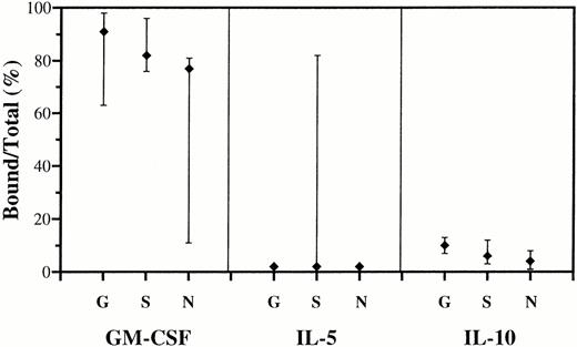
There was no saturable binding of 125I-labeled E coli-derived IL-1ra, IL-2, IL-3, IL-4, or G-CSF to 10 mg/mL of at least 15 different batches of IgG from three manufacturers. In contrast, GM-CSF, IL-5, and IL-10 bound at variable degrees to individual batches of IgG (Fig 1 and Table1). Binding of IL-5 was only detected in 1 of 15 IgG preparations. Papain or pepsin treatment of IgG showed that the binding of GM-CSF, IL-5, and IL-10 occurred predominantly or exclusively to the Fab part of IgG, because cytokines bound to intact IgG but not to enzymatically treated IgG were retained on protein A columns.
Saturable binding of GM-CSF, IL-5, and IL-10 to IgG. Five to 9 batches of Gammagard (G), Sandoglobulin (S), and Nordimmun (N) were tested in parallel at 4 mg/mL IgG and 0.1 to 0.2 ng/mL125I-cytokines: E coli-derived GM-CSF and IL-10, and Sf-21 cell-derived IL-5. The results are shown as median percentage (ranges) of displaceable binding divided by the total amount of 125I-cytokine in the assay. The background binding was <3%.
Saturable binding of GM-CSF, IL-5, and IL-10 to IgG. Five to 9 batches of Gammagard (G), Sandoglobulin (S), and Nordimmun (N) were tested in parallel at 4 mg/mL IgG and 0.1 to 0.2 ng/mL125I-cytokines: E coli-derived GM-CSF and IL-10, and Sf-21 cell-derived IL-5. The results are shown as median percentage (ranges) of displaceable binding divided by the total amount of 125I-cytokine in the assay. The background binding was <3%.
Cytokine Binding Avidities and Capacities of Pharmaceutic IgG Preparations
| Cytokine . | Gammagard . | Sandoglobulin . | Nordimmun . | |||
|---|---|---|---|---|---|---|
| Kav (pmol/L) . | Bmax (μmol/mol) . | Kav (pmol/L) . | Bmax (μmol/mol) . | Kav (pmol/L) . | Bmax (μmol/mol) . | |
| GM-CSF | 6 | 5 | 8 | 1.3 | 8 | 1.3 |
| 11 | 2.8 | 6 | 1.4 | 7 | 1 | |
| 4 | 4.2 | 6 | 1.1 | 9 | 1.3 | |
| 7 | 2.9 | 9 | 0.24 | |||
| IL-5 | 15 | 1.3 | ||||
| IL-10 | 108 | 0.12 | 80 | 0.1 | 380 | 0.3 |
| 351 | 0.4 | |||||
GM-CSF binding was tested with individual batches of 0.2 to 10 mg IgG/mL (giving similar binding activity). For IL-5 and IL-10, the assays were performed with 2 and 15 to 20 mg IgG/mL, respectively.
Binding Avidities and Capacities
As shown in Table 1, all IgG batches bound GM-CSF with high activities (Kav ≈ 10 pmol/L) but with variable binding capacities. IL-5 bound with similar avidity to the single positive IgG preparation, and IL-10 bound with over ten times lower avidities. The highest binding capacities, 2 to 5 μmol/mol IgG, were obtained with GM-CSF, the lowest with IL-10.
It is unlikely that the IgG preparations contained molecules that interfered with the cytokine Ab. Thus, when mixing low activity batches, or low and high activity batches, GM-CSF bound as the sum of activities contributed by the individual IgG preparations. There was also no effect on the binding of IL-5 when mixing the positive batch with either of seven negative ones. Taken together, this suggests that individual batches of IgG contained highly variable amounts of cytokine Ab.
Binding Specificity
Using 1,000 to 10,000 times excess amounts of unlabeled cytokines, there was no cross-binding to IgG between the eight cytokines tested, and IL-1α, IL-6, or IFNα2A. Hence, IgG bound specifically to GM-CSF, IL-5, and IL-10, respectively.
Effect of cytokine glycosylation.
Since nonglycosylated and variously glycosylated recombinant GM-CSF and IL-5 competed differently for their respective Ab (Table2), we investigated the binding of native cytokines in supernatants of in vitro stimulated human MNC. Complete suppression of binding to 125I-labeled recombinant cytokines was obtained with supernatants containing RIA reactivity for GM-CSF at 35 kD and IL-5 at 50 kD (in agreement with the reported sizes of the native, glycosylated cytokines12 13).
Quantitation of Native and Recombinant GM-CSF and IL-5 by Different Immunoassays
| Cytokine . | IgG Batch . | Assay . | Native Cytokine (ngeq/mL) . | Recombinant Cytokine (ngeq/ng) . | ||
|---|---|---|---|---|---|---|
| Reference = E coli GM-CSF: | Yeast | CHO | ||||
| GM-CSF | 1 | RIA | 19 | 0.5 | 0.5 | |
| 1 | F/T v. T | 27 | ||||
| 2 | RIA | 19 | 0.3 | 0.03 | ||
| 2 | F/T v. T | 28 | ||||
| 3 | RIA | 20 | 0.3 | 0.1 | ||
| 4 | RIA | 18 | 0.2 | 0.05 | ||
| ELISA | 27 | 0.74 | 0.6 | |||
| Reference = Sf-21 IL-5: | SW 25 | T ni | E coli | |||
| IL-5 | 5 | RIA | 2.4 | 1 | 0.1 | 0.05 |
| 5 | F/T v. T | 2.8 | ||||
| ELISA | 1.6 | 0.66 | 0.1 | 0.04 | ||
The activities of native and recombinant cytokines were measured by RIA and F/T versus T using different IgG batches and, also, with ELISA using MoAb from the kit manufacturer. Results are mean of triplicates, SDs were <10%.
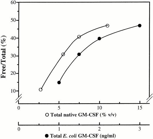
We also quantitated the supernatant contents of native GM-CSF and IL-5 using the same IgG batch for RIA and for absorption of both native, recombinant, and reference recombinant cytokines (Fig 2 and Table2). Native GM-CSF tested with only 40% higher values in the F/T versus T assay compared with those in RIA (Table 2), and the relative binding of native and E coli GM-CSF (the reference preparation) was independent of the IgG preparation used. The table also shows similar binding of native IL-5 and recombinant IL-5 expressed by the insect cell line Sf-21 (the reference preparation), but not by T ni cells or E coli-derived IL-5. Absorption experiments with 20 mg/mL IgG and 3 ng/mL of native and E coli IL-10 (the reference preparation) showed <30% binding even to maximum capacity IgG batches (binding of 0.6 to 2 ng IL-10/20 mg IgG, N = 3). Because of the sensitivity limit, it was not possible to analyze lower concentrations of IL-10. Since the reference preparations bound identically or only slightly stronger than the native cytokines, it is likely that the results shown in Fig 1 and Table 1 are valid approximations of the binding characteristics of IgG to native GM-CSF, IL-5, and IL-10.
Binding of native and recombinant GM-CSF to IgG. F/T versus T plots obtained by absorption to IgG (2 mg/mL) of native (○) and E coli-derived (•) GM-CSF. Total and free GM-CSF were quantitated by ELISA. Results are shown as means of duplicates.
Binding of native and recombinant GM-CSF to IgG. F/T versus T plots obtained by absorption to IgG (2 mg/mL) of native (○) and E coli-derived (•) GM-CSF. Total and free GM-CSF were quantitated by ELISA. Results are shown as means of duplicates.
Characterization of GM-CSF Binding to IgG
Because of their prevalence and strong binding, the Ab to GM-CSF were further characterized.
GM-CSF–IgG complex formation and binding stability.
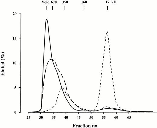
Complexes with molecular weights ≥600 kD were formed when125I-GM-CSF reacted with increasing concentrations of IgG (Fig 3). These complexes were highly stable. Using IgG levels that bound approximately 40 pmol/L GM-CSF, more than 85% of bound 125I-GM-CSF was retained after 8 hours at 37°C in presence of excess unlabeled cytokine. Similar results were obtained at 4°C.
Sephacryl S-300 HR molecular size elution profile of125I-GM-CSF bound at variable IgG concentrations.125I-GM-CSF, 0.25 ng/mL, was incubated at 37°C for 18 hours with 10 mg/mL ( ), 5 mg/mL (–––), and 0.5 mg/mL (-------) of the same IgG batch. Similar findings were obtained with IgG from all three manufacturers.
Sephacryl S-300 HR molecular size elution profile of125I-GM-CSF bound at variable IgG concentrations.125I-GM-CSF, 0.25 ng/mL, was incubated at 37°C for 18 hours with 10 mg/mL ( ), 5 mg/mL (–––), and 0.5 mg/mL (-------) of the same IgG batch. Similar findings were obtained with IgG from all three manufacturers.
Inhibition of binding to GM-CSF-R+ cells by IgG.
In contrast to what was seen using the human cell line HL-60, variable amounts of 125I-GM-CSF associated to PMN when incubated with different IgG batches, but with constant levels of125I-GM-CSF and anti–GM-CSF-R MoAb. This variation disappeared if F(ab′)2 fragments of IgG were used (Table3). Consequently, there was a significant binding of GM-CSF–IgG complexes to PMN and this binding depended on the Fc part of the IgG molecules (possibly to Fcγ-RII and Fcγ-RIII on PMN14,15).
Influence of IgG and F(ab′)2 on the Binding of 125I-GM-CSF to PMN
| Batch . | Competitor . | Competitor . | ||
|---|---|---|---|---|
| None . | GM-CSF . | MoAb to GM-CSF-R . | ||
| None | None | 278 | 17 | 24 |
| 1 | IgG | 230 | 20 | 131 |
| 1 | F(ab′)2 | 97 | 18 | 21 |
| 2 | IgG | 327 | 20 | 284 |
| 2 | F(ab′)2 | 42 | 22 | 21 |
| 3 | IgG | 265 | 21 | 20 |
| 3 | F(ab′)2 | 252 | 19 | 20 |
Three batches of IgG, 3.6 mg/mL, were tested in parallel with the corresponding F(ab′)2 fragments. The results are shown as mean cpmbound (N = 2) to 2 × 10⁶ PMN incubated with 125I-GM-CSF at 2,000 cpm/200 4 μL. Cpmfree in supernatants of samples added only IgG or F(ab′)2 were: batch 1, 232 and 264; batch 2, 125 and 112; batch 3, 1,602 and 1,570.
To avoid misinterpretation of IgG-induced interference with GM-CSF-R binding, background binding to PMN was assessed in the presence of excess anti–GM-CSF-R MoAb. Now, IgG suppressed the binding of125I-GM-CSF to both HL-60 cells and human PMN in a dose dependent manner, and complete blockade could be achieved with the IgG preparations (data not shown).
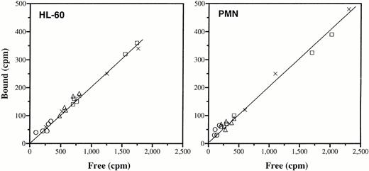
To further evaluate the interaction of IgG with cellular binding of GM-CSF, the amounts of cytokine bound to GM-CSF-R were plotted as a function of the free cytokine concentrations in the absence or presence of 15 different batches of IgG. As shown in Fig4, simple binding competition was observed with different IgG preparations, because the amount of specifically bound 125I-GM-CSF depended solely on the free concentration of the cytokine.
Effect of IgG on the binding of GM-CSF to GM-CSF-R. Data are expressed as specific 125I-GM-CSF bound to cells versus free cytokine—in the absence of IgG (X), or presence of Gammagard (○), Sandoglobulin (▵), or Nordimmun (□). Five different batches of IgG from each manufacturer were tested in parallel. HL-60 cells were incubated with 3,900 cpm/200 μL and 1.25 mg/mL of IgG, or with decreasing concentrations of the tracer alone. PMN were treated as the HL-60 cells, except that they were coincubated with 4 mg/mL of IgG at 2,900 cpm/200 μL. The background binding was assessed with excess Ab to GM-CSF-R. Data are means of duplicates and representatives of two to five experiments.
Effect of IgG on the binding of GM-CSF to GM-CSF-R. Data are expressed as specific 125I-GM-CSF bound to cells versus free cytokine—in the absence of IgG (X), or presence of Gammagard (○), Sandoglobulin (▵), or Nordimmun (□). Five different batches of IgG from each manufacturer were tested in parallel. HL-60 cells were incubated with 3,900 cpm/200 μL and 1.25 mg/mL of IgG, or with decreasing concentrations of the tracer alone. PMN were treated as the HL-60 cells, except that they were coincubated with 4 mg/mL of IgG at 2,900 cpm/200 μL. The background binding was assessed with excess Ab to GM-CSF-R. Data are means of duplicates and representatives of two to five experiments.
Low prevalence of anti–GM-CSF IgG in healthy individuals.
Less than 15% of total 125I-GM-CSF bound in a nonsaturable manner to sera of 50 healthy individuals. Because of recoveries exceeding 95% in individual sera (N = 15), there was no interference with the GM-CSF binding activity of IgG. Consequently, 1,258 plasma samples were tested for GM-CSF binding to IgG; 4 were positive. The Kavs were from 11 to 70 pmol/L and the Bmax values were from 5 to 320 μmol GM-CSF/mol IgG. The anti–GM-CSF activities of these plasma samples were fully recovered in the total plasma pool.
Recovery of infused anti–GM-CSF IgG in vivo.
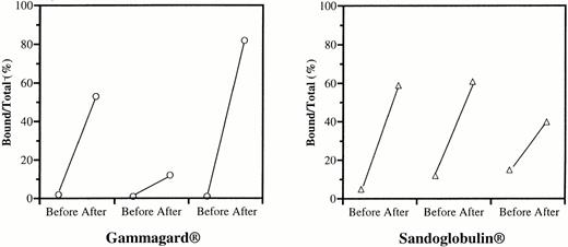
We finally investigated Ab to GM-CSF in sera of two patients, who over several years received monthly injections of IgG at high dosage. As shown in Fig 5, IgG infusion increased the binding of 125I-GM-CSF to serum. Furthermore, serum samples did not interfere with 125I-GM-CSF binding to the infused IgG. To estimate the in vivo recovery, we calculated the increase in total serum IgG following infusion. Similar results were obtained when calculated on the basis of specific Ab to GM-CSF (14 ± 3 mg/mL, N = 6) and total IgG (13 ± 2 mg/mL). These data strongly suggest that the infused Ab to GM-CSF were fully available and functional in vivo.
GM-CSF binding to serum of patients treated with IgG. Two patients with systemic lupus erythematosus and mixed connective tissue disease, respectively, received 60 g of either Gammagard or Sandoglobulin over 3 days at intervals of 1 month. Sera were collected immediately before and after three consecutive series of infusions. Data are means of duplicates and expressed as percent saturable binding of 125I-GM-CSF to IgG relative to the total amount of tracer using 4% (vol/vol) serum and 2,500 cpm/100 μL of125I-GM-CSF.
GM-CSF binding to serum of patients treated with IgG. Two patients with systemic lupus erythematosus and mixed connective tissue disease, respectively, received 60 g of either Gammagard or Sandoglobulin over 3 days at intervals of 1 month. Sera were collected immediately before and after three consecutive series of infusions. Data are means of duplicates and expressed as percent saturable binding of 125I-GM-CSF to IgG relative to the total amount of tracer using 4% (vol/vol) serum and 2,500 cpm/100 μL of125I-GM-CSF.
DISCUSSION
The occurrence of IgG Ab against certain cytokines in healthy and diseased individuals is increasingly realized.4,16-22Natural Ab have been reported against recombinant human IL-1β, IL-2, IL-8 and tumor necrosis factor (TNF)α by some23-30 but not by others.4,18,31 Besides differences in assay sensitivities, the discrepancies in detecting cytokine Ab may be related to the different assay methods. Saturation binding analysis is generally the preferred method of testing these Ab, and the binding must be shown to occur exclusively to the Fab part of the Ab.4,32 These criteria are fulfilled in previous studies of IL-1α, IL-6, and IFNα binding to human IgG,5-7 33 and they were used in the present study for the detection of anti-cytokine Ab in serum or plasma samples, and in pooled human IgG.
Specific Ab against GM-CSF, IL-5, and IL-10 were detected in pooled human IgG, and none were found against five other cytokines, including IL-2. In agreement with our findings of Ab to GM-CSF in 4 of 1,258 plasma samples (this study) and none against IL-10 in 50 sera,4 other studies have shown less than 2% of sera weakly positive for anti–GM-CSF Ab or anti–IL-10 Ab.16,20In contrast, IgG Ab directed against IL-1α and IL-6 have been reported in up to 30% and 20% of healthy individuals, respectively.4
Despite a much less frequent occurrence in sera of healthy individuals, the avidities of anti–GM-CSF Ab in pharmaceutically prepared IgG were in the same range as those reported for IL-1α Ab; the binding capacities were generally one to five times higher.5 This observation makes the Ab binding of GM-CSF the dominant cytokine binding activity presently identified in pooled IgG.5,7Higher activities of anti–IFNα Ab than those detected in individual sera have also been observed in human IgG.7 Simple addition of Ab activities contributed by individual donors to a pool of IgG may not be found in situations where increased clonality of an Ab specificity or interference with anti-idiotypic Ab has developed.3 There is, however, no evidence of anti-idiotypic Ab blocking the activity of any anti-cytokine Ab. In this study, for example, none of several individual sera or IgG preparations interfered with the binding of GM-CSF to IgG. Ab to GM-CSF, administered to patients during high-dose IgG therapy, were also fully recovered and available for reaction, in agreement with similar observations for Ab to IL-1α, IL-6, and IFNα infused with pooled IgG.34 Finally, a similar degree of polyclonality was detected by molecular size chromatography of GM-CSF–IgG complexes from different IgG batches containing variable amounts of these Ab.
We found 0.3% of individual plasma samples positive for anti–GM-CSF Ab, but these samples had binding capacities up to 100 times higher than, and avidities similar to, the ones usually detected in the IgG preparations. In a pool of nearly 1,000 plasma samples, the anti–GM-CSF Ab activity was the sum of the activities contributed by these few positive specimens. Hence, the anti–GM-CSF Ab activities in pharmaceutical IgG preparations are most likely contributed by only a few highly positive donors. This would explain the variable levels of anti–GM-CSF Ab activities in different preparations of pooled normal IgG; differences between the donor populations and manufacturing procedures might contribute to this.
Recombinant proteins may differ from their native counterparts in their binding characteristics.10,35,36 The naturally occurring Ab identified with 125I-labeled recombinant cytokines in the present and previous studies have all been shown to cross-bind the corresponding native cytokine.6,7 37 To evaluate the relative binding to Ab of recombinant and native cytokine, we compared the results obtained by RIA with those from absorption experiments using the same Ab. This showed that IgG bound similarly to native (glycosylated) and to nonglycosylated GM-CSF. On the other hand, IgG bound in a similar fashion to native (glycosylated) and to glycosylated recombinant IL-5, but not to nonglycosylated IL-5.
Taken together, the data show that native GM-CSF binds to IgG with high avidity (Kav ≈ 10 pmol/L) and with capacities of 2 to 5 μmol/mol IgG. Hence, IgG at 10 mg/mL would have the potential to bind >90% of GM-CSF, if present at concentrations up to 2 ng/mL (calculated according to Svenson et al5). Native IL-10 cannot be expected to bind significantly to IgG, whereas strong binding of IL-5 may occur in sporadic IgG preparations.
It is uncertain from previous studies whether anti–GM-CSF Ab induced by treatment of patients with E coli- or yeast-expressed GM-CSF react with endogenous GM-CSF.14,36 38 In contrast, our study demonstrates binding of native GM-CSF to anti–GM-CSF Ab in pooled normal IgG. Any differences in this regard between therapy-induced and naturally occurring anti–GM-CSF Ab would have to be resolved by binding and cross-binding analyses.
An agonistic effect in vivo of cytokine Ab has been proposed.39 This has been underlined by experience with in vitro neutralizing MoAb to cytokines, which have shown both inhibition and enhancement of cytokine activities in vivo.40 The agonistic effect of in vitro neutralizing MoAb has been explained by a decreased clearance of the cytokine if present in monomeric immune complexes, combined with low concentrations of the MoAb that favor redistribution of the cytokine from the immune complexes to cellular cytokine receptors. A carrier function of anti–GM-CSF Ab in pooled IgG is considered unlikely, however, because of their high avidity, stability, and capacity to form large immune complexes with GM-CSF and block binding to GM-CSF receptors.
GM-CSF stimulates the formation and function of PMN and monocytes.41 Maturation of monocytes to macrophages and dendritic cells and MHC class II expression are augmented by GM-CSF, and it is a strong adjuvant during immunizations with weak immunogens such as tumor cells and soluble proteins and peptides.41-45GM-CSF is induced by and potentiates the release of cytokines such as IL-1 and TNFα, both known to be centrally involved in acute and chronic inflammatory reactions.19,41 Indeed, treatment with Ab to GM-CSF increases the survival of endotoxin-challenged mice and, in combination with Ab to IL-3, mice with experimental cerebral malaria.46 47 Therefore, Ab to GM-CSF in pooled human IgG may be of benefit in the treatment of immunoinflammatory disorders, but their presence could reduce the efficacy of IgG preparations used in the prevention of infection. Isolation of different, naturally occurring anti-cytokine Ab and the use of IgG preparations selectively enriched or depleted of Ab to specific cytokines are attractive ways to further analyze the therapeutic potential of natural Ab to cytokines.
ACKNOWLEDGMENT
Drs Børge Thing Mortensen (Rigshospitalet, Copenhagen), Kendall A. Smith (Cornell Medical College, New York, NY), Satwant Narula (Schering-Plough, Kennilworth, NJ), Jan de Vries (DNAX, Palo Alto, CA), and Steven Gillis (Immunex Corporation, Seattle, WA) are thanked for generous gifts of cytokines and cytokine receptors. Amersham DK and Novo-Nordisk kindly donated radiolabeled cytokines and Nordimmun, respectively. Dr Charlotte Lundsgaard (Copenhagen County Hospital in Glostrup) helped with the sampling of patient sera, and Susanne Meldgaard provided excellent technical help.
Supported by Rigshospitalet University Hospital, the Danish Cancer Society, the Danish Biotechnology Program, the Danish Rheumatism Association.
Address reprint requests to Klaus Bendtzen, MD, DMSc, IIR 7521, Rigshospitalet, Tagensvej 20, DK-2200 N, Copenhagen, Denmark.
The publication costs of this article were defrayed in part by page charge payment. This article must therefore be hereby marked “advertisement” in accordance with 18 U.S.C. section 1734 solely to indicate this fact.

This feature is available to Subscribers Only
Sign In or Create an Account Close Modal