Abstract
The integrin IIbβ3 mediates platelet aggregation through its fibrinogen and adhesive protein-binding properties. Particular interest concerns the role of the cytoplasmic domains of IIb and β3. We now report the molecular analysis of IIbβ3 from a patient with a Glanzmann’s thrombasthenia-like syndrome for whom the principal characteristics are an approximate 50% total platelet content of IIbβ3 but with a much lower proportion in the surface pool (Hardisty et al, Blood 80:696, 1992). Polymerase chain reaction (PCR) single-strand conformational polymorphism and DNA sequencing showed a heterozygous mutation giving rise to amino acid substitution R995 to Q in the GFFKR sequence of the cytoplasmic domain of IIb. Reverse transcriptase-PCR and polymorphism analysis only detected mRNA for the mutated allele of the IIb gene and a single allele of the β3 gene in his platelets, suggesting other unidentified defects. Site-directed mutagenesis followed by transient expression of the mutated IIb together with wild-type β3 in Cos-7 cells resulted in a markedly decreased expression of the complex at the cell surface when compared with cells transfected with wild-type IIb and β3. Flow cytometry with PAC-1 and a stable Chinese hamster ovary–transfected cell line showed that the mutated receptor was not locked into a high activation state, although it became so in the presence of the activating antibody, anti-LIBS6. This is the first reported natural mutation in the highly conserved GFFKR sequence of the IIb cytoplasmic domain.
GLANZMANN’S THROMBASTHENIA is an inherited bleeding disorder in which quantitative or qualitative defects of the fibrinogen receptor, αIIbβ3(the platelet glycoprotein [GP] IIb-IIIa complex), result in the absence of the aggregation response after platelet activation.1-3 A member of the integrin family, αIIbβ3 is formed through the noncovalent association of two subunits encoded by different genes, both of which are localized on chromosome 17.4 Binding studies with monoclonal antibodies (MoAbs) showed that about 50,000 copies of this receptor are present at the surface of resting platelets.5An internal pool of about the same size is associated with membranes of the surface-connected canalicular system, α-granules, and dense granules.6,7 After activation of platelets by physiological agonists such as adenosine diphosphate, a conformational change in the surface receptor enables binding of fibrinogen (or other adhesive proteins) and aggregation.8 With strong agonists such as thrombin, the internal pool is also mobilized, providing more αIIbβ3 complexes to participate in platelet-platelet interactions.9
Transcription of the αIIb and β3 genes occurs in megakaryocytes and the two proteins are assembled and then glycosylated in the endoplasmic reticulum. Endoproteolytic cleavage of αIIb in the Golgi apparatus leads to the separation of heavy and light chains which remain disulfide bonded.10 It is the light chain that carries the transmembrane and cytoplasmic domains of αIIb. Post-translational modifications, including glycosylations and the association of the two glycoproteins into a heterodimeric complex, are necessary for cell-surface expression of the receptor.10,11Molecular abnormalities described in patients with Glanzmann’s thrombasthenia have provided information on structural domains essential for αIIbβ3 complex formation and expression. For example, two amino acid substitutions adjacent to the first and the fourth calcium binding sites of αIIbaltered the conformation of the complex and prevented its transport to the golgi apparatus,12,13 whereas a mutation located between the second and the third calcium binding site greatly slowed the maturation kinetics.14 Further information on the functional domains of the complex have been provided by studies on variant forms of the disease. Thus, mutations in the extracellular domain of β3 are associated with a failure of the constituted complex to bind fibrinogen when stimulated and a mutation in the cytoplasmic domain of β3 leads to defective “inside-out” signaling.15-17
We now report the molecular analysis of αIIbβ3 from a patient with an atypical form of Glanzmann’s thrombasthenia. As we previously reported, the patient’s platelets were characterized by (1) a total platelet αIIbβ3 content of less than 50% of normal levels and (2) an altered partition of the residual αIIbβ3 receptor within the different platelet membrane systems.18 Only approximately 15% of the normal levels of this receptor was found at the platelet surface, although the internal pool remained substantial. The residual receptor was functional and aggregation was observed after in vitro activation of platelets by physiological agonists, although the kinetics were slow and only small aggregates formed. The patient’s platelets did not bind fibrinogen spontaneously. Among the abnormalities that we have found for this patient is a heterozygous point mutation, G to A located at nucleotide 3078 of the cDNA (numbering according to Frachet et al19), leading to an R995 to Q amino acid substitution in the αIIb cytoplasmic domain. Site-directed mutagenesis and transient expression in Cos-7 cells showed that the mutation was likely to be directly involved in the low surface expression on his platelets. In view of the purported role of the cytoplasmic domains of αIIb and β3 in controlling the activation state of the complex,20,21 we also prepared a stable transfected Chinese hamster ovary (CHO) cell line expressing the mutated receptor. Flow cytometry showed that the cells only bound appreciable amounts of PAC-1, a marker of the activated complex,22 in the presence of an αIIbβ3-activating antibody.
This is the first time that a mutation in the cytoplasmic domain of αIIb has been reported in Glanzmann’s thrombasthenia. The interest of the R995 to Q substitution and the reduced cell-surface expression of αIIbβ3 is reinforced by its location in the GFFKR sequence which is conserved in the cytoplasmic domain of all integrin α subunits.
MATERIAL AND METHODS
Case Report
The case report and specific features that characterize the platelets of patient A.P., a young Italian man, were given by Hardisty et al.18 His father (F.P.) and mother (M.P.) were also studied. There is no evidence of consanguinity and no family history of bleeding. However, the patient has suffered from occasional bleeding since early childhood although blood transfusion has never been performed. Bleeding mostly occurred after knocks or small trauma and was not spontaneous. As detailed earlier, the main features of A.P.’s platelets are a total αIIbβ3 content that is about half that observed with normal platelets yet with a surface expression that is sparse compared with the size of the internal pool.18 The platelet aggregation response is poor but not absent and this is why he was originally referred to as having a “thrombasthenia-like” syndrome. His unactivated platelets did not bind fibrinogen spontaneously. Platelets from his parents aggregated normally, although the number of αIIbβ3 receptors, as determined in MoAb-binding studies, was lower than is usually seen: approximately 28,000 for his father’s platelets and approximately 20,000 for his mother’s platelets.18 Control donors were adult members of our hospital staff. Informed consent was obtained.
Polymerase Chain Reaction–Single-Strand Conformational Polymorphism (PCR-SSCP) and DNA Sequencing
Genomic DNA from the propositus, both parents and healthy volunteers, was isolated from whole blood using the QIAmp blood kit (Qiagen, Chatsworth, CA) according to the manufacturer’s instructions. A total of 43 pairs of oligonucleotides allowed the amplification of each exon together with the intronic splicing signals. After 30 cycles of amplification, an aliquot of each product was loaded onto a 2% agarose gel.23 After electrophoresis, bands were visualized under ultraviolet light after ethidium bromide coloration.
SSCP analysis was performed using the Pharmacia Phast System and precast minigels (Pharmacia-Biotech, Saint-Quentin en Yvelines, France) as we previously described.23-25 The oligonucleotide primers for amplifying exon 3 of the β3 gene and exons 21 and 26 of the αIIb gene are given elsewhere.23,24 Genotyping for the HPA-1 and HPA-3 alloantigen systems was performed using an Allele-Specific Restriction Assay (ASRA) as previously described (also see below).23 24The primers used to amplify exon 30 of the αIIb gene were5′CAGCAAATCATCTGTATACCCT3′ (sense, hybridizing in intron 29) and5′CCCAAAGCTTGGAGGCAACT3′ (antisense, hybridizing downstream of exon 30). DNA amplification products exhibiting an altered migration pattern in the SSCP analysis were directly sequenced using a fentomol DNA sequencing kit (Promega-France, Lyon, France). To ensure that those mutations detected in heterozygous form were not artifacts of the sequencing methodology, we cloned the corresponding amplification products into the pGem-T vector (Promega-France) and sequenced 12 independent clones using the T7 Sequencing kit (Pharmacia-Biotech).
Transfection of Cos-7 Cells and Transient Expression of Recombinant αIIbβ3
Site-directed mutagenesis was performed as before,25 using the pALTER-1 Vector system (Promega-France) in which we had previously cloned wild-type cDNA for αIIb. A CTTCAAGCAGAACCGGC oligonucleotide was used to generate the αIIb mutation (the mutated base is indicated in bold case). After selection, full-length cDNAs were entirely sequenced to be certain that only the desired mutations were present. Wild-type or mutated cDNAs for αIIb and β3 were subcloned between EcoRI and HindIII sites of the eukaryotic expression vector pSM as we previously constructed.25 Cos-7 cells, cultivated in Dulbecco’s modified Eagle’s medium (Life Technologies, Cergy-Pontoise, France) supplemented with 7% fetal calf serum, were transfected as follows: 3 × 10⁵ cells per 3.5-cm petri dish were seeded 24 hours before transfection which was performed during 4 hours with 3 μg of one plasmid or 1.5 μg of each plasmid (one carrying αIIb cDNA and the other β3 cDNA) and Transfectam (Promega-France) according to the manufacturer’s conditions. Surface membrane expression of αIIbβ3 was analyzed by flow cytometry and total cell expression by Western blotting, both after 72 hours of culture.
In some experiments, Cos-7 cells were also cotransfected with pSV-β plasmid (Promega) coding for β-galactosidase. Transfection efficiency was measured on a fraction of the cells using an anti–β-galactosidase MoAb (Promega) after cell permeabilization using the Fix and Perm Cell Permeabilization Kit (Caltag Laboratories, Burlingame, CA) and flow cytometry.
Flow cytometry.
Cos-7 cells were harvested after a 10-minute incubation at room temperature after the addition of phosphate-buffered saline (PBS) containing 5 mmol/L EDTA. The cells were washed twice with PBS-0.1% (wt/vol) bovine serum albumin (BSA) and counted. Volumes (0.5 mL) containing 400,000 cells in PBS-0.1% BSA was then incubated for 30 minutes under saturating conditions with EDU-3, an MoAb specific for the αIIbβ3 complex (a gift from Dr Villela, Barcelona, Spain); SZ22 (Immunotech, Marseille, France), specific for αIIb; or XIIF9 (prepared by our laboratory), specific for β3. Cells were washed twice with PBS-0.1% BSA before being incubated for 30 minutes in 0.5 mL PBS-BSA containing fluorescein isothiocyanate (FITC)-conjugated F(ab)2 antibody to mouse IgG (Silenius Laboratories, Hawthorn, Australia) and analyzed after another round of centrifugation using a Becton-Dickinson FACscan (Becton-Dickinson, Le Pont de Claix, France).25 In each experiment 10,000 cells were counted across the laser. The limit of positivity was defined as the maximum observed fluorescence when Cos-7 cells were transfected by the expression vector alone.
Western blotting.
Transfected Cos-7 cells were obtained as described above, sedimented, and resuspended in 50 μL of 10 mmol/L Tris-HCl, pH 7, containing 150 mmol/L NaCl, 3 mmol/L EDTA, and 2% (wt/vol) sodium dodecyl sulfate (SDS). Samples (50 μg) were loaded onto a 7% polyacrylamide gel and the proteins separated by electrophoresis before transfer to nitrocellulose membrane as described.26 The membranes were saturated for 2 hours with 5% (wt/vol) nonfat dry milk in 20 mmol/L Tris-HCl, 150 mmol/L NaCl, 0.05% (vol/vol) Tween 20, pH 7. After five washes in washing buffer (0.5% nonfat dry milk, 20 mmol/L Tris-HCl, 150 mmol/L NaCl, 0.05% Tween 20, pH 7), membranes were incubated for 2 hours with the murine MoAb XIIF9, specific for β3, or SZ 22, specific for αIIb. After five washes, membranes were incubated for 1 hour with peroxydase-coupled goat antibody to murine IgG (Jackson ImmunoResearch, West Grove, PA) and bound MoAb revealed using a chemiluminescence procedure (Amersham-France, Courtaboeuf, France). In each experiment, 1 μg of SDS-soluble proteins from platelets of a normal donor were electrophoresed as a control.
Transfection of CHO Cells and Evaluation of PAC-1 Binding
Wild-type and mutated αIIb cDNAs were subcloned into the pCDN(−) expression vector (Invitrogen, San Diego, CA) betweenEcoRI and HindIII restriction sites. A previously characterized CHO cell line stably expressing the β3subunit was transfected using pCDN wild-type or mutated αIIb cDNA using lipofectamine as previously described.27 Briefly, after neomycin selection, cell membrane expression of αIIbβ3 was assessed using MoAbs P2 and EDU-3, both specific for the αIIbβ3 complex. A stable cell line maximally expressing the mutated αIIbβ3 was subcloned. The affinity state of the complex was analyzed in these cells using FITC-labeled PAC-1 (Becton-Dickinson), a murine IgM specific for the activated complex.22 GP IIb-IIIa expression was controlled using PerCP-conjugated anti-β3(Becton-Dickinson) detected with an FL3 threshold gate as recommended by the manufacturer. In each experiment, 4 × 10⁵cells were incubated for 45 minutes in the presence of FITC–PAC-1 (15 μL of the solution provided by the manufacturer) with or without 2 μmol/L of the MoAb anti-LIBS6 (a generous gift of Dr Mark Ginsberg, Scripps Research Institute, La Jolla, CA) as described.28Anti-LIBS6 is an activating MoAb that binds directly to the complex. Binding of PAC-1 was then assessed by flow cytometry, and 5,000 cells were analyzed. Results for the subcloned cell line expressing the mutated complex were compared with those obtained for CHO cells transfected with wild-type αIIbβ3. The specificity of PAC-1 binding was also assessed in the presence of 1 mmol/L RGDS peptide (Sigma Chemical Co, St Louis, MO). On occasion, results were expressed as the activation index (AI) defined as 100 × (Fo − Fr)/(FoLIBS6 − Fr LIBS6), where Fo is the median fluorescence intensity (MFI) of PAC-1 binding and Fr is the MFI of PAC-1 binding in the presence of RGDS. Fo LIBS6 is the MFI of PAC-1 binding in the presence of 2 μmol/L of this antibody and Fr is the MFI of PAC-1 binding in the presence of anti-LIBS6 and the competitive inhibitor.20
Reverse Transcriptase (RT)-PCR on Platelet mRNA
RT-PCR was performed on platelet lysates from the patient, his father, and his mother prepared according to a procedure we recently described.29 Genotyping of HPA-1 (PlA) and HPA-3 (Bak) systems was performed on PCR products amplified as already described30,31 and using HpaII and FokI restriction enzymes, respectively. A TaqI polymorphism was determined using the TaqI restriction enzyme.32Mutations located in exon 30 of the αIIb gene were determined both after direct sequencing of amplification products and after cloning and sequencing amplification products from cDNAs. In this case, 12 independent clones were sequenced for each family member using a T7 sequencing kit (Pharmacia-Biotech).
RESULTS
PCR-SSCP on Genomic DNA Together With Sequence Analysis
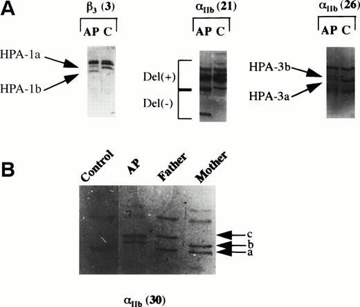
We designed 43 pairs of oligonucleotides allowing the specific amplification of all exons including splice sites of both the αIIb and β3 genes, and developed a nonradioactive SSCP procedure to analyze each amplification product. For the patient (A.P.), 4 out of 43 DNA amplification products showed a SSCP profile different to that observed for the control. Three of the observed differences in the SSCP patterns were caused by known polymorphisms (Fig 1A). The first of these involved exon 3 of the β3 gene and was due to the HPA-1 polymorphism.23 As summarized in Table 1, the SSCP patterns for this exon showed that the patient and his father were heterozygous HPA-1a/HPA-1b, whereas his mother was homozygous HPA-1a/HPA-1a. ASRA performed usingHpaII confirmed these genotypes. In the same manner, the differences observed for the SSCP patterns for amplification products carrying exons 21 and 26 of the αIIb gene (Fig 1A) were caused by two polymorphisms that we have previously described as being bilaterally linked: HPA-3 and a 9-bp deletion (Del) in intron 21.24 SSCP analysis showed that both A.P. and his mother were heterozygous HPA-3a/HPA-3b and Del−/Del+,whereas his father was homozygous for the HPA-3a and Del− polymorphisms (Table 1). Genotyping for HPA-3 was confirmed using the FokI restriction enzyme.
PCR-SSCP analysis of the IIband β3 genes. PCR amplification products were submitted to SSCP analysis by electrophoresis on the minigels of the PhastSystem apparatus and the migrated products detected by silver staining. (A) Only the amplification product for exon 3 of the β3 gene of patient A.P. exhibited a different migration profile to the control (C); this corresponded to the HPA-1 genetic determinant, the control being HPA-1a/HPA-1a and the patient being HPA-1a/HPA-1b. For the IIb gene, three amplification products had different patterns when compared with the control. For amplification product 21, this corresponded to a previously described polymorphism (Noted Del+ and Del−) in intron 21. Amplification product 26 corresponded to the HPA-3 system, the control being HPA-3a/HPA-3a and the patient being HPA-3a/HPA-3b. (B) SSCP analysis of amplification product 30 of the IIbgene showed a previously undescribed pattern. Illustrated are the patterns obtained for the patient (A.P.), a control, his father, and his mother. Band a was present for the control and the two parents, band b for A.P. and his mother, and band c for A.P. and his father.
PCR-SSCP analysis of the IIband β3 genes. PCR amplification products were submitted to SSCP analysis by electrophoresis on the minigels of the PhastSystem apparatus and the migrated products detected by silver staining. (A) Only the amplification product for exon 3 of the β3 gene of patient A.P. exhibited a different migration profile to the control (C); this corresponded to the HPA-1 genetic determinant, the control being HPA-1a/HPA-1a and the patient being HPA-1a/HPA-1b. For the IIb gene, three amplification products had different patterns when compared with the control. For amplification product 21, this corresponded to a previously described polymorphism (Noted Del+ and Del−) in intron 21. Amplification product 26 corresponded to the HPA-3 system, the control being HPA-3a/HPA-3a and the patient being HPA-3a/HPA-3b. (B) SSCP analysis of amplification product 30 of the IIbgene showed a previously undescribed pattern. Illustrated are the patterns obtained for the patient (A.P.), a control, his father, and his mother. Band a was present for the control and the two parents, band b for A.P. and his mother, and band c for A.P. and his father.
Summary of the Polymorphism Analysis as Performed for Patient (A.P.) and His Parents
| Polymorphisms . | Patient (A.P.) . | Mother (M.P.) . | Father (F.P.) . | |||
|---|---|---|---|---|---|---|
| DNA . | cDNA . | DNA . | cDNA . | DNA . | cDNA . | |
| HPA-1 | a/b | a | a | a | a/b | a/b |
| TaqI | −/+ | − | − | − | + | + |
| HPA-3 | a/b | a | a/b | a | a | a |
| Exon 30 | 2/3 | 3 | 1/2 | 1 | 1/3 | 1/3 |
| Del | −/+ | NA | −/+ | NA | − | NA |
Analysis of different polymorphisms on DNA was performed using ASRA, SSCP, or DNA sequencing. The detection of the polymorphisms on RNA was performed using the same methods after RT-PCR. HPA-1 andTaqI polymorphisms are carried on the β3 gene and HPA-3, exon 30 polymorphisms, and Del are carried on the αIIb gene. For TaqI: − refers to the absence of digestion and + to digestion by the TaqI restriction enzyme. For Exon 30: 1 refers to the wild-type published sequence; 2 to the silent mutation, C to T, in the codon for Val990; and 3 to the G to A mutation responsible for the Arg995 to Gln amino acid substitution. Del: − and + refer, respectively, to the absence or presence of a 9-bp deletion in intron 21 of the αIIb gene. This 9-bp deletion is observed in HPA-3b individuals only.
Abbreviation: NA, not available for study on cDNA because this polymorphism is located in an intron.
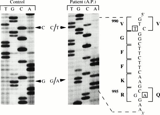
In contrast to these results, the pattern observed for the amplification product carrying exon 30 of the αIIb gene of the propositus was new to us (Fig 1B). Up to now, no polymorphism has been reported in this region of the αIIb gene. As is shown in Fig 1B, not only was the SSCP pattern for this amplification product different for the patient, but it was also different for both parents. In summary, band a was present for the control and for both parents but absent for the patient (A.P.), band b was present only for the propositus and his mother, and band c was restricted to the propositus and his father (F.P.). Direct DNA sequencing of PCR products showed that the differences between A.P. and the control were in fact due to two different base pair substitutions. The patient was heterozygous for both substitutions, the first, a C to T change in position 3064 of the cDNA, and the second, a G to A change in position 3078 of the cDNA (Fig 2). These mutations were detected in a heterozygous state in his parent’s DNA; the first was possessed by his mother and the second by his father (data not shown). Although the C to T transition did not induce an amino acid change, the G to A transition inherited from his father led to an amino acid substitution, R to Q in position 995 of the αIIbprotein. Significantly, this amino acid substitution was located in the GFFKR sequence conserved in all cytoplasmic tails of integrin α subunits (see Discussion).
Direct DNA sequencing of amplification product 30 of the IIb gene for the control and for the patient (A.P.). Two detected heterozygous mutations are indicated in open squares and the corresponding amino acid sequence and the substitution R995to Q are noted with bold letters.
Direct DNA sequencing of amplification product 30 of the IIb gene for the control and for the patient (A.P.). Two detected heterozygous mutations are indicated in open squares and the corresponding amino acid sequence and the substitution R995to Q are noted with bold letters.
Expression of Recombinant DNA in Cos-7 Cells
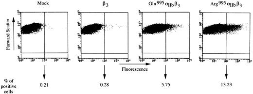
The G to A change responsible for the R to Q substitution in position 995 was introduced into wild-type αIIb cDNA by site-directed mutagenesis. After DNA sequencing to ensure that only this alteration was present, and subcloning into the eukaryotic expression vector pSM, Cos-7 cells were cotransfected with wild-type β3 cDNA and wild-type or mutated αIIb cDNA. Cell-surface expression of the complex was analyzed by flow cytometry 72 hours later. Using EDU3, an MoAb specific for the αIIbβ3 complex, the percentage of positive Cos-7 cells was 13% when cotransfected with the two wild-type cDNAs and 5.6% for cells cotransfected with wild-type β3 cDNA and mutated αIIb cDNA (Fig3). In the illustrated experiment, the MFI for positive cells expressing complexes composed of mutated αIIb and wild-type β3 was 192, a value that was considerably lower than the MFI of 422 observed for cells expressing the wild-type complex (these results were typical of those obtained in three separate experiments). Similar findings were also obtained using XIIF9, an MoAb specific for the β3 subunit (data not shown). Nevertheless, because the definition of the limit of positivity was established as the maximum observed fluorescence when Cos-7 cells were transfected by the expression vector alone, we cannot exclude that some cells expressing a low surface αIIbβ3density in the transfection experiments remained in this zone. Results were comparable when using preparations of DNA from three different clones, one of them from an independent construction. To eliminate the possibility of a different transfection efficiency, we used an internal standard to control this parameter. Using pSV-β coding for β-galactosidase and MoAb against this protein, flow cytometry showed similar transfection efficiencies in permeabilized cells expressing wild-type or mutated complexes. Therefore, surface αIIbβ3 expression seems to be directly influenced by the R995 to Q amino acid substitution in the αIIb subunit.
Detection of the IIbβ3complex at the Cos-7 cell surface using flow cytometry. Surface expression of the IIbβ3 complex was analyzed using EDU-3, an MoAb specific for the complex, and an anti-mouse IgG coupled to FITC as described in Materials and Methods. Fluorescence intensity (log) is on the abscissa and forward scatter constitutes the ordinate. The limit of positivity is represented by a vertical bar and was defined as described in Materials and Methods. The percentage of positive cells corresponded to the percentage of cells where fluorescence was above this limit and was determined for 10,000 cells analyzed for each transfection.
Detection of the IIbβ3complex at the Cos-7 cell surface using flow cytometry. Surface expression of the IIbβ3 complex was analyzed using EDU-3, an MoAb specific for the complex, and an anti-mouse IgG coupled to FITC as described in Materials and Methods. Fluorescence intensity (log) is on the abscissa and forward scatter constitutes the ordinate. The limit of positivity is represented by a vertical bar and was defined as described in Materials and Methods. The percentage of positive cells corresponded to the percentage of cells where fluorescence was above this limit and was determined for 10,000 cells analyzed for each transfection.
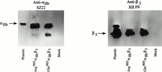
Western blotting was performed on lysates from transfected Cos-7 cells to further ensure that the lower expression of the mutated complex at the cell surface was not caused by a lower synthesis of one of the two proteins. As shown in Fig 4, αIIb and β3 subunits were present at approximately the same level in Cos-7 cells after cotransfection of wild-type β3 cDNA and wild-type or mutated αIIb cDNA. The low molecular weight product detected with SZ22 for mutated αIIb was not observed in all experiments and was not observed when Western blotting was performed on the patient’s platelets.18
Analysis of IIb and β3proteins in Cos-7 cells by immunoblotting. Fifty micrograms of proteins from each cell lysate transiently cotransfected with wild-type β3 and wild-type or mutated IIb as indicated were separated on 7% polyacrylamide gels without disulfide reduction and transferred to nitrocellulose membrane. One microgram of protein from normal platelets was used as a positive control; mock refers to cells transfected with expression vector alone. An MoAb specific for IIb (SZ22) and an MoAb specific for β3 (XIIF9) were used.
Analysis of IIb and β3proteins in Cos-7 cells by immunoblotting. Fifty micrograms of proteins from each cell lysate transiently cotransfected with wild-type β3 and wild-type or mutated IIb as indicated were separated on 7% polyacrylamide gels without disulfide reduction and transferred to nitrocellulose membrane. One microgram of protein from normal platelets was used as a positive control; mock refers to cells transfected with expression vector alone. An MoAb specific for IIb (SZ22) and an MoAb specific for β3 (XIIF9) were used.
PAC-1 Binding to Transfected CHO Cells
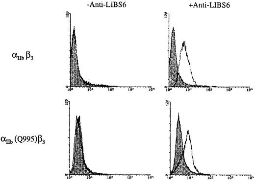
A CHO cell line stably expressing the β3 subunit was transfected using pCDN wild-type or mutated αIIb cDNA. After neomycin selection, evaluation of the surface expression of αIIbβ3 again showed a low surface expression of the mutated complex (data not shown). Replating under conditions of limiting dilution was followed by the selection of a clone that expressed the mutated αIIbβ3complex in readily measurable levels. Binding of PAC-1 to these cells and to a stable CHO cell line containing wild-type αIIband β3 is shown in Fig 5. Little or no binding was observed to either cell line, indicating that the mutated complex was not in a high-affinity state. At the same time, greater than 95% of the cells bound PerCP-conjugated anti-β3 or either of the MoAbs P2 or EDU-3 (anti–GP IIb-IIIa complex, data not shown). In contrast, incubation of cells expressing both the wild-type and the mutated complex with the activating MoAb, anti-LIBS6, induced the binding of PAC-1 (Fig 5). In other words, the mutated complex was only able to bind PAC-1 after activation. This result is in agreement with the previously reported observation that platelets of patient A.P. did not spontaneously bind fibrinogen but bound this adhesive protein after activation.18 The anti–LIBS6-induced binding of PAC-1 was not observed in the additional presence of 1 mmol/L RGDS, a competitive inhibitor of PAC-1 for αIIbβ3 (not illustrated). Hughes et al20 have expressed the activation state of αIIbβ3 as the AI (see Materials and Methods). Typical results for the CHO line expressing αIIbβ3 with αIIb (R995Q) showed the AI to be 23, whereas that obtained for the wild-type αIIbβ3 was 8. The ratio for AI 995Q/AI wild-type was therefore of the order of 3, whereas the corresponding ratio given by Hughes et al20 for 995A/wild-type was of the order of 10 (see Discussion).
Determination of the activation state of the mutated IIbβ3 complex in a stable CHO cell line using flow cytometry. Cells stably transfected with wild-type β3 and wild-type IIb or wild-type β3 and mutated IIb were incubated with FITC–PAC-1 in the presence or absence of 2 μmol/L anti-LIBS6 as detailed in Materials and Methods. Log of the fluorescence is on the abscissa and the number of cells examined is on the ordinate. A total of 5,000 cells were analyzed. PAC-1 binding was assessed in the presence (░) or absence (□) of 1 mmol/L RGDS peptide as shown. Note that PAC-1 binding requires the presence of the activating MoAb and was inhibited by RGDS.
Determination of the activation state of the mutated IIbβ3 complex in a stable CHO cell line using flow cytometry. Cells stably transfected with wild-type β3 and wild-type IIb or wild-type β3 and mutated IIb were incubated with FITC–PAC-1 in the presence or absence of 2 μmol/L anti-LIBS6 as detailed in Materials and Methods. Log of the fluorescence is on the abscissa and the number of cells examined is on the ordinate. A total of 5,000 cells were analyzed. PAC-1 binding was assessed in the presence (░) or absence (□) of 1 mmol/L RGDS peptide as shown. Note that PAC-1 binding requires the presence of the activating MoAb and was inhibited by RGDS.
RT-PCR on Platelet RNA
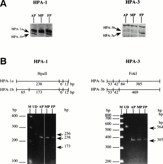
The mutation leading to the R to Q amino acid substitution was present in a heterozygous state in the patient’s genome and was unlikely on its own to account for a surface expression of 15% of the complex on his platelets.18 Because we failed to find any other abnormality using SSCP, we next analyzed platelet mRNA. Our strategy was to look for the genetic markers of αIIbβ3 (as revealed using genomic DNA) in mRNA isolated from platelets of the patient and his parents. By using RT-PCR and ASRA, we were able to determine if both alleles of each gene were effectively expressed or at least present at a detectable level. Using random oligonucleotides, we reverse transcribed platelet RNA and with specific oligonucleotides for αIIb and for β3 we amplified the appropriate regions of both cDNAs. Figure 6 shows that mRNA carrying the genetic determinant for HPA-1b and encoded by the β3allele inherited from his father was undetectable in platelets from A.P. although present in his father’s platelets. Analysis of theTaqI polymorphism carried on the β3 gene confirmed this result (Table 1). On its own, this would explain the lower total content of αIIbβ3 complexes in the patient’s platelets as compared with those of his father.18 However, mRNA carrying the HPA-3b determinant and coded by the αIIb allele inherited from his mother’s genome was also not detected in RNA isolated from platelets of the patient. In this case, the corresponding mRNA was absent from his mother’s platelets. Study of a second polymorphism carried by the same allele, a silent C to T mutation in exon 30, confirmed these findings (Table 1). Sequencing also showed that mRNA coded by the mutated αIIb allele from his father was exclusively present in the platelets of A.P. Thus, the data which are summarized in Table 1suggest that A.P. may have inherited from his mother an unexpressed allele for αIIb, the other αIIb allele coming from his father being mutated and giving rise to a R to Q substitution in the highly conserved cytoplasmic GFFKR domain. The low total αIIbβ3 complex content in A.P.’s platelets are therefore due to a combination of factors, one or more of which remain to be elucidated.
HPA-1 and HPA-3 typing on DNA and on mRNA from platelets from patient A.P., his mother (M.P.), and his father (F.P.). (A) SSCP analysis was performed on genomic DNA fragments. (B) Amplified cDNA was subjected to restriction enzyme digestion, HpaII for HPA-1 andFokI for HPA-3. The predicted size of the products is indicated. Analysis was performed on 10% polyacrylamide gels stained using EtBr (see arrows). M, size markers; UD, undigested fragment.
HPA-1 and HPA-3 typing on DNA and on mRNA from platelets from patient A.P., his mother (M.P.), and his father (F.P.). (A) SSCP analysis was performed on genomic DNA fragments. (B) Amplified cDNA was subjected to restriction enzyme digestion, HpaII for HPA-1 andFokI for HPA-3. The predicted size of the products is indicated. Analysis was performed on 10% polyacrylamide gels stained using EtBr (see arrows). M, size markers; UD, undigested fragment.
DISCUSSION
We have described the molecular analysis of αIIb and β3 from a patient with an atypical form of Glanzmann’s thrombasthenia. In their initial report on this patient, Hardisty et al18 showed that the density of αIIbβ3 complexes on his platelets was strongly decreased (approximately 15% of the normal level). However, thrombin stimulation was followed by the appearance on the surface of a substantial internal pool of αIIbβ3. Although a residual aggregation response was observed with physiological agonists, the kinetics were slower and the size of the aggregates much smaller than usual. Fibrinogen-binding experiments confirmed that the residual complexes were functional. This point is perhaps reinforced by the presence of fibrinogen in the α-granules of the patient’s platelets, a presence presumed to be caused by the αIIbβ3-dependent internalization of plasma fibrinogen.33 To define the genetic defect responsible for the disease, we performed an extensive analysis of the exons of the αIIb and β3 genes using nonradioactive PCR-SSCP methodology developed in our laboratory. We found that a heterozygous G to A mutation at nucleotide 3078 of αIIbcDNA, leading to an R995 to Q amino acid substitution in αIIb, was sufficient to decrease surface αIIbβ3 expression by approximately 50% when mutated αIIb was coexpressed with wild-type β3 in Cos-7 cells. Because this mutation was heterozygous in the patient, intriguing questions were raised relating to the extent of the observed decrease of αIIbβ3 on the surface of his platelets (approximately 15% of normal levels) and the altered repartition of the integrin within the surface and internal membrane systems.
In view of this unusual phenotype, we performed RT-PCR on platelet extracts for the patient and both parents to determine if both alleles of the two genes were correctly expressed. First, we found that mRNA corresponding to the αIIb allele carrying the HPA-3b determinant and coded for by DNA from his mother and the propositus was not detected in the platelets of either subject. Secondly, we failed to find mRNA for the HPA-1b–containing β3 allele in the platelets of the patient despite the fact that SSCP and genomic analysis indicated that mRNA for this allele was present in platelets from his father. It would be of interest to serologically type the patient’s platelets for the expression of the HPA-1a and HPA-1b alloantigens using specific alloantibodies, but the very low level of αIIbβ3 at the cell surface makes this difficult. At the present time, we tentatively conclude that additional and so far unidentified DNA abnormalities contribute to the low or absent expression of two of the alleles. Such abnormalities could affect noncoding regions of the αIIb or β3genes (promotor sequences, for example). Alternatively, they may have been missed by the SSCP procedure which is known to have less than a 100% detection rate, particularly when the size of the amplified products reaches 300 bp or more.34 We also cannot exclude the possibility of exon skipping within one or more alleles, an example of which has been recently described.35 Whatever the cause, it should be emphasized that only the mRNA coding for the mutated αIIb was detected in A.P. platelets, suggesting that Q995 predominated in the αIIbβ3complexes that were present.
In our original report, we showed that platelets from the patient’s father (F.P.) expressed approximately 28,000 αIIbβ3 molecules as measured using radiolabeled AP-2 (anti-αIIbβ3) and Tab (anti-αIIb) in direct binding studies.18 This compared with a mean value of 37,000 molecules when αIIbβ3 expression was assessed on platelets from a series of 27 normal subjects using AP-2. Such a value for the father is compatible with the presence of a wild-type αIIb allele which leads to a normal cell-surface expression and a Q995 αIIb mutated allele leading to a reduced expression. The platelets of the mother expressed approximately 20,000 molecules of αIIbβ3,18 a value compatible with the presence of two alleles coding for β3 and one allele coding for αIIb. Nevertheless, comparing the surface αIIbβ3 expression of individuals within a large population of normal donors is made difficult by the fact that numbers differ widely and that overlap can occur with obligate heterozygotes for type I Glanzmann’s disease.36Because the only expressed allele for αIIb in the platelets of the propositus was mutated, the abnormal surface expression compared with the internal compartment would suggest a role for the GFFKR domain in protein trafficking. Significantly, on the basis of our results, a subject with a homozygous expression of the Q995 αIIb mutated allele and two normally expressed β3 alleles would possess platelets with sufficient surface-expressed αIIbβ3 to support platelet aggregation and for a bleeding diathesis not to be observed (as is the situation for heterozygotes in classic Glanzmann’s thrombasthenia). Thus, the bleeding syndrome in A.P. may be related to the special combination of defects in his case.
Much of the interest in the described amino acid substitution resides in its location in the highly conserved GFFKR sequence found in the cytoplasmic tail of all α subunits of human integrins and in all steroid hormone receptors (consensus sequence K×FF[K/R]R). Burns et al37 showed that overexpression of calreticulin, a calcium-binding protein present in the endoplasmic reticulum and also in the nucleus, inhibited glucocorticoid response–mediated transcriptional activation of a glucocorticoid-sensitive reporter gene and concluded that the binding of this protein to the GFFKR sequence maintained the receptor in a low-affinity state. These observations are relevant, because O’Toole et al38 showed that a deletion mutant in which αIIbβ3lacked the bulk of the cytoplasmic tail of the αΙΙbsubunit (they retained two amino acids adjacent to the transmembrane domain) was in a high activation state able to bind ligand (fibrinogen) directly when expressed in a CHO cell line. O’Toole et al39 then showed that replacement of the αIIbcytoplasmic tail by the α5 cytoplasmic tail gave rise to a αIIbβ3 chimeric receptor in a high activation state. This suggested that factors other than the GFFKR sequence per se influenced the shift from a low to a high activation state. Nonetheless, it was suggested that the GFFKR sequence could influence the interaction between the α- and β-subunit transmembrane peptides, and ultimately, the conformation of the extracellular domains. Kassner et al40 showed that replacement of the cytoplasmic tail of the α4 integrin by a cytoplasmic tail from other α subunits did not alter the affinity state and that the effects of any change were influenced by the cell type. In contrast, Utsumi et al41 showed that truncation of the α4 integrin subunit before the GFFKR sequence abolished α4β1 cell-surface expression. Kassner et al42 then showed that a deletion just after this sequence did not prevent expression but induced a much less active receptor. It was suggested that the affinity state may be regulated by the five to seven amino acids after the GFFKR sequence.
In terms of these data, patient A.P., who represents the first example of a mutation in the GFFKR region being detected in an inherited disease, represents a natural model for studying the role of this sequence in controlling the activation state of αIIbβ3. Initial experiments with Cos-7 cells transfected with αIIbβ3 containing αIIb (R995Q) confirmed that the mutation results in a lower cell-surface expression of the complex. Then, experiments with a stable CHO cell line transfected with αIIbβ3 containing αIIb(R995Q) showed that the mutated complex was not in a high activation state but remained functional in that activation could be induced by the anti-LIBS6 antibody. Recently, two opposing studies were published on this subject. Low et al21 showed that transfection of Epstein-Barr virus–transformed B lymphocytes with wild-type β3 and αIIb truncated proximal to the GFFKR region led to a reduced expression of the mutated αIIbβ3 complex. These authors further showed (1) that the αIIb tail was not required for αIIbβ3 function and (2) that substitution of the GFFKR sequence by AAAAA resulted in an almost total loss of αIIbβ3 cell-surface expression because of an impairment in the ability of αIIb to associate with β3. Nevertheless, replacement of GFFKR by AAAAA gave rise to a complex which enabled the transfected lymphocytes to more easily adhere to fibrinogen when expressed at the lymphocyte cell surface. In contrast, Hughes et al20 reported that substitution of F, F, or R in the GFFKR sequence of the αIIb cytoplasmic tail by alanine resulted in a high-affinity receptor when transiently expressed in CHO cells. The same result was obtained when D723 of the β3 subunit was substituted by an A. Thus, the authors suggested that a salt bridge between D723 and R995 participates in the regulation of the activation state of the complex although the literature suggests that the situation is more complicated. The amino acid substitution that we have found did not give rise to a high activation state receptor. This may be because of the nature of the amino acid change. Indeed, the glutamine residue, an uncharged residue having a polar nature, could induce a modified interaction between the αIIb and β3 cytoplasmic tails and/or between cellular factors giving rise to receptor not locked in a high activation state but more easily activatable. This would fit with the results that we have obtained when using stable CHO cell lines. Here, the activation index was higher for 995Q than for the wild-type but was much weaker than that observed by others when the 995R was muted to 995A.20 As a result, the patient’s platelets would not be expected to spontaneously bind fibrinogen, and this was the case.18
In view of our results, it is reasonable to consider that cell-surface expression and a high activation state of αIIbβ3 are not independent processes. Natural mutations giving rise to a constitutive high activation state receptor may be lethal if fully expressed, and give rise to a permanent thrombotic state. Because cell-surface expression of one subunit is dependent on the presence of the other, mutations inducing modifications in subunit association, even in cytoplasmic domains,21 can affect the efficiency of membrane expression. In the case of αIIbβ3, this could be the result of natural selection to impair a normal cell membrane expression of high affinity receptors. It is of interest to compare our results with those reported for another variant of Glanzmann’s thrombasthenia in which a role for the cytoplasmic domain of the β3 subunit was highlighted. Chen et al17 showed that in the variant termed Lariboisière I, substitution of Ser752 to Pro in the cytoplasmic tail of the β subunit resulted in a receptor that was locked in a low affinity state. Significantly, Ylänne et al43 showed that this effect was mainly caused by the presence of the proline, which induced a β-turn in the protein. Other recent work has established that a novel 111-residue polypeptide, designated β3-endonexin, interacts with the cytoplasmic tail of β3 (although not when the S752 → P mutation is present) and is a potential modulator of the affinity state of αIIbβ3.44 It would seem that both cytoplasmic domains have key roles.
Recently, Coppolino et al45 highlighted that calreticulin is essential for integrin-mediated calcium signaling and cell adhesion. Because calreticulin is also a calcium-binding protein, it is tempting to speculate that this protein can play a major role in the activation state of integrins by modifying the salt bridge between the two subunits. Another candidate protein reacting with the GFFKR domain of αIIb has also been described.46 In a general context, Briesewitz et al47 showed that functional specificity was under the control of the α subunit of the α1β1 integrin and that the assembly of the two subunits was dependent on both cytoplasmic domains. Further experiments are planned to determine the effect of the mutation that we have described in the GFFKR region on its capacity (1) to bind potential regulatory proteins, (2) influence the stability of the interaction of αIIb with β3, and (3) to modify intracellular trafficking of αIIbβ3within cells. With this goal, transfection experiments are planned in cells showing megakaryocyte-like features but lacking endogenous αIIb.
In conclusion, to the best of our knowledge, this is the first time that an amino acid substitution in the highly conserved cytoplasmic GFFKR sequence of an integrin α subunit has been reported in a human genetic disease.
Supported by fundings from the CNRS, Université de Bordeaux II, the Conseil Régional d’Aquitaine, and the Ministère de l’Enseignement Supérieur et de la Recherche (ACC-SV N°9). O.P. was a recipient of a postdoctoral fellowship from the Société Française d’Hématologie.
Address reprint request to F. Bourre, PhD, UMR 5533 CNRS, Hôpital Cardiologique, Avenue de Magellan, 33604 Pessac, France.
The publication costs of this article were defrayed in part by page charge payment. This article must therefore be hereby marked “advertisement” in accordance with 18 U.S.C. section 1734 solely to indicate this fact.

This feature is available to Subscribers Only
Sign In or Create an Account Close Modal