To the Editor:
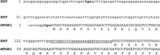
In an attempt to identify potential new genes homologous to ETO,1,2 we screened the dbEST database (http://www.ncbi.nlm.nih.gov./dbEST/index.html) using the entire ETO cDNA sequence as a probe (EMBL accession no. X79990). Among the ESTs identified, we selected two overlapping clones (clone IDs 43629 and 274508) and sequenced them to completion. A putative translation initiation site was identified by the presence of a strong Kozak consensus sequence,3 followed by a 1,725-bp open reading frame (ORF) coding for a putative protein of 575 amino acids (aa) (GenBank accession no. AF068266). We named this gene EHT (ETO Homologous on chromosome Twenty; see mapping data below). The putative EHT protein is closely homologous to ETO/MTG8 (≈65% identity) and Nervy, an ETO Drosophila homolog (≈24% identity) showing the four conserved domains found in MTG8 and Nervy.4 To find EST clones that could represent the EHT 5′UTR, we screened the dbEST data bank with the 5′ end of clone 274508, and identified an EST clone (GenBank accession no.AA635096) encompassing the EHT ATG codon, preceded by an in-frame stop codon 129 bp upstream (Fig 1, top). Therefore, we considered this sequence the putative 5′UTR of EHT gene. While this work was in progress, the cloning of a similar ORF, named MTGR1 (myeloid translocation gene-related protein 1) was reported5; it presents an ATG codon, immediately preceded by a stop codon and with no clear Kozak sequence, upstream to and in-frame with the EHT ATG (see Fig 1, top), leading to the coding of additional 29 aa with respect to EHT ORF. Interestingly, Calabi and Cilli have recently deposited in the dbEST data bank a 5′ UTR MTGR1 sequence (accession no. AF052212) that is identical to the putative EHT 5′ UTR (GenBank clone AA635096). Overall, these data suggest the presence of two alternative 5′ ends in the MTGR1/EHT gene, similar to ETO/MTG8 gene.1 2 The existence in nature of EHT ORF was confirmed by direct sequencing of a reverse transcriptase-polymerase chain reaction (RT-PCR) amplified fragment encompassing the coding sequence from HL60 mRNA (data not shown).
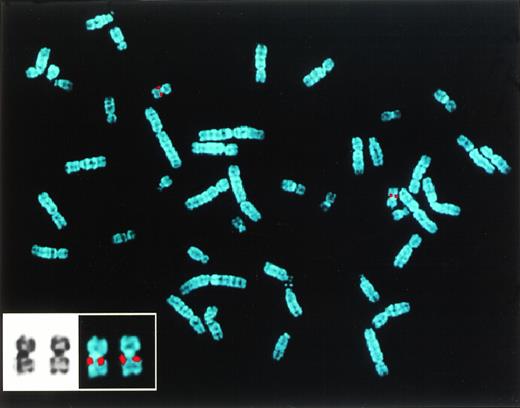
(Top) Sequence comparison between the 5′ end of EHT and MTGR1 (italics) cDNAs. Translated sequences are shown in capital letters; a 27-bp nucleotide stretch upstream of the EHT ATG which is similar to the MTGR1 is underlined. The stop codons upstream of ATGs are in bold letters. (Bottom) Mapping by FISH of EHT gene on chromosome 20q11.
(Top) Sequence comparison between the 5′ end of EHT and MTGR1 (italics) cDNAs. Translated sequences are shown in capital letters; a 27-bp nucleotide stretch upstream of the EHT ATG which is similar to the MTGR1 is underlined. The stop codons upstream of ATGs are in bold letters. (Bottom) Mapping by FISH of EHT gene on chromosome 20q11.
To map the EHT gene, we generated by PCR a probe from the specific 3′ UTR of the EHT gene and screened a human placenta cosmid library (Clontech, Palo Alto, CA), isolating a specific cosmid clone subsequently used as probe in fluorescence in situ hybridization (FISH) analysis on normal human metaphase spreads. By FISH we mapped the gene on chromosome 20q11 (Fig 1, bottom); the mapping was further refined by PCR of DNA from somatic cell hybrids (kindly provided by Mariano Rocchi, University of Bari, Italy) on 20q11.2-20q11.3 (data not shown).
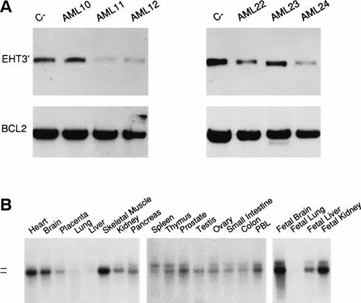
Cytogenetic studies have shown that this region is deleted in ≈10% cases of polycythemia vera (PV),6,7 ≈5% myelodysplastic syndromes (MDS), and in ≈3% of acute myeloid leukemias (AML).8-11 We performed Southern blot analysis on 40 cases of AML (9 M0, 8 M1, 6 M2, 9 M4, 8 M5) and found gene deletion apparently in four cases (1 M0, 1 M1, 2 M4) (10%) (Fig2A), a frequency higher than that found by conventional cytogenetics. However, the assessment of the deletion frequency needs the analysis of a more representative series. Furthermore, as no RNA was available from hemizygous deleted patients, we were not able to test whether EHT may be a candidate tumor suppressor gene of the region by mutation analysis of the retained allele.12
(A) Analysis of EHT gene in AML cases by quantitative Southern blot: cases AML11, 12, 22, and 24 show a loss of signal with respect to the control probe (PFL-1 probe for Bcl-2 gene). Because the percentage of leukemia cells in all cases is greater than 70%, this finding is consistent with an homozygous deletion. The probe (EHT3′) was specific for the 3′ UTR of EHT. (B) EHT gene expression analysis. Poly-A+ Northern blot filters (Clontech), were hybridized with the EHT3′ probe. The two dashes indicate the molecular weight (9.5 and 7.5 kb). The two transcripts were not represented in all of the tissues at the same level; in the testis, heart, brain, and skeletal muscle, the lower transcript was particularly expressed.
(A) Analysis of EHT gene in AML cases by quantitative Southern blot: cases AML11, 12, 22, and 24 show a loss of signal with respect to the control probe (PFL-1 probe for Bcl-2 gene). Because the percentage of leukemia cells in all cases is greater than 70%, this finding is consistent with an homozygous deletion. The probe (EHT3′) was specific for the 3′ UTR of EHT. (B) EHT gene expression analysis. Poly-A+ Northern blot filters (Clontech), were hybridized with the EHT3′ probe. The two dashes indicate the molecular weight (9.5 and 7.5 kb). The two transcripts were not represented in all of the tissues at the same level; in the testis, heart, brain, and skeletal muscle, the lower transcript was particularly expressed.
We investigated the expression pattern of EHT by means of Northern blot analysis using the 3′ UTR specific probe. Two transcripts of ≈9.5 and 7.5 kb (Fig 2B) were detected; the difference in size of the two transcripts may be due to the use of different polyadenylation signals and/or alternative 5′UTR regions, as in the case of the MTGR8 gene.13 In normal fetal and adult human tissues, the two transcripts were expressed ubiquitously, although at different levels (Fig 2B); in tumoral cell lines, low levels of expression were detected in hematopoietic cell lines of lymphoid and myeloid origin and melanoma cells, while higher levels of expression were found in the SW480 carcinoma cell line, but apparently absent in the A549 lung carcinoma cell line (data not shown).
As far as normal MTGR1/EHT function is concerned, Kitabayashi et al5 showed the direct interaction of MTGR1 and the AML1-MTG8 fusion protein, leading to an enhancement of cell proliferation mediated by granulocyte colony-stimulating factor (G-CSF) in a murine myeloid model (L-G cell line). This suggests that MTGR1 has an oncogenic rather than a tumor suppressor activity. Nevertheless, when MTGR1 is transfected alone in L-G cells, the proliferative response to G-CSF was lower than in the normal control, thus suggesting a possible negative growth-control in normal cells.
In conclusion, the data presented here and those previously reported suggest that the MTGR1/EHT gene may represent a new candidate for the tumor suppressor gene supposed to be involved in the deletion of the 20q11 region in myeloid tumors. Further studies are necessary to rule out this hypothesis.
ACKNOWLEDGMENT
A partial cDNA sequence of the EHT gene was deposited by us in the GenBank database under accession number AF039200, on 18-DEC-1997, before the MTGR1 delivery date (January 22, 1998). This work was supported by a grant from Associazione Italiana Ricerca sul Cancro (AIRC) to A.N.


This feature is available to Subscribers Only
Sign In or Create an Account Close Modal