Abstract
Extracellular signal-regulated kinase (ERK) is an important intermediate in signal transduction pathways that are initiated by many types of cell surface receptors. It is thought to play a pivotal role in integrating and transmitting transmembrane signals required for growth and differentiation. Constitutive activation of ERK in fibroblasts elicits oncogenic transformation, and recently, constitutive activation of ERK has been observed in some human malignancies, including acute leukemia. However, mechanisms underlying constitutive activation of ERK have not been well characterized. In this study, we examined the activation of ERK in 79 human acute leukemia samples and attempted to find factors contributing to constitutive ERK activation. First, we showed that ERK and MEK were constitutively activated in acute leukemias by in vitro kinase assay and immunoblot analysis. However, in only one half of the studied samples, the pattern of ERK activation was similar to that of MEK activation. Next, by semiquantitative reverse transcriptase-polymerase chain reaction (RT-PCR) and immunoblot analysis, we showed hyperexpression of ERK in a majority of acute leukemias. In 17 of 26 cases (65.4%) analyzed by immunoblot, the pattern of ERK expression was similar to that of ERK activation. The fact of constitutive activation of ERK in acute leukemias suggested to us the possibility of an abnormal downregulation mechanism of ERK. Therefore, we examined PAC1, a specific ERK phosphatase predominantly expressed in hematopoietic tissue and known to be upregulated at the transcription level in response to ERK activation. Interestingly, in our study, PAC1 gene expression in acute leukemias showing constitutive ERK activation was significantly lower than that in unstimulated, normal bone marrow (BM) samples showing minimal or no ERK activation (P = .002). Also, a significant correlation was observed between PAC1 downregulation and phosphorylation of ERK in acute leukemias (P= .002). Finally, by further analysis of 26 cases, we showed that a complementary role of MEK activation, ERK hyperexpression, and PAC1 downregulation could contribute to determining the constitutive activation of ERK in acute leukemia. Our results suggest that ERK is constitutively activated in a majority of acute leukemias, and in addition to the activation of MEK, the hyperexpression of ERK and downregulation of PAC1 also contribute to constitutive ERK activation in acute leukemias.
EXTRACELLULAR signal-regulated kinase (ERK) is a signaling molecule common to pathways that regulate the proliferation and differentiation in diverse cell types including hematopoietic cells.1,2 In blood cells and their precursors, the activation of ERK has been shown to be involved in the proliferation and cellular response of various hematopoietic cytokines, including steel factor, granulocyte-macrophage colony-stimulating factor, interleukin-3 (IL-3), and IL-5.3-6 In response to a wide range of cytokine and growth factor stimuli, activated ERK is translocated into the nucleus and activates a number of nuclear transcription factors7,8 that lead to a dramatic recruitment and activation of a large group of cellular regulatory processes. As far as the constitutive activation of ERK is concerned, the importance of the activation of ERK by sequential upstream kinases, referred to as the ERK cascade, has been generally accepted. On stimulation by a variety of growth factors, the protein kinases, Raf and MEK kinase (MEKK), MAPK/ERK kinase (MEK), and ERK, are successively activated by phosphorylation.9 10
Recently, a constitutively active mutant of MEK has been shown to transform NIH3T3 cells,11,12 and some reports have shown constitutive activation of ERK in human malignancies, including acute leukemia.13-15 Although previous studies showed constitutive activation of ERK in human malignancies, detailed mechanisms underlying such activation have not been well characterized. A previous study with acute leukemia showed that samples with constitutively activated ERK also showed elevated MEK activity.13 This result supports the hypothesis that MEK activation is necessary for ERK activation. However, Oka et al14 reported that some discrepancy between ERK and MEK activation was observed in renal cell carcinoma and suggested the possibility of the presence of other mechanisms for constitutive activation of ERK. Also, in previous studies with human malignancies,13,14 no cases or only a small percent showed mutation of ras, an upstream regulator of ERK cascade despite a high frequency of constitutive activation of ERK. These results indicate the possibility of other regulatory mechanisms, which may be critical in the constitutive activation of ERK. In this respect, it is a notable finding that hyperexpression of ERK, as a possible additional mechanism of constitutive ERK activation, was found in breast cancer.15
In general, the extent of protein phosphorylation is balanced by antagonism of kinase and phosphatase. Therefore, recently cloned dual-specificity protein-tyrosine phosphatases (PTPases) that exhibited dual catalytic activity toward phosphotyrosine and phosphothreonine in substrate proteins may play a pivotal role in the regulation of the ERK signaling pathway.16-18 Phosphatase of activated cells (PAC1), a member of the ERK phosphatase family, predominantly expressed in hematopoietic tissues, exhibits stringent substrate specificity for ERK in vitro.18-21 The kinetics of gene expression and nuclear localization and the ability of PAC1 to inactivate ERK are all consistent with a role of this phosphatase in the compensatory inactivation of the stimulated ERK signaling pathway.
Based on these observations, we attempted to find molecular mechanisms underlying constitutive ERK activation in acute leukemias. In this study, we have examined the relationship between ERK activation and MEK activation and also whether the level of ERK expression contributes to the activation of ERK. Finally, we have studied the regulation of PAC1, a phosphatase induced by the activation of ERK.
MATERIALS AND METHODS
Cells.
We used 79 programmed-frozen leukemia and 10 normal bone marrow (BM) samples, which were separated by Ficoll-Hypaque. Acute leukemias included 67 acute myelocytic leukemias (AMLs; one M0, 10 M1, 12 M2, 12 M3, 22 M4, 10 M5 in the French-American-British classification) and 12 acute lymphoblastic leukemias (ALLs).22
Purification of CD34+ cells.
To investigate the adequacy of unselected normal marrow cells for comparison with immature leukemic cells, we also studied CD34+ cells, the early hematopoietic stem/progenitor cells, from one normal marrow donor. CD34+ cells were purified by immunomagnetic bead methods using anti-CD34 monoclonal antibody (Miltenyl Biotech Inc, Bergisch Gladbach, Germany). The purity of isolated CD34+ cells was more than 90% by flow cytometry (FACScan; Becton Dickinson, Lincoln Park, NJ).
Cell cultures.
K562 cells (American Type Culture Collection Certified Cell Lines [ATCC CCL] 243) were maintained in RPMI 1640 medium containing 10% fetal bovine serum supplemented with 100 U/mL penicillin, 100 μg/mL streptomycin, 2 mmol/L L-glutamine, 1 mmol/L sodium pyruvate, and 1 mmol/L nonessential amino acids.
In vitro ERK assay.
For kinase assay and immunoblotting, samples were rapidly thawed and washed twice with phosphate-buffered saline containing 1 mmol/L sodium orthovanadate. The viability assay was performed by the trypan blue exclusion test. All acute leukemia samples showed over 90% viable blast cells. Whole cell lysates were extracted as reported previously.23 Concisely, the cells were lysed in lysis buffer (50 mmol/L β-glycerophosphate, 1 mmol/L sodium orthovanadate, 20 mmol/L HEPES [pH, 7.4], 2 mmol/L EDTA, 1 mmol/L dithiothreitol, 1% Triton X-100, 1 mmol/L phenylmethylsulfonyl fluoride [PMSF], 2 μg/mL leupeptin, 25 μg/mL aprotinin, 10% glycerol). After incubating for 30 minutes at 4°C, lysates were centrifuged at 25,000g for 20 minutes. The protein concentration was determined with aid of a Bio-Rad Protein Assay Kit (Bio-Rad Laboratories, Richmond, CA). The activity of ERK was assayed by measuring the kinase activity for the synthesized peptide containing PLS/TP, a recognition sequence for ERK1/2 (Amersham, Buckinghamshire, UK).24 Per manufacturer’s information, this peptide contains no other phosphorylation sites and is much more specific for ERK1/2 than the commonly used substrate, myelin basic protein. Reaction mixture containing 30 μg of protein was spotted onto phosphocellulose paper (Amersham) and washed in 75 mmol/L phosphoric acid. The radioactivity on the filter was measured by densitometry scanning (BAS2500; FUJIX, Tokyo, Japan).
Immunoblot analysis.
The lysates containing the same amounts of protein were subjected to 10% sodium dodecyl sulfate-polyacrylamide gel electrophoresis (SDS-PAGE) and transferred electrophoretically onto immobilon-P membrane (Millipore Corp, Bedford, MA). The transfer efficiency was confirmed by Ponceau-S staining. Blots were blocked in 5% nonfat dried milk in 20 mmol/L Tris-HCl (pH 7.6), 137 mmol/L NaCl, and 0.05% Tween 20 and then probed with a phospho-specific ERK1/2 antibody that recognizes Thr202/Tyr204 phosphorylated ERK1/2, a phospho-specific MEK1/2 antibody that recognizes Ser217/221 phosphorylated MEK1/2 or a total ERK1/2 antibody that recognizes total ERK1/2 (New England Biolabs, Beverly, MA). Also, we examined the immunoblot analysis using α-tubulin (Oncogene, Cambridge, MA) as a protein loading control to adjust the difference in protein loading amounts for individual samples. Immunoreactive bands were detected by horseradish peroxidase (HRP)-conjugated secondary antibody using enhanced chemiluminescence reagents (Amersham). The signal intensity of the autoradiogram was quantified by using densitometry scanning and the value of the phosphorylation or expression of each molecule divided by the α-tubulin expression was used for comparison with the phosphorylation or expression of each molecule.
Semiquantitative RT-PCR.
Total RNA from leukemia and normal BM cells was extracted with commercial kit (RNeasy Mini Kit; QIAGEN, Hilgen, Germany). First-strand cDNA was synthesized in 20 μL reaction mixture containing 1 μg of total RNA, 1 mmol/L of each deoxynucleotide triphosphate (dNTP), 20 U of avian myeloblastosis virus (AMV) reverse transcriptase (Boehringer Mannnheim, Mannheim, Germany), 1.6 μg of oligo-dT primer. The 50-μL PCR reaction mixture contained cDNA derived from 100 ng of total RNA, 1.25 U Taq DNA polymerase, 0.2 mmol/L of each dNTP, 0.2 μmol/L of each primer, 1.5 mmol/L MgCl2. The sequences of the primers were: ERK-225: sense 5′-TCTGTAGGCTGCATTCTGGC-3′; antisense 5′-GGCTGGAATCTAGCAGTC-3′/PAC118: sense 5′-TTGCCCTACCTGTTCCTGGG-3′; antisense 5′-GTCTCAAACTGCAGCAGCTG-3′/β-actin26: sense 5′-GTGGGGCGCCCCAGGCACCA-3′; antisense 5′-GTCCTTAATGTCACGCACGATTTC-3′. PCR was performed with a DNA thermal cycler (Perkin Elmer-Cetus, Norwalk, CT) under the following conditions: denaturation at 94°C for 1 minute, primer annealing at 60°C for ERK2 and β-actin or at 64°C for PAC1 for 1 minute and then chain elongation at 72°C for 2 minutes. Optimal conditions for RT-PCR to quantitate the expression of each gene were determined as described previously,27 and we performed the amplification at 25 cycles for ERK2 and β-actin or at 22 cycles for PAC1. Ten microliters of PCR products were separated on 1% agarose gels containing 0.05 μg/mL of ethidium bromide and examined. We analyzed the levels of ERK-2 and PAC1 gene expression using densitometry scanning.
Statistical analysis.
Student’s t-test was used for comparison of in vitro ERK activity and expression level of each gene by RT-PCR between leukemia and normal BM samples. Linear correlation between expression levels of ERK from RT-PCR and those from immunoblot analysis was determined by calculating Pearson’s correlation coefficient. Fisher’s exact probability test was used for all 2 × 2 tables. Data were analyzed with SPSS statistical software package (SPSS Inc, Chicago, IL). A P value less than .05 was considered to be statistically significant.
RESULTS
Constitutive activation of ERK in acute leukemias.
To determine whether ERK is activated in acute leukemia, we first examined the activity of ERK by a commercial kit with synthesized peptide for ERK1/2. The ERK activity in 79 leukemia samples (16.6 densitometric unit ± 1.2 standard error [SE]) was significantly greater than that in normal BM samples (5.2 densitometric unit ± 1.2 SE) (P = .003). When the kinase activity was analyzed according to the type of acute leukemia, AML samples (18.6 densitometric unit ± 1.3 SE) showed statistically higher activity than ALL samples (10.2 densitometric unit ± 1.7 SE) (P = .03). For AML, the average activity of ERK was lower in the M3 subtype than in other subtypes (P < .05). The increased ERK activity was confirmed by immunoblot analysis using phospho-specific ERK antibody in 26 leukemia samples. By immunoblot analysis, the phosphorylation of the ERK2 was predominant than that of ERK1 in acute leukemia samples (Fig 1), and thus we defined ERK phosphorylation as the intensity of the phosphorylated ERK2 band. Human K562 cells, a chronic myeloid leukemia cell line in blast crisis, showed high level of constitutive ERK activation (Fig 1). Therefore, ERK was arbitrarily defined to be constitutively active when the value of the ERK phosphorylation divided by the α-tubulin expression in the leukemia sample was greater than one third the value measured in the K562 cells, because the phosphorylation of normal BM samples was minimal or not detected. Activation of ERK in acute leukemias was detected in 19 of the 26 leukemia samples analyzed (73.1%) (Table 1) (Fig 1A, C, and D). Unselected normal BM cells are heterogeneous and may not be representative of acute leukemia because it is possible that ERK activation observed in acute leukemia is simply due to enrichment of immature progenitor cells, not to the leukemic process. Therefore, to determine whether unselected normal BM cells are adequate for comparison with immature leukemic cells and activation of ERK in acute leukemias is not simply due to enrichment of immature progenitor cells, we examined if the changes observed in acute leukemias occurred in normal immature BM cells. From one normal BM donor, we isolated CD34+ cells, the early hematopoietic stem/progenitor cells, by immunomagnetic bead methods and examined immunoblot analysis. By immunoblot analysis, the intensity of ERK phosphorylation in CD34+ cells was not different from that in normal BM cells (Fig 1A). Thus, these results suggest to us that unselected marrow cells may be adequate for comparison with leukemic cells and the results observed in acute leukemias are not simply associated with enrichment of immature progenitor cells.
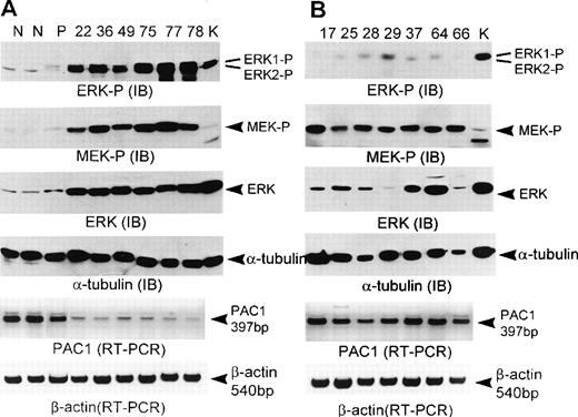
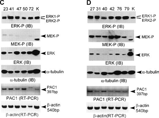
Complementary role of MEK activation, ERK hyperexpression, and PAC1 downregulation in determining the activation of ERK in human acute leukemias. Normal unselected BM (N), purified CD34+ (P), and acute leukemia samples were subjected to immunoblot (IB) and RT-PCR analysis as described in Materials and Methods. For immunoblot analysis, lysates containing the same amount of protein were applied to each lane; 25 μg of protein for phosphorylated ERK1/2 (ERK-P); 50 μg of protein for phosphorylated MEK1/2 (MEK-P); 25 μg of protein for total ERK1/2 (ERK); 25 μg of protein for -tubulin. In all immunoblot analyses, K562 samples (K) were loaded together as the evidence of same exposure time in autoradiogram. Separated proteins were transferred to the immobilon-P membrane and stained with phospho-specific ERK1/2 antibody or phospho-specific MEK1/2 antibody or total ERK1/2 antibody or -tubulin antibody, respectively. In immunoblot analysis of total ERK using 25 μg protein, the ERK1 band was not visualized because ERK1 detected by total ERK1/2 antibody was much less abundant than ERK2. For RT-PCR analysis, PCR was performed with PAC1 primer for 22 cycles, with β-actin primers for 25 cycles. PCR products derived from 20 ng of total RNA were applied to each lane. Leukemia samples above the lanes from (A) to (D) correspond with those in Table 1.
Complementary role of MEK activation, ERK hyperexpression, and PAC1 downregulation in determining the activation of ERK in human acute leukemias. Normal unselected BM (N), purified CD34+ (P), and acute leukemia samples were subjected to immunoblot (IB) and RT-PCR analysis as described in Materials and Methods. For immunoblot analysis, lysates containing the same amount of protein were applied to each lane; 25 μg of protein for phosphorylated ERK1/2 (ERK-P); 50 μg of protein for phosphorylated MEK1/2 (MEK-P); 25 μg of protein for total ERK1/2 (ERK); 25 μg of protein for -tubulin. In all immunoblot analyses, K562 samples (K) were loaded together as the evidence of same exposure time in autoradiogram. Separated proteins were transferred to the immobilon-P membrane and stained with phospho-specific ERK1/2 antibody or phospho-specific MEK1/2 antibody or total ERK1/2 antibody or -tubulin antibody, respectively. In immunoblot analysis of total ERK using 25 μg protein, the ERK1 band was not visualized because ERK1 detected by total ERK1/2 antibody was much less abundant than ERK2. For RT-PCR analysis, PCR was performed with PAC1 primer for 22 cycles, with β-actin primers for 25 cycles. PCR products derived from 20 ng of total RNA were applied to each lane. Leukemia samples above the lanes from (A) to (D) correspond with those in Table 1.
Constitutive Activation of ERK Cascade in Acute Leukemias
| Case . | Type . | ERK Activation . | MEK Activation . | ERK Hyperexpression . | PAC1 Downregulation . |
|---|---|---|---|---|---|
| 17 | M5 | − | +† | − | − |
| 22 | M3 | +* | + | +‡ | +1-153 |
| 23 | M2 | + | − | + | + |
| 25 | M2 | − | + | − | − |
| 27 | M3 | + | + | − | + |
| 28 | M1 | − | + | − | + |
| 29 | M4 | − | + | − | − |
| 31 | M3 | + | + | − | + |
| 36 | M4 | + | + | + | + |
| 37 | M4 | − | + | + | − |
| 40 | M4 | + | + | − | + |
| 41 | M5 | + | − | − | + |
| 42 | M5 | + | + | − | + |
| 44 | M5 | + | + | + | − |
| 47 | M1 | + | − | + | + |
| 49 | M4 | + | + | + | + |
| 50 | M3 | + | − | + | − |
| 64 | M1 | − | + | + | − |
| 65 | M2 | + | + | + | − |
| 66 | M5 | − | + | − | − |
| 72 | M3 | + | − | + | + |
| 75 | L2 | + | + | + | + |
| 76 | L2 | + | + | − | + |
| 77 | M2 | + | + | + | + |
| 78 | M4 | + | + | + | + |
| 79 | M3 | + | + | − | + |
Defined as positive when the value of the ERK2 phosphorylation divided by the α-tubulin expression was greater than one third the value of K562 cell by immunoblot analysis.
Defined as positive when the value of the MEK phosphorylation divided by the α-tubulin expression was greater than twofold the mean value of normal BM samples by immunoblot analysis.14
Defined as positive when the value of the total ERK expression divided by the α-tubulin expression as twofold the mean value of normal BM samples by immunoblot analysis.14
Defined as positive when the value of the PAC1 expression was less than one half the mean value of normal BM samples by semiquantitative RT-PCR.
Relationship between ERK activation and MEK activation in acute leukemias.
To detect the activation of MEK in acute leukemia, we examined the phosphorylation of MEK using phospho-specific MEK antibody and determined whether the activation of ERK is accompanied by the activation of MEK. Normal BM samples and purified CD34+cells exhibited minimal MEK phosphorylation (Fig 1A). MEK was considered to be constitutively active when the value of the MEK phosphorylation divided by the α-tubulin expression in the leukemia sample was greater than twofold the mean value measured in the normal BM samples.14 Activation of MEK in acute leukemias was detected in 21 of the 26 cases analyzed (80.8%) (Table 1) (Fig 1A, B, and D). In 14 of 26 cases (53.8%), the pattern of ERK activation was similar to that of MEK activation. Although ERK activation seemed to be associated with MEK activation, this relationship was not statistically significant (P = .28) (Table 2).
Correlation Between ERK Activation and MEK Activation
| ERK Activation . | No. . | MEK Activation . | |
|---|---|---|---|
| Positive† . | Negative . | ||
| Positive* | 19 | 14 | 5 |
| Negative | 7 | 7 | 0 |
| Total | 26 | 21 | 5 |
| ERK Activation . | No. . | MEK Activation . | |
|---|---|---|---|
| Positive† . | Negative . | ||
| Positive* | 19 | 14 | 5 |
| Negative | 7 | 7 | 0 |
| Total | 26 | 21 | 5 |
P = .28.
Defined as positive when the value of the ERK2 phosphorylation divided by the α-tubulin expression was greater than one third the value of K562 cell by immunoblot analysis.
Defined as positive when the value of the MEK phosphorylation divided by the α-tubulin expression was greater than twofold the mean value of normal BM samples by immunoblot analysis.14
Relationship between ERK activation and ERK expression in acute leukemias.
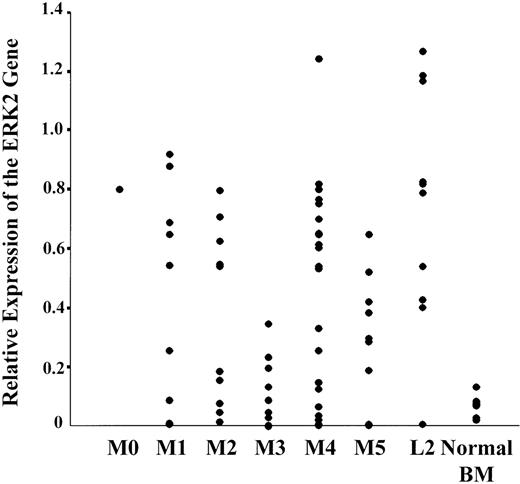
To examine the expression level of ERK in acute leukemia, we examined the ERK expression by semiquantitative RT-PCR and immunoblot analysis. For RT-PCR analysis, we examined the expression of ERK2, the more predominant ERK isoform in acute leukemia samples. Human K562 cells constitutively expressed the ERK2 gene and showed 10-fold higher ERK2 gene expression than normal BM samples. Therefore, we used the K562 cells as a positive control, and the value of the ERK2 gene expression divided by the β-actin gene expression was used for comparison with the expression of the ERK2 genes, with the value of gene expression in K562 cells defined as 1.00. Figure 2 is a schematic representation of the various expressions of the ERK2 gene in 79 acute leukemias. The average levels of ERK2 gene expression in acute leukemia samples (0.43 ± 0.01 SE) were significantly higher than those in normal BM samples (0.10 ± 0.01 SE) (P = .03). When the expression levels of ERK2 gene were analyzed according to the type of acute leukemia, there was no significant difference between AML and ALL. For AML, the average levels of ERK2 gene expression were lower in the M3 subtype than in other subtypes (P < .05). When correlation was examined to confirm that the quantitation of ERK gene expression by RT-PCR reflected the amount of ERK protein in the same leukemia samples, a strong positive correlation was observed between the levels of ERK2 gene expression quantified by RT-PCR and the levels of total ERK protein by immunoblot analysis (Pearson’s correlation coefficient, 0.437; P = .01).
Relative expression of the ERK2 gene in acute leukemia and normal BM samples. The value of ERK2 gene expression in individual samples was obtained as described in the text (defined in K562 cells as 1.00).
Relative expression of the ERK2 gene in acute leukemia and normal BM samples. The value of ERK2 gene expression in individual samples was obtained as described in the text (defined in K562 cells as 1.00).
To examine whether the increased amount of total ERK protein contributed to constitutive ERK activation in acute leukemias, we studied the relationship between ERK expression and ERK activation in 26 cases by immunoblot analysis. The expression of ERK in normal BM samples and purified CD34+ cells was minimal (Fig 1A). Hyperexpression of ERK protein was considered to have occurred when the value of the total ERK expression divided by the α-tubulin expression in the leukemia sample was greater than twofold the mean value measured in the normal BM samples.14 Hyperexpression of ERK in acute leukemias was detected in 14 of the 26 leukemia samples analyzed (53.8%) (Table 1) (Fig 1A and C). In 17 of 26 cases (65.4%), the pattern of ERK expression was similar to that of ERK activation. Although ERK activation appeared to be associated with total ERK, this relationship was not statistically significant (P = .19) (Table 3).
Correlation Between Activation and Expression of ERK
| ERK Activation . | No. . | ERK Hyperexpression . | |
|---|---|---|---|
| Positive3-151 . | Negative . | ||
| Positive3-150 | 19 | 12 | 7 |
| Negative | 7 | 2 | 5 |
| Total | 26 | 14 | 12 |
| ERK Activation . | No. . | ERK Hyperexpression . | |
|---|---|---|---|
| Positive3-151 . | Negative . | ||
| Positive3-150 | 19 | 12 | 7 |
| Negative | 7 | 2 | 5 |
| Total | 26 | 14 | 12 |
P = .19.
Defined as positive when the value of the ERK2 phosphorylation divided by the α-tubulin expression was greater than one third the value of K562 cell by immunoblot analysis.
Defined as positive when the value of the total ERK expression divided by the α-tubulin expression as twofold the mean value of normal BM samples by immunoblot analysis.14
Downregulation of ERK signaling pathway is compromised in acute leukemias.
The constitutive activation of ERK suggested to us the possibility of an abnormal downregulation mechanism of ERK. PAC1, a member of the ERK phosphatase family, was seen as a maximally expressed PTPase in the hematopoietic tissue and induced in response to ERK activation.18-21 Therefore, to determine whether abnormal ERK downregulation mechanisms were present, we examined PAC1 gene expression by semiquantitative RT-PCR because PTPases, including PAC1, are principally upregulated at the transcription level in response to ERK activation.28 Interestingly, the average levels of PAC1 gene expression in acute leukemias (0.23 ± 0.01 SE) were significantly lower than those in normal BM samples (0.58 ± 0.01 SE) (P = .002). The CD34+ cells showed a level of PAC1 expression similar to normal BM samples (Fig 1A). Then, to determine whether the downregulation of PAC1 contributed to the constitutive activation of ERK, we compared PAC1 expression with ERK activation in each leukemia sample. PAC1 was considered to be downregulated when the value of expression in the leukemia sample was less than one half of the mean value measured in the normal BM samples. Downregulation of PAC1 in acute leukemias was detected in 17 of the 26 cases analyzed (65.4%) (Table 1) (Fig 1A, C, and D). Furthermore, in 22 of 26 cases (84.6%), PAC1 downregulation was significantly correlated with ERK activation (P = .002) (Table 4).
Correlation Between ERK Activation and PAC1 Downregulation
| ERK Activation . | No. . | PAC1 Downregulation . | |
|---|---|---|---|
| Positive4-151 . | Negative . | ||
| Positive4-150 | 19 | 16 | 3 |
| Negative | 7 | 1 | 6 |
| Total | 26 | 17 | 9 |
| ERK Activation . | No. . | PAC1 Downregulation . | |
|---|---|---|---|
| Positive4-151 . | Negative . | ||
| Positive4-150 | 19 | 16 | 3 |
| Negative | 7 | 1 | 6 |
| Total | 26 | 17 | 9 |
P = .002.
Defined as positive when the value of the ERK2 phosphorylation divided by the α-tubulin expression was greater than one third the value of K562 cell by immunoblot analysis.
Defined as positive when the value of the PAC1 expression was less than one half the mean value of normal BM samples by semiquantitative RT-PCR.
DISCUSSION
The identification of constitutively activated signaling molecules involved in transducing an oncogenic signal in leukemia cells is likely to shed light on the mechanism of leukemogenesis. In this study, we focused on the analysis of ERK, which is a key kinase in intracellular signal transduction pathways for cell proliferation and differentiation. Our study showed a high frequency of activation of ERK in human acute leukemias, but we did not detect any, or only minimal, phosphorylation of ERK in normal BM cells. This is in agreement with a recent report that the constitutive activation of ERK occurs frequently in acute leukemia cells.13 Also, when we examined the activation of JNK (c-Jun NH2-terminal linases), a member of MAPK family, on the seven cases (22, 49, 72, 75, 77, 78, and 79), which showed constitutive ERK activation, none of these samples showed JNK activation (data not shown). This result led us to the conclusion that the ERK activation in acute leukemias is the ERK-specific event among various MAPK pathways. Until now, the activated status of ERK has been known to be predominantly determined by a highly conserved ERK cascade.9,10 Actually, in a previous study examining limited numbers of leukemia samples,13 ERK activation was accompanied by MEK activation. However, in our study examining 26 leukemia samples, about one half of the leukemia samples showed a relationship between ERK activation and MEK activation, but in the remaining samples, any relationship of activation between both kinases was not observed. Such a discrepancy was also observed in a previous study with renal cell carcinoma.14 Therefore, although MEK is the only activator, which is responsible for ERK activation,29 30 our results suggest that the constitutive activation of ERK observed in acute leukemia cells is unlikely to simply reflect phosphorylation of the protein by upstream kinases.
In our study, RT-PCR and immunoblot analysis showed a markedly increased expression of ERK in acute leukemia, compared with normal BM cells. Although there was no statistical significance, the pattern of ERK expression was similar to that of ERK phosphorylation in 17 of 26 cases (65.4%) examined by immunoblot analysis. These findings are consistent with a previous study of breast cancer, which demonstrated that the activity and expression of ERK was elevated in both primary and metastatic lesions.15 Whereas posttranslational ERK modification has been extensively studied, relatively little is known about altered ERK transcription. The variation of ERK mRNA levels in different organs suggests that a tissue-specific transcription factor may play an important role in the regulation of ERK expression.31 Also, in the cloning study of a murine ERK1 gene, the promoter of ERK1 contains consensus binding sites for many transcription factors, including AP-1, AP-2, Sp1, CTF-NF1, Myb, p53, NF-IL-6, and Ets-1.32 Genetic strategies should be relatively straightforward in determining which transcription factors, including tissue-specific factors, are important for ERK expression and if there are mutations in the promoter of the ERK gene, which might account for the hyperexpression of ERK in acute leukemia. Although the molecular basis for ERK hyperexpression must be determined, the increased amount of ERK protein available for phosphorylation by activated upstream kinases in acute leukemia could increase the amount of activated ERK and potentiate the proliferative capacity of leukemia cells.
ERK activation of acute leukemia samples in our study was considered to be constitutive because the response of ERK to a wide variety of extracellular stimuli showed transient phosphorylation and was followed by rapid dephosphorylation.2,31 Furthermore, MEK activation and ERK hyperexpression were not sufficient to explain the complete mechanism of constitutive activation of ERK in acute leukemia. These results raised the question of the abnormality of the downregulation mechanism of ERK activation. In the cell, a dynamic balance exists between phosphorylation and dephosphorylation, resulting from the interplay between protein kinase and protein phosphatase. Therefore, PTPases may play a distinct role in the regulation of the ERK signaling pathway. PAC1, a member of the MAPK phosphatase family, was described as a maximally expressed PTPase in the hematopoietic tissue.18-21 Abundant data indicate that constitutive activation of the ERK cascade increases the expression of PAC1, providing a pivotal role for this phosphatase in compensatory inactivation of the stimulated ERK signaling pathway because PAC1 exhibits stringent substrate specificity for ERK and constitutive PAC1 expression inactivates ERK.19,33-35 Furthermore, ERK phosphorylation and activity are modulated by amounts of PAC1 in transfection study using PAC1, thus highlighting a role for the MAPK cascade.19 Because PAC1 is principally upregulated at the transcription level in response to ERK activation,27 we chose to study the PAC1 gene expression. Surprisingly, PAC1 gene expression in acute leukemia samples showing constitutive ERK activation was below one half that of the basal, unstimulated, normal BM samples showing minimal or no ERK activation. Considering the constitutively activated status of ERK in acute leukemia samples, downregulation of the PAC1 gene in acute leukemia samples is an attractive finding, and this result suggests that induction of the PAC1 gene is definitely downregulated in acute leukemia. In this respect, our data provide the first evidence for downregulation of phosphatase induction in response to activation of ERK in human malignancies. Taken together, a significant correlation between PAC1 downregulation and ERK activation suggests that no matter what is stimuli for activating ERK, dysregulated switch-off mechanism for activated ERK contributes to the constitutive activation of ERK in acute leukemia. Current research in our laboratory is adopting a genetic strategy aimed at the introduction of PAC1 into leukemic cells showing constitutive ERK activation and downregulation of PAC1, which may allow us to determine whether the inhibition of constitutively activated ERK by overexpression of PAC1 influences the growth of leukemic cells.
Furthermore, we found the complementary roles of MEK activation, ERK hyperexpression, and PAC1 downregulation in determining the activation of ERK by the analysis of individual cases. We attempted to classify 26 cases into four groups. In group 1 (Fig 1A), 6 cases showed strong ERK phosphorylation by synergistic effect of MEK activation, ERK hyperexpression, and PAC1 downregulation. In group 2 (Fig 1B), 7 cases showed MEK activation, but not ERK activation, and 6 of 7 cases showed PAC1 expression similar to normal BM samples. Considering the role of MEK as a sole activator of ERK, inactivated ERK in these six cases may be the result of the preferential dephosphorylation of ERK by normally functional PAC1 under the affect of still-activated MEK. In group 3 (Fig 1C), 5 cases did not show MEK activation, although ERK was activated. These findings suggest the possibility of the presence of alternative ERK activators other than MEK1/2. However, another type of MEK has been not identified in vivo. Also, certain leukemic oncogene products have been not reported to activate ERK independent of MEK. Recently, a gain-of-function mutation of mammalian ERK2 (D319N ERK2) was demonstrated.36 This mutant was shown to have an increased sensitivity to lower levels of signaling in vivo as a result of decreased sensitivity to phosphatase, but such mutant has not as yet been found in human malignancy. However, on the basis of our results, four of these five cases showed PAC1 downregulation. These findings suggest that the downregulated status of PAC1 may be insufficient for dephosphorylation of ERK, and therefore, the ERK was maintained in an activated status despite dephosphorylation of MEK after withdrawal of stimuli. Also, interestingly, K562 cells showed the mechanism of ERK activation similar to this group. In group 4 (Fig 1D), seven cases showed ERK activation, but not ERK hyperexpression. All cases in this group showed PAC1 downregulation, and six cases showed MEK activation. Therefore, in this group, ERK may be constitutively activated by a combined effect of MEK activation and PAC1 downregulation despite minimal ERK expression. Although this aspect calls for further investigation, our present findings support the belief that the complementary role of the three different regulatory mechanisms could contribute to determining the constitutive activation of ERK in acute leukemia.
In conclusion, ERK and MEK were constitutively activated in a majority of human acute leukemias. Based on our data, in addition to activation by sequential upstream kinases, the hyperexpression of ERK and the downregulation of PAC1 are thought to contribute to constitutive ERK activation. Furthermore, the complementary relationship of these regulatory mechanisms may play an important role in constitutive ERK activation in acute leukemia.
The publication costs of this article were defrayed in part by page charge payment. This article must therefore be hereby marked “advertisement” in accordance with 18 U.S.C. section 1734 solely to indicate this fact.
REFERENCES
Author notes
Address reprint requests either to Seong-Cheol Kim, MD, Division of Hematology-Oncology, Department of Internal Medicine, Yonsei University College of Medicine, Seodaemun-Gu, Shinchon-Dong 134, Seoul 120-752, Korea; e-mail: seockim@chollian.net; or to Won-Jae Lee, PhD, Laboratory of Immunology, Medical Research Center, Yonsei University College of Medicine, CPO Box 8044, Seoul, Korea; e-mail:wjlee1@yumc.yonsei.ac.kr.


This feature is available to Subscribers Only
Sign In or Create an Account Close Modal